Anatomic Variant of Retrogressive Palmaris Longus: A Case Report
2 Department of Anaesthesiology, All India Institute of Medical Sciences, Rishikesh, Uttarakhand, India
Received: 04-Apr-2022, Manuscript No. ijav-22-4703; Editor assigned: 06-Apr-2022, Pre QC No. ijav-22-4703 (PQ); Reviewed: 25-Apr-2022 QC No. ijav-22-4703; Revised: 27-Apr-2022, Manuscript No. ijav-22-4703(R); Published: 30-Apr-2022, DOI: 10.37532/1308-4038.15(4).186
Citation: Suyashi S. Anatomic Variant of Retrogressive Palmaris Longus: A Case Report. Int J Anat Var. 2021;15(4):169-170.
This open-access article is distributed under the terms of the Creative Commons Attribution Non-Commercial License (CC BY-NC) (http://creativecommons.org/licenses/by-nc/4.0/), which permits reuse, distribution and reproduction of the article, provided that the original work is properly cited and the reuse is restricted to noncommercial purposes. For commercial reuse, contact reprints@pulsus.com
Abstract
An interesting case of unusual unilateral variant of palmaris longus tendon of forearm was noticed by us. We found two bellies of palmaris longus as well as their different insertions. These observations will help in understanding morphological variations of this muscle and its clinical implications. Palmaris longus is a fusiform muscle in the superficial flexor group of muscles of forearm. It originates from medial epicondyle of humerus by common flexor tendon. We found PL having one origin i.e. from medial epicondyle from common tendinous origin of flexor muscles and then it divided to form two bellies having two long tendons distally. Understanding of presence or absence or anomalies of PL is not only important for medical professionals but also for evolutionary biologists. Awareness of anatomy and variations of flexor tendons is important for health care practitioners for correct diagnosis and management of pain, disease and trauma of forearm and hand.
Keywords
Variation, Palmaris longus, Volkmann’s ischaemic contracture, Tendon graft
Introduction
The Palmaris longus muscle (PL) is a fusiform muscle. It lies in the superficial flexor group of muscles of forearm. It originates from medial epicondyle of humerus by common flexor tendon (along with flexor digitorum superficial muscle (FDS), flexor carpi ulnar is muscle (FCU) and flexor carpi radial is muscle (FCR)). After removing skin, subcutaneous tissue and fascia of anterior compartment of forearm, PL can be seen, lying superficial to FDS and between FCU and FCR muscle. It is directed downwards and outwards [1]. It has short muscular belly (up to mid forearm level) and then it takes the form of long and slender tendon. It then crosses in front of flexor retinaculum and is continuous with the central part of palmar Apo neurosis [2]. It is innervated by median nerve [3]. Anatomical awareness of structures of forearm and their relations is essential for clinicians and surgeons. As there are several patients of distal neuropathies, so this study will be useful for medical professionals for the correct diagnosis and treatment. We found a case of PL having two bellies and two insertions. Such a case has never been reported in India.
Case Report
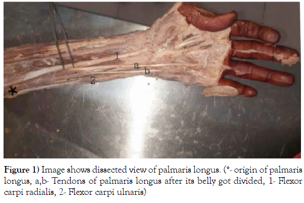
We found an interesting case of unusual unilateral variant of Palmaris longus tendon of forearm in male cadaver of Indian origin, during routine cadaveric dissection sessions of MBBS students in Department of Anatomy, All India Institute of Medical Sciences, Jodhpur. During dissection of flexor compartment of forearm after removing skin and superficial fascia, PL was seen. This structure was traced from origin to insertion. Due to its anatomical location and tendinous insertion, it was identified as PL. We found PL having one origin i.e. from medial epicondyle from common tendinous origin of flexor muscles and then it divided to form two bellies having long tendon distally. Medial and lateral tendons were inserted medially into the fourth slip and medial aspect of third slip of FDS respectively (Figure1). Both the bellies were innervated by branches of median nerve.
Discussion
Important observations were done by us in our case report. We found two bellies of PL as well as their different insertions. These observations will help in understanding morphological variations of PL muscle and its clinical implications.
Embryologically, during 7th week of intrauterine life, forearm flexor muscles develop from a mesodermal condensation of dorsolateral cells of somite’s which then migrate into the limb bud. Further these mesenchyme cells undergo division and form superficial and deep group of flexor muscles of forearm. During development if an additional rift is present in the superficial forearm flexor mass, then this develops into additional tendon of PL [4]. Morphogenetically, development of tendon and muscle of PL was regulated by HOX gene [5].
PL is a muscle with short belly and long tendon, so it is phylogenetically considered as a retrogressive muscle. And its absence in humans is common. Going back to evolution, PL was well developed in the species who used their forelimbs for walking and weight bearing. So, it was well developed in mammals [6]. While according to some other researches, PL is well developed in species with a significantly higher ratio of upper limb weight to body weight. This ratio is very low in humans and hence PL is less developed and also its function is accessory. As the species evolved, forelimbs too evolved to a prehensile organ. Long flexor muscles of forearm began to undergo partial atrophy caudocranially [7]. And intrinsic muscles of hand took over the functions of PL. This resulted in partial degradation of PL and was replaced by fibroaponeurotic palmar aponeurosis. Further with the evolution of bipedal gait, degeneration of PL continued. PL finally evolved as a muscle with small belly and long tendon. Its function became rudimentary. Now it functions as accessory flexor for wrist and metacarpophalangeal joints. It helps in slight amplification of palmar grip. It acts as an anchor of palmar fascia. It tauts the skin and palmar fascia of hand. And therefore, shear the forces to palmar aponeurosis in distal direction. It may assist in thumb abduction movement.
PL has great clinical importance. It is used for cosmetic and reconstructive purposes. Plastic surgeons use it for tendon transplantation.
Otorhinolaryngologists and ophthalmologists use this tendon as graft for surgeries like lip augmentation, ptosis correction and treatment of facial paralysis [8]. It is preferred as tendon donor because it is easily approachable (because of its superficial location) and it satisfies the criteria of required diameter, length and availability and can be used without leading to any complication or malfunctioning. It also helps in recognizing median nerve during surgeries.
Variants of PL can cause Volkmann’s ischemic contracture, Carpal tunnel syndrome, Guyon’s syndrome and Dupuytren’s contracture due to compactness of tendons n nerves in anterior compartment of forearm. This leads to pain and numbness in the region of median nerve in hand and ape-thumb like deformity. Person having PL variation have pain in wrist on doing repetitive hand movement (like mechanics) due to median nerve compression. Anomalies of PL makes electro-myographical studies of median nerve at wrist and in endoscopic wrist procedures difficult [9]. According to some studies variants of PL can increase the pinch strength of fingers of hand [10].
Conclusion
Knowledge of variations of PL variations is crucial not only for the anatomists but also plastic surgeons, pain physicians, radiologists and orthopedists for correct diagnosis and treatment of disease or trauma of forearm and hand. Understanding of presence or absence or anomalies of PL is not only important for medical professionals but also for evolutionary biologists.
REFERENCES
- Natsis K, Levva S, Totlis T. Three-headed reversed palmaris longus muscle and its clinical significance. Ann Anat. 2007; 189(1): 97-101.
- Standring S. Gray’s Anatomy: The Anatomical Basis of Clinical Practice. 40th ed. London: Elsevier; 2008:847.
- Yammine K. Clinical prevalence of palmaris longusagenesis: a systematic review and meta-analysis. Clin Anat. 2013;26(6):709-18.
- Iqbal S, Iqbal R, Iqbal F. A bitendinous palmaris longus: Aberrant insertions and its clinical impact- A case report. 3 J Clin Diagnostic Res. 2015; 9(5): 3-5.
- Angelini Junior LC, Angelini FB, de Oliveira B. Use of the tendon of the palmaris longus muscle in surgical procedures: Study on cadavers. Acta Ortop Bras. 2012; 20(4): 226-9.
- Sharma DK, Shukla CK, Sharma V. Clinical assessment of absence of palmaris longus muscle and its association with gender, body Sides, handedness and other neighboring anomalies in a population of central India. J Anat Soc India. 2012; 61:13-20.
- Kumar P. Duplication of palmaris longus muscle. Int J Anat Var. 2013; 6:207–9.
- Ioannis D, Anastasios K, Konstantinos N. Palmaris longusmuscle’s prevalence in different nations and interesting anatomical variations: Review of the literature. J Clin Med Res. 2015; 7(11): 825-30.
- Park MJ, Namdari S, Yao J. Anatomic variations of the palmaris longus muscle. Am J Orthop. 2010; 39: 89-94.
- Cetin A, Genc M, Sevil S, Coban YK. Prevalence of the palmaris longusmuscle and its relationship with grip and pinch strength: A study in a Turkish pediatric population. Hand (NY). 2013; 8(2):215-20.
Google Scholar, Indexed at, Crossref
Google Scholar, Indexed at, Crossref
Google Scholar, Indexed at, Crossref
Google Scholar, Indexed at, Crossref
Google Scholar, Indexed at, Crossref


