Anatomical variation in the origin of superior thyroid artery
2 Department of Radiology, Akdeniz University, Antalya, Turkey
3 Faculty of Nursing, Akdeniz University, Antalya, Turkey
Citation: Sindel M, Özgür O, Aytaç G, et al. Anatomical variation in the origin of superior thyroid artery. Int J Anat Var. 2017;10(3):56-7.
This open-access article is distributed under the terms of the Creative Commons Attribution Non-Commercial License (CC BY-NC) (http://creativecommons.org/licenses/by-nc/4.0/), which permits reuse, distribution and reproduction of the article, provided that the original work is properly cited and the reuse is restricted to noncommercial purposes. For commercial reuse, contact reprints@pulsus.com
Abstract
The Superior Thyroid Artery (STA) is the dominant arterial supply of the thyroid gland, larynx and the neck. Awareness of variations in STA is crucial for invasive operations in the neck region. We reported a case that STA derives from the Common Carotid Artery (CCA). A patient with possible transient ischemic attack had an digital substraction angiography. Angiography revealed superior thyroid artery derived from the common carotid artery in the left.
Keywords
Superior thyroid artery; Angiography; Variation
Introduction
The superior thyroid artery arises from the anterior surface of the ECA as a first branch, just below the level of the greater cornu of the hyoid bone and lies anterior and inferior. It gives the superior infrahyoid, sternocleidomastoid, superior laryngeal, cricothyroid, glandularis anterior, posterior and lateralis branches. Than it enters the superior pole of the thyroid gland anteromedially. The superior thyroid artery is generally considered to be always present. It was reported to arise from the common carotid in 18% of cases, the point of division of the common carotid in 36%, or from the external carotid in 36% of cases [1]. The common carotid artery has no branches and anatomic variations of this artery are rare. One of these variatios is the superior thyroid artery which arises from the common carotid artery. Knowledge of the variations of the superior thyroid artery is very important for neck dissections, surgeries, catheterizations, aneurysms, carotis endarterectomy surgeries.
Case Report
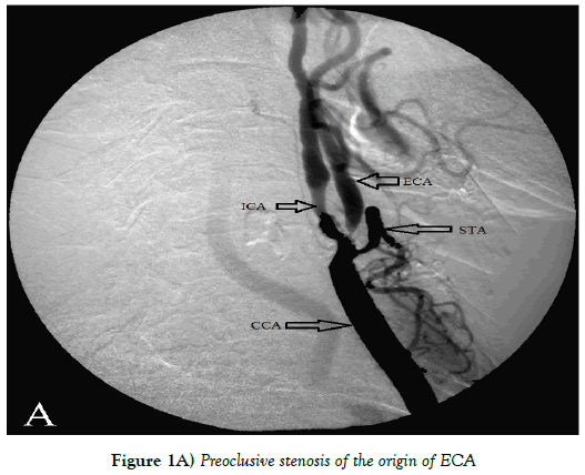
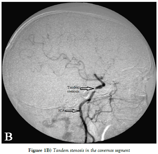
A 74 year-old female with possible transient ischemic attack visited Akdeniz University Hospital and underwent digital substraction angiography for evaluation of cerebrovascular disease. Angiography showed a stenosis in the distal part of left common carotid artery and proksimal parts of the internal and external carotid arteries with an ulcerated plaque. 95% preoclusive stenosis of the origin of ECA is detected (Figure 1A). ICA was narrowed and had tandem stenosis in the cavernos segment (Figure 1B). ICA continued with narrowed middle cerebral artery. Vertebro-basilar system was normal. The anterior part of the brain was supplied by the right internal carotid system mostly.
Figure 1A: Preoclusive stenosis of the origin of ECA
Figure 1B: Tandem stenosis in the cavernos segment
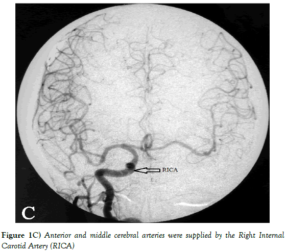
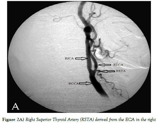
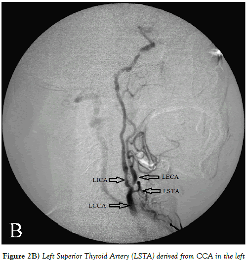
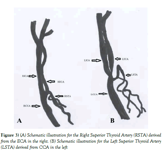
Angiographic examination taken from right common carotid artery catheterization revealed both anterior cerebral arteries and middle cerebral arteries were supplied by the right common carotid artery (Figure 1C). It was viewed that left anterior cerebral artery and left middle cerebral artery both were filled by anterior comminican artery. Angiography and schematic illustration revealed superior thyroid artery derived from the ECA in the right (Figures 2A and 3A), CCA in the left (Figures 2B and 3B). Superior thyroid artery was protected because it derived before the oclusion.
Figure 2a: Right Superior Thyroid Artery (RSTA) derived from the ECA in the right
Figure 2b: Left Superior Thyroid Artery (LSTA) derived from CCA in the left
Discussion
The anatomical variations of superior thyroid artery have been described in the literature. Knowledge of these variations is crucial for decreasing morbidity during the surgeries [2]. Lucev showed in 47.5%, Hollinshead in 16%, Banna and Lasjaunias in 10%; STA was arising from CCA [2-4]. Ongeti reported in 13.1% of 46 cadavers STA derived from CCA [5]. Lo studied with 36 cadavers and they showed in 1.5% STA derived from CCA [6]. Origin of STA is predictable, arising from ECA in more than 70% cases but like all the previous studies and like our case STA could arise from CCA. Awareness of this could be important during invasive surgeries of this area. Gupta evaluated 25 angiograms, including 14 rights and 11 left. On the right side, STA was noted to arise from ECA in 10 (71.5%), bifurcation of CCA in 3 (21.5%), and CCA in 1 (7%) patient. Left STA was seen to arise from ECA in 8 (72.5%), bifurcation of CCA in 2 (18.5%), and ICA in 1 (9%) patient [7]. Adachi reported that in their study STA derived from CCA in 4.7% in the right, in 22% in the left [8]. Anagnostopoulou studied with 68 cadavers. They showed that in their study STA derived from CCA in 19.1% in the right, in 25% in the left [9]. Ozgur studied in 20 Anatolian cadavers. In this study they reported STA originated from CCA in right and left respectively 15%, 55% [10].
In our case STA was arising from CCA before the preoclusive stenosis with ulcerated plaque, only in the left. Except this because of the oclusion frontal part of the brain was supplied by the right internal carotid system mostly. Knowledge of possible anatomic and pathological variations of the STA is very important for safe catheterization and approaches in neck area.
Author’s Contribution
Ozhan Ozgur and Timur Sindel developed the idea and protocol, collected the medical records of cases. Gunes Aytac, Muzaffer Sindel and Ozlem Kastan contributed to development, abstracted data, reviewed published articles and prepared and wrote the manuscript. All authors contributed to the work presented in this paper.
REFERENCES
- Bergman RA, Afifi KA, Miyauchi R. Illustrated encyclopedia of human anatomic variation. 2015.
- Hollinshead WH. Head and Neck, in anatomy for surgeons. Hoeber Harper International: Philadelphia. 1996;564-76.
- Banna M, Lasjaunias P. The arteries of the lingual thyroid: angiographic findings and anatomic variations. AJNR Am J Neuroradiol. 1990;11:730-2.
- Lucev N. Variations of the great arteries in the carotid triangle. Otolaryngol Head Neck Surg. 2000;122:590-1.
- Ongeti KW, Ogeng'o JA. Variant origin of the superior thyroid artery in a Kenyan population. Clin Anat. 2012;25:198-202.
- Lo A. Anatomical variations of the common carotid artery bifurcation. ANZ J Surg. 2006;76:970-2.
- Gupta P. Variations in superior thyroid artery: A selective angiographic study. Indian J Radiol Imaging. 2014;24:66-71.
- Adachi B. The arterial system of Japanese, in Head and Neck. Kenkyusha. 2013;58-62.
- Anagnostopoulou S, Mavridis I. Emerging patterns of the human superior thyroid artery and review of its clinical anatomy. Surg Radiol Anat. 2014;36:33-8.
- Ozgur Z. Clinically relevant variations of the superior thyroid artery: an anatomic guide for surgical neck dissection. Surg Radiol Anat. 2009;31:151-9.


