Anatomical variation of Jugulo-subclavian venous confluence
Received: 12-Oct-2018 Accepted Date: Oct 30, 2018; Published: 09-Nov-2018
Citation: Donkor YO, Maalman RSE, Ayamba AM. Anatomical variation of Jugulo-subclavian venous confluence. Int J Anat Var. Dec 2018;11(4):125-126.
This open-access article is distributed under the terms of the Creative Commons Attribution Non-Commercial License (CC BY-NC) (http://creativecommons.org/licenses/by-nc/4.0/), which permits reuse, distribution and reproduction of the article, provided that the original work is properly cited and the reuse is restricted to noncommercial purposes. For commercial reuse, contact reprints@pulsus.com
Abstract
Variations of superficial veinous dranaige are very common in the head and neck regions. The jugular veins display disparities in its development and pathway. During a routine dissection study with the medical students of an adult male cadaver (head and neck regions) in the Anatomy laboratory, School of Medicine, a variation of the jugular veins communicating with subclavian vein to form jugulo-subclavian trunk/confluence draining into brachiocephalic vein was found on right side of the body. It was studied and photographed. The jugular veins joined in the cervical region and pass under the posteromedial border of clavicle to communicate with the subclavian vein as brachiocephalic vein. No abnormality was detected on the left cervical region. These venous vessels; the subclavian and jugular are preferred sites for long term central venous access. Therefore knowledge of these venous variations is not only of importance to anatomists but surgeons and clinicians performing catheterization and surgical procedures in the region of the neck, vascular or reconstructive surgery. This prevents inadvertent injury during surgery and venepuncture at the neck region.
Keywords
Subclavian vein; External jugular vein; Internal jugular vein; Jugulo-subclavian; Venous confluence; Brachiocephalic vein; Variation
Introduction
The word jugular means neck. The cervical region is composed of both superficial and deep veins draining the region. Superficial veins lie within the superficial fatty layer (camper’s) where as deep veins lie to the deep fascia of the muscle layer. Superficial veinous drainage of the neck drain into external jugular veins (EJV) and deep veinous drainage of the neck drain into both subclavian vein (SCV) and internal jugular vein (IJV). The jugular veins exit the skull bilaterally through the jugular foramina. External jugular vein drains into the subclavian vein which unites with IJV to form brachiocephalic veins (BV) that drains into the superior vena cava (SVC). [1,2]. Jugular veinous system display a wide range of embryological variations in terms of development, pathways and tributaries [3].
Case Report
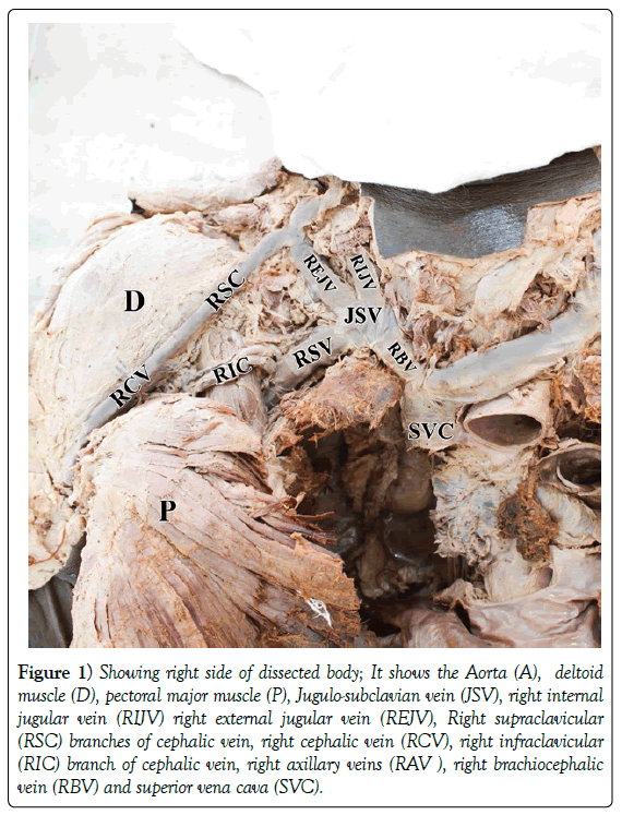
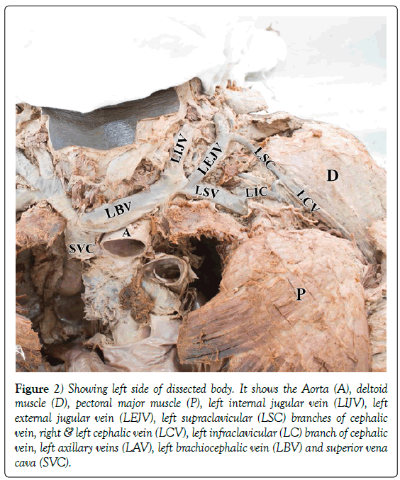
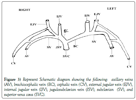
During routine dissection for undergraduate medical students in the Department of Anatomy, School of Medicine, University of health and Allied Sciences, UHAS, Volta Region, Ghana, an anatomical variation of jugulo-subclavian venous (JSV) confluence was observed at the right side of the neck region in a middle aged male cadaver (Figures 1 and 2). The neck region was carefully dissected and the veins neatly separated. In addition, a sketch (Figure 3) was made for clearer view. The anomalous confluence was studied in detail and photographed (Figure 1). EJV communicates with IJV as they course superficially to the sternocleidomastoid muscle to join with subclavian vein forming jugulo-subclavian venous (JSV) confluence draining into right brachiocephalic vein. No anomaly was observed on the left side (Figure 2).
Figure 1) Showing right side of dissected body; It shows the Aorta (A), deltoid muscle (D), pectoral major muscle (P), Jugulo-subclavian vein (JSV), right internal jugular vein (RIJV) right external jugular vein (REJV), Right supraclavicular (RSC) branches of cephalic vein, right cephalic vein (RCV), right infraclavicular (RIC) branch of cephalic vein, right axillary veins (RAV ), right brachiocephalic vein (RBV) and superior vena cava (SVC).
Figure 2) Showing left side of dissected body. It shows the Aorta (A), deltoid muscle (D), pectoral major muscle (P), left internal jugular vein (LIJV), left external jugular vein (LEJV), left supraclavicular (LSC) branches of cephalic vein, right & left cephalic vein (LCV), left infraclavicular (LC) branch of cephalic vein, left axillary veins (LAV), left brachiocephalic vein (LBV) and superior vena cava (SVC).
Discussion
Superficial veins of the neck show a broad spectrum of embryological variations with respect to their formation, course and termination [4]. The embryological development of the veinous system of the, face, scalp and neck is still ambiguous and its variations are still not clearly understood [5]. The jugular vein is clinically important in view of its significant anatomical variations.
Normal anatomical orientation of the EJV directly opens into subclavian vein which then meets the IJV to form brachiocephalic vein (Figure 3). In the present cause, EJV joins and anastomosed with IJV before terminating with subclavian vein (Figure 3). The development of veins of the scalp, face and neck has not been clearly understood with paucity of information on the course [5].
The EJV has been one of the common veins reported to have variations to it formation and pathway. It has been reported that EJV pathway drains into either internal jugular veins or subclavian veins [6]. In a dissection study of 50 cadavers, 100 EJV, 60% cases of EJV drains into jugulo-subclavian venous confluence, 36% of external jugular vein drains into SCV at a length away from internal jugular while 4% drains into IJV [2].
Cornelius and Penelope [7] and Skandalakis [8] have reported that superficial venous system of EJV in its course generally communicates with the superior part of the IJV and drains through series of tributaries in the neck region. The clavicle is encircled by anastomosing circular veins consisting of EJV joining with IJV superiorly and subclavian inferiolaterally forming the jugulosubclavain trunk terminating into the brachiocephalic vein inferiomedially [5].
Clinically, injury to the clavicle resulting in fracture may damage jugulosubclavian venous, leading to profuse bleeding [9]. Masses of hematomas formed are likely to cause inflammation which will block and shunt blood supply to th neck region. Therefore, the location of JSV and it topographical relationships with bigger vessels like the brachiocephalic vein and superior vena cava may also intenisfy the danger of critical emergency suitation during head and neck surgical procedure [10]. Furthermore, during cardiac catherization procedure there is a possibility that unexpected diversion of catheter through the EJV may take place in such instance.
Access to the central venous system is highly recommended if peripheral access fails. The suitable location for central venous access is subclavian vein, internal jugular and external jugular veins [1].
Conclusion
The present case report revealed internal jugular vein (IJV) external jugular vein (EJV) and subclavian (SCV) confluence of the jugulo subclavian venous draining into brachiocephalic vein are extremely uncommon and has got tremendous clinical significance for physicians and surgeons. This is because the external jugular vein (EJV) is usually preferred for numerous surgical procedures, in vitro diagnostic and treatment protocol. In addition, EJV is mostly used in vascular or reconstructive surgery in the face and neck region. Knowledge about these unexpected variations prevents inadvertent injury. Finally, this case report will inform and guide surgeons during catheterization and cannulation during surgery and emergencies.
Acknowledgement
We are grateful to the technicians (Mr. Bewu and Mr. Ahoga) in the Department of Basic Medical Science, Anatomy Laboratory, School of Medicine, UHAS, Ho, for their cordial help in preparing the laboratory and cadevers for dissection. Our final appreciation goes to the class of 2021 medical Students School of Medicine, UHAS, for their zeal, eager and cooperation during dissection.
REFERENCES
- Dhivya S, Chitra S. External jugular vein communicating with internal jugular vein-a case report. J Med Dental Sci. 2013;30:5512-6.
- Deslaugiers B, Vaysse P, Combes JM, et al. Contribution to the study of the tributaries and the termination of the external jugular vein. Surg Radiol Anat. 1994;16:173-7.
- Prakash R, Prabhu LV, Kumar J, et al. Variations of jugular veins: Phylogenic correlation and clinical implications. Southern Med J. 2006;99:1146-8.
- Tuli A, Choudhry R, Agarwal S, et al. Facial vein draining into external jugular vein in humans: Its variations, phylogenetic retention and clinical relevance. Surg Radiol Anat. 2003;25:36-41.
- Choudhry R, Tuli A, Choudhry S. Facial vein terminating in external jugular vein: An embryonic interpretation. Surg Radiol Anat. 1997;19:73-7.
- Bergman RA, Afifi AK, Miyauchi R. Illustrated encyclopedia of human anatomic variation: Opus II: cardiovascular system: veins: Head, neck and thorax. 2011.
- Cornelius R, Penelope GR. Hollinshead’s Textbook of Anatomy. Lippincott-Raven publishers. 1997;81.
- John ES. Skandalakis’ Surgical Anatomy. Paschalidis Medical Publications. 2004;37.
- Anastasopoulos N, Paraskevas G, Apostolidis S, et al. Three superficial veins coursing over the clavicles: A case report. Surg Radiol Anat. 2015;37:1129-31.
- Patil RA, Rajgopal L, Lyer P. Absent external jugular vein: Ontogeny and clinical implications. Int J Anat Var. 2013;6:103-5.


