Anatomical Variations in Sternocleidomastoid: A Series of 3 Cases
Received: 23-Jul-2020 Accepted Date: Nov 12, 2020; Published: 19-Nov-2020, DOI: 10.37532/1308-4038.20.13.31
This open-access article is distributed under the terms of the Creative Commons Attribution Non-Commercial License (CC BY-NC) (http://creativecommons.org/licenses/by-nc/4.0/), which permits reuse, distribution and reproduction of the article, provided that the original work is properly cited and the reuse is restricted to noncommercial purposes. For commercial reuse, contact reprints@pulsus.com
Abstract
Sternocleidomastoid is an important muscle of head and neck lying in close proximity to the vital structures like spinal accessory nerve, external jugular vein, carotid sheath with its contents, greater auricular nerve, facial and lingual veins, cervical plexus, vagus nerve, ansa cervicalis etc. It originates as two heads, one from the superolateral part of manubrium sterni in form of a tendon (sternal head) and another from medial one-third of superior surface of clavicle which is a musculotendinous structure (Clavicular head). The muscle is inserted into lateral aspect of mastoid process with the help of thick tendon and into lateral half of superior nuchal line by a thin aponeurosis.
Keywords
Ectopic opening; Lacrimal canaliculi
Introduction
Sternocleidomastoid is an important muscle of head and neck lying in close proximity to the vital structures like spinal accessory nerve, external jugular vein, carotid sheath with its contents, greater auricular nerve, facial and lingual veins, cervical plexus, vagus nerve, ansa cervicalis etc. It originates as two heads, one from the superolateral part of manubrium sterni in form of a tendon (sternal head) and another from medial one-third of superior surface of clavicle which is a musculotendinous structure (Clavicular head). The muscle is inserted into lateral aspect of mastoid process with the help of thick tendon and into lateral half of superior nuchal line by a thin aponeurosis. We report a series of 3 cases where variations were noted along both origin and insertion of SCM muscle [1].
Case Report
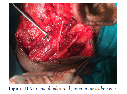
During a neck dissection of a 49-year-old male patient who was a case of well differentiated squamous cell carcinoma of left buccal mucosa in the Department of Oral and Maxillofacial surgery, A.C.P.M Dental College and Hospital, Dhule, a rare variation was found towards the end of insertion of SCM muscle. Skin, superficial fascia, platysma, deep fascia was removed and the muscles of neck were exposed [2]. The variation that was encountered was an unusual split towards the insertion end of SCM just above the point where lesser occipital nerve communicates with greater auricular nerve. Normally, the muscle along its insertion is a continuous band which gets inserted into lateral aspect of mastoid process and the occipital bone. This unusual split gave rise to a triangular space superiorly and two bellies which are assumed to be inserted into mastoid and occipital bone separately. The exact attachments were not exposed as it was not the need of the operative procedure. Also, it can always be suspected that the same variation could be bilateral [3].
The boundaries of the so formed triangle can be marked as it is bounded anteroinferior by carotid triangle and its contents, superiorly by the mastoid process and part of occipital bone and posteroinferiorly by occipital part of posterior triangle of neck. Superficially, this space is related to skin, superficial fascia,platysma, external jugular vein, part of posterior division of retromandibular and posterior auricular veins, some part of lesser occipital nerve. Deep relations include superior part of carotid sheath along with its contents, posterior belly of digastric, internal jugular vein [4].
All the variations of SCM can be attributed to the unusual splitting in the mesoderm of the posterior sixth branchial arch during organogenesis. It is however also possible that such variations are evolutionary in nature. In most cases, these variations go unnoticed and do not produce any symptoms. But this may cause functional deficits as the muscle lies in close proximity to various neurovascular structures. SCM has many vital structures under its andthus provides physical protection to them. Thus, all these variations may or may not just compromise this protective function which is more relevant in this case.
Case Report (2)
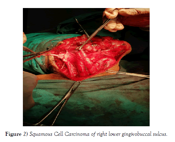
During a routine neck dissection of a 55 year old male patient who was a diagnosed case of Well Differentiated Squamous Cell Carcinoma of right lower gingivobuccal sulcus in the Department of Oral and Maxillofacial Surgery, A.C.P.M Dental College and Hospital, Dhule, a variation in the anatomy of SCM that was encountered was an additional clavicular head which originated from superior surface of middle third of clavicle as a fresh belly and blended with the fibres of normal clavicular head of the muscle.
The size of the extra clavicular head was almost the half of normal clavicular head [5].
The presence of additional heads of SCM have previously been reported unilaterally as well as bilaterally. Some have also reported separate cleido-occipital muscle. The presence of additional bellies bilaterally has been reported by Nayak et al. (2006) and Ramesh et al. (2007). The knowledge of variations of sternocleidomastoid
muscle is important for head and neck surgeons. It is also useful for the plastic surgeons. The sternocleidomastoid muscle can be used in several ways during surgery. Conley & Gullane (1980) have explained various uses of the muscle such as a). its use along with a part of clavicle to reconstruct mandible, b). reconstruct mandibular defects, c). transportas a myocutaneous flap for reconstruction to protect carotid and innominate arteries.
The additional head reported by us may not have any functional advantage on the movement of the neck. Since it covers the movement of the neck. Since it covers the lower part of the neck, it might cause difficulties in the surgeries in that region. It may also interfere in invasive techniques. Plastic surgeons can make best use of this Plastic surgeons can make best use of this [6].
Case Report (3)
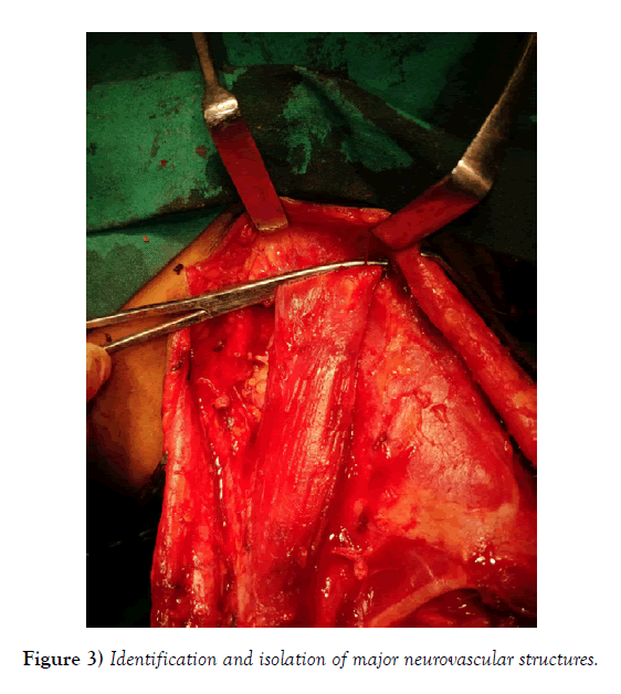
During a routine neck dissection of a 39 year old male who was a diagnosed case of well differentiated squamous cell carcinoma of left buccal mucosa, left lower lip, alveolus, in the Department of Oral and Maxillofacial Surgery, A.C.P.M Dental College, Dhule, a rare variation was encountered in the anatomy of Sternocleidomastoid of right side as there was no division of the muscle towards its end of origin into separate sternal and clavicular heads which is a normal feature of the muscle. Such a division normally produces a lesser supraclavicular fossa housing important neurovascular structures like lower part of Internal Jugular Vein and Subclavian vein.
The absence of such a split would result in complete obliteration of the lesser supraclavicular fossa thereby altering the normal anatomy of the same. The major neurovascular structures present beneath may be positioned abnormally at least expected sites. Now such a phenomenon will significantly cause trouble in routine procedures like I.J.V cannulation where lesser supraclavicular fossa is taken as a guide to perform the procedure. Also, surgeons may accidently encounter such a variation in anatomy which maylead to great difficulties during the procedure in identification and isolation of major neurovascular structures.
Financial support and sponsorship
No funding was received for this study
Conflicts of Interest and Informed Consent
There is no conflict of interest. This article does not contain any studies with animals performed by any of the authors. All procedures performed in studies involving human participants were in accordance with the ethical standards of the institutional and/or national research committee and with the 1964 Helsinki declaration and its later amendments or comparable ethical standards. Informed consent was obtained from all individual participants included in the study.
REFERENCES
- Bergman RA, Thomson SA. Compendium of Anatomic Variation, In:Muscles,Urban and Schwarzenber, Baltimore,1988.
- Stranding S, Berkovits BKB, Hackney CM, et al. TheAnatomical basis of clinical practice. 39th Churchill and Livingstone, Edinburg, 2005.
- Cherian SB, Nayak S. A case of unilateral third head of sternocleidomastoid muscle. Int J Morphol. 2008;26:99-101.
- Saxena A, Prasad A, Sood K. Unilateral four heads of sternocleidomastoid muscle: A rare case report. Eur J Anat. 2013;17:186-9.
- Rao R,Vishnumaya G, Shetty P, et al. Variation in the origin of Sternocleidomastoid Muscle. A case reports. Int J Morphol. 2007;25.
- Anil A, Kastamoni Y, Anil F, et al. Variation of bilateral multiheaded sternocleidomastoid muscle. Gazi Med J. 2017;28:56-7.


