Anatomical Variations of the Circle of Willis - Evaluation with Computed Tomographic Angiography
2 MD Additional professor, Department of Radiodiagnosis, IGIMS, Bihar, India
3 MD, Additional Professor, Department of Anatomy, IGIMS,Bihar, India
4 MS, Senior Resident, Department of Anatomy, ESIC MCH, Aryabhatt Knowledge University Bihar, India
5 MD, Additional Professor, Department of Radiodiagnosis, IGIMS, Bihar, India
Received: 01-Jul-2022, Manuscript No. ijav-22-5129; Editor assigned: 04-Jul-2022, Pre QC No. ijav-22-5129 (PQ); Accepted Date: Jul 21, 2022; Reviewed: 18-Jul-2022 QC No. ijav-22-5129; Revised: 21-Jul-2022, Manuscript No. ijav-22-5129 (R); Published: 28-Jul-2022, DOI: 10.37532/1308-4038.15(7).209
Citation: Kumari P, Prasad U, Kumar B, Nitisha, Kumar A, et al. Anatomical Variations of the Circle of Willis - Evaluation with Computed Tomographic Angiography. Int J Anat Var. 2022;15(7):194-203.
This open-access article is distributed under the terms of the Creative Commons Attribution Non-Commercial License (CC BY-NC) (http://creativecommons.org/licenses/by-nc/4.0/), which permits reuse, distribution and reproduction of the article, provided that the original work is properly cited and the reuse is restricted to noncommercial purposes. For commercial reuse, contact reprints@pulsus.com
Abstract
Objective: To study anatomical variations and anomalies of the Circle of Willis using computed tomographic angiography.
Material and methods: Observational and analytical study done in 100 patients from Novemeber 2019 to September 2021. Computed tomographic data of 100 patients was collected from the department of radiodiagnosis, Indira Gandhi Institute of Medical Sciences Patna along with the interpretation of senior consultant radiologist and data interpretated.
Conclusion: Among the 100 cases normal Circle of Willis were seen in 33% and variations seen in 67% of cases. We observed 25 different types of variations some of which are isolated and and some in combination with others. A wide variability in the posterior arterial component of the Circle of Willis was observed.
Keywords
Corcle of willis; Variations; Angiography; Computed tomography
INTRODUTION
Brain receives blood from two internal carotid and two vertebral arteries, connected by a central anastomosis i.e. circulus arteriosus cerebri, which is located in the subarachnoid space within the interpeduncular fossa at the base of brain. This polygonal arterial circle called ‘Circle of Willis’ was named on the honour of English doctor, Sir Thomas Willis (27 January 1621 - 11 November 1675), who played an important part in the history of anatomy and neurology. He was a pioneer in research into the anatomy of the brain, muscles and nervous system. The anatomy of the brain and nerves, as described in Thomas Willis’s book, Cerebri anatome cui accessit Nervorum descriptio et usus (1664; “Anatomy of the Brain, with elaborate Description of the Nerves and Their Function”), was the most complete and accurate account of the nervous system to that time [1].
The internal carotid artery (ICA) bifurcates at the medial end of the Sylvian fissure into the anterior cerebral artery (ACA) and the middle cerebral artery (MCA). The ACA courses anteromedially to cross over the optic nerve and optic chiasma to communicate with the opposite side of the anterior cerebral artery through the anterior communicating artery (ACOM). The segment which extends from origin to the junction with anterior communicating artery, is referred to as the A1 segment [2]. The anterior communicating (ACOM) artery is situated above the optic chiasma2 and below the rostrum of the corpus callosum. The anterior communicating artery (ACOM) is a small connecting artery between the paired anterior cerebral arteries and provides an important anastomotic channel for collateral circulation through the Circle of Willis [3] (CW). The important perforating arteries supplying the optic chiasm, infundibulum, subcallosal area, and preoptic area of the hypothalamus may arise from the anterior communicating artery and also from fenestrated anterior communicating artery. Internal carotid artery also gives rise to anterior choroidal (Ach), ophthalmic arteries and posterior communicating (PCOM) at the base of the brain. From each internal carotid artery (ICA), a posterior communicating artery arises at the anterior perforated substance and runs back through the interpeduncular cistern to join the ipsilateral posterior cerebral artery (PCA).
The left and right vertebral arteries come together at the level of the pons to form the midline basilar artery. This basilar artery lies in a shallow depression, the basilar sulcus, on the anterior/ ventral surface of the pons in the prepontine cistern [4]. It continues superiorly to terminate in the interpeduncular cistern by dividing into the right and left posterior cerebral artery [5].
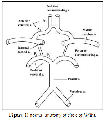
Circle of Willis comprises of:
Anterior circulation
• Left and right internal carotid arteries (ICA).
• Horizontal pre-communicating (A1) segments of the left and right anterior cerebral arteries (ACA).
• Single anterior communicating artery (ACOM).
Posterior circulation
• Left and right posterior communicating arteries (PCOM).
• Horizontal pre-communicating (P1) segments of left and right posterior cerebral arteries (PCA).
• Single basilar artery (tip) [6].
NOTE: although some consider the PCOM to be anterior circulation (Figure 1).
The anterior cerebral arteries travel toward the anteromedial aspects of the interhemispheric fissure of the brain, supply the midline regions of the frontal lobe, parietal lobe and cingulate cortex as well as the corpus callosum [7] through their central and cortical branches. The posterior cerebral arteries supply the occipital lobe of the brain, the inferior aspect of the temporal lobes, midbrain, thalamus and choroid plexus of the third and lateral ventricles [7] through their central and cortical branches. Circle of Willis give rise to several small perforating (central) arteries, many of which enter into the brain through the anterior and posterior perforated substances, which are areas of grey matter located at the base of the forebrain and the interpeduncular fossa respectively. The perforating arteries supply structures located on the ventral surface of the brain such as the optic chiasm, pituitary gland, mammillary bodies and pineal gland. Additionally, they provide blood supply for the deep structures of the brain including the internal capsule, basal ganglia and thalamus [7].
• Perforating branches are
• Medial lenticulostriate arteries (from the A1 segment of ACA)
• Thalamoperforating and thalamogeniculate arteries (from the basilar tip, proximal PCAs and PCOMs)
• Perforating branches (from the ACOM)7
This arterial anastomosis assists to slow down the blood before it reaches the brain and helps in collateral circulation and therefore essential for the maintenance of a stable and constant blood flow to the brain. Also the pulsations of these arteries help in drainage of the cerebrospinal fluid in the interpeduncular cistern [8].
The anatomy of circulus arteriosus cerebri / Circle of Willis is considered typical if all the component vessels (pre-communicating segments of anterior and posterior cerebral arteries and anterior and posterior communicating arteries) are present; they are neither duplicated nor triplicated; contributing vessels are arising from its typical sources and the external diameter is not less than 1 mm [9]. Cerebral arteries with external diameter of less than 1 mm were considered hypoplastic, whereas for the communicating arteries it was considered hypoplastic if the external diameter was less than 0.5 mm [10]. Ideal distribution of blood to the brain depends largely on the morphology, patency and size of the component vessels of circle of Willis8. Many variations have been reported in arteries forming the circle in their formation, development, origin and external diameter. Different anomalies such as absence, fenestration, hypoplastic and accessory vessels have been observed [8]. Any changes in its morphology may lead to the appearance of variable severity of different syndromes of vascular insufficiency [9].
Circle of Willis in which none of the arterial segment missing is considered as complete circle and circle with even single arterial segment absent is classified as incomplete CW. A complete Circle of Willis CW is seen only in 20-25% of individuals [6].
Anatomical variations in Circle of Willis are possibly determined genetically, develop during intrauterine life and persist even after birth. Variations in the arteries of Circle of Willis are common in their origin, termination and distribution. The probable reason for the variations may be due to persistence of the vessels that should normally disappear or disappearance of the vessels that should normally persist or the formation of new vessels due to hemodynamic factors. In spite of these arterial variations the brain function may not be affected due to the collateral circulation and compensation from the arteries of the other side [8].
There is a large variety of configurations of the Circle of Willis among both normal and diseased populations in different ethnic groups [11].
Circle of Willis shows many variations in their shape, size, symmetry and calibre which has been reported by many authors. The common variations are –Hyperplasia, hypoplasia, fenestration, trifurcation, absence and duplication of single or multiple arteries.
Posterior circulation anomalies are more common than anterior circulation variants and are seen in nearly 50% of anatomical specimens3.
Unilateral /bilateral absent internal carotid arteries are rare congenital anomaly. If one internal carotid artery is absent, intrasellar intercarotid communicating arteries are common and there’s a high incidence of associated aneurysms [6].
DEFINITION OF COMMON VARIATIONS OF CIRCLE OF WILLIS
• Fenestration is the term used for an arterial lumen that divides into distinctly separate lumens with distal convergence (they may or may not share an adventitial layer).
• Duplication is used for two distinct arteries with separate origins which converge distally to form single trunk.
• Absence and hypoplasia are used for absence and small size of the vessel, respectively.
• Trifurcation of the anterior cerebral artery is used for presence of three anterior cerebral artery (ACA) A2 segments. Third artery represents persistence of the embryonic median callosal artery.
• Bihemispheric anterior cerebral artery is used for hypoplasia of one of the anterior cerebral arteries (ACA) A2 segment. Bilateral major arterial supply is from the other, dominant A2 segment.
• Azygos anterior cerebral artery is used for single midline anterior cerebral artery (ACA) A2 segment that represents persistence of the median callosal artery.
• Infundibulum is used for cone-shaped dilatation at the origin of posterior communicating artery and AChA that are smaller than 2 mm. Those arteries arise from the apex of a cone-shaped dilatation.
• Foetal origin of the posterior cerebral artery (PCA) is used when posterior communicating artery is prominent with ipsilateral the same size or hypoplastic PCA P1 segment (partial type foetal PCA) or absent posterior cerebral artery P1 segment (full type foetal PCA).
Anterior circulation variations include
• Duplication of anterior communicating artery.
• Hypoplasia / absence of A1 segment.
• Absent / fenestrated ACOM.
• Fenestration of A1 and A2 segment.
• Trifurcation of anterior cerebral artery (persistence of medial callosal artery).
• Azygos anterior cerebral artery (persistence of single midline medial callosal artery), this may be associated with congenital anomaly like holoprosencephaly.
• Bi-hemispheric anterior cerebral artery [unilateral hypoplasia of A2 Segment of anterior cerebral artery].
Posterior circulation variations are
• Hypoplasia of one or both PCOM
• Origin of PCA from the ICA with absent/hypoplastic P1 segment (fetal /embryonic PCA)
• Infundibular dilatation of the PCOM origin
• Unilateral/ bilateral absence of posterior communicating artery
• Fetal origin of (PCA)
• Fenestration of (PCA)
Chen Chi [12] et al studied the circle of Willis in magnetic resonance angiography of brain in 153 healthy Chinese people and classified Circle of Willis according to its integrity into four types:
1) Type I- an intact circle.
2) Type II- a complete anterior circulation but an incomplete posterior circulation.
3) Type III- an incomplete anterior circulation but a complete posterior circulation.
4) Type IV- an incomplete anterior and posterior circulation.
SIGNIFICANCE OF THE STUDY
The study will provide information regarding the importance of knowing the variations in the configuration or branching pattern of the Circle of Willis in a selected group representing the population of Bihar India. The knowledge about variations of the Circle of Willis is clinically important as it plays an important role in cerebral hemodynamic. It also provides relevant data on these variations for its possible implications like neurovascular surgery in maintaining constant blood supply of brain.
AIMS & OBJECTIVES
1) To study anatomical variations and anomalies of the Circle of Willis
using computed tomographic angiography.
2) To find out the most common variation in Bihar.
3) To find out the prevalence of other variations of Circle of Willis.
4) To compare the result with previous studies of different authors.
METHODOLOGY
This study was done in the Department of Anatomy Indira Gandhi Institute of Medical Science, Patna, Bihar, India. The study was started after approval of the institutional ethical committee. This was an observational and analytical type of study. Total number of patients included in this study was 100 and duration of study was from November 2019 to September 2021. Computed tomographic angiography data of 100 patients collected from the Department of Radiodiagnosis, Indira Gandhi Institute of Medical Sciences, Patna, along with the interpretation by senior consultant radiologist. Patients evaluated by computed tomographic angiography of brain for various reasons were included in the study. Large and acute subarachnoid /suprasellar bleed which causes vasospasm of major blood vessels and so hamper in anatomical detail of Circle of Willis and significant space occupying lesion encroaching the anatomy of Circle of Willis were excluded from this study. The brains with gross morphological abnormalities in computed tomographic angiography (CTA) were excluded from this study. External diameter of various arteries that take part in the formation of circulus arteriosus cerebri were taken and studied. Photographs were developed from screenshot of picture archiving and communication (PACS) computer. The data was analysed with Microsoft office excel software, classified and compared with the previous literatures. We assessed the anatomy of the circle of Willis and classified our findings to describe the results of our analyses. We classified our circles according to Chen Chi [12] classification into 4 types
REVIEW OF LITERATURE
Normal Circle of Willis formation is found in 23-30% in various studies while majority of the circle shows variations. Several studies have reported a range of variations in the anatomy of the Circle of Willis as a whole.
1. Alpers et al [10] (1959), Riggs and Rupp et al [13] (1963), Raja Reddy et al [14] (1972) and Fetterman and Moran [15] (1941) had reported that hypoplastic vessels were more commonly occurring in the posterior communicating artery followed by the posterior cerebral artery.
2. Lazorthes et al [16] (1979), stated that the variations in the caliber of the segments were due to amplitude of neck movements in the later years of life. Bilateral PCOM hypoplastic variants were the most common in Lazorthes et al, Riggs and Rupp et al13 and El Khamlichi et al [17] works. More than 50% of the cases in their studies were Type 1 (complete circle with no hypoplasia), Type 4 (one hypoplastic PCOM), Type 6 (both hypoplastic PCOM) and Type 7 (hypoplastic PCOM and hypoplastic ACOM). The foetal form of the PCA (P1 smaller than PCOM on the same side) was seen in 26% of cases on the right side and 28% on the left.
3. Kamath [18] and Fawcett et al [19] (1981), observed that the hypoplasia was more commonly seen in the posterior cerebral artery than in the posterior communicating arteries. They observed that an abnormal narrowing of vessels was a commoner occurrence on the right side than on the left side in their series, and therefore they concluded that this may be related in some way to the need for a better blood supply to the left hemisphere.
4. Hillen B et al [20] (1987) found in their study that variants with unilateral and bilateral hypoplasia of posterior communicating artery (PCOM) were the most common in their study, similar to previous works. No aplasia of the pre-communicating part of the anterior cerebral artery (A1), the pre-communicating part of the posterior cerebral artery (P1) and anterior communicating artery (ACOM) had been seen.
5. C. Macchi et al [21] (1996) studied Magnetic Resonance Angiography (MRA) of the Circle of Willis in 100 healthy subjects. They found that 41% of the circles were classical, 21% having hypoplasia of the posterior communicating artery (PCOM), while in 13% the posterior cerebral artery (PCA) was found to originate from the internal carotid artery. In 9% of these cases, three anterior cerebral arteries (ACA) were present. In 3% the anterior communicating artery (ACOM) could not be identified, while the left posterior communicating artery (PCOM) was hypoplastic. In 2% the absence of a posterior communicating artery (PCOM) was associated with the origin of a posterior cerebral artery (PCA) from the internal carotid artery.
6. M J Krabbe-Hartkamp et al [22] (1998) studied circle of Willis of 150 volunteers who were grouped consistent with the age and underwent three-dimensional time-of-flight MR angiography of the arterial circle at 1.5 T. The anatomic variants of the anterior and posterior parts of the circle were determined separately, the completeness of the whole circle was assessed, and therefore the diameters of all component vessels were measured.111 (74%) subjects demonstrated a whole anterior a part of the circle, 78 (52%) demonstrated an entire posterior a part of the circle, and 63 (42%) demonstrated a wholly complete circle of Willis (complete anterior and posterior parts of the circle combined). The presence of a wholly complete circle of Willis was slightly higher in younger persons and in women. Most vessel diameters were smaller in women, aside from the diameter of the posterior artery. Statistically significant differences were found in vessel diameters between the younger and also the older age groups.
7. SB Pai et al [2] (2005) studied the microsurgical anatomy of anterior circulation of Circle of Willis in Indian population. They found that variations were more common in the anterior communicating artery (ACOM) and distal part of anterior cerebral artery (DACA) i.e A2 segments rather than the A1 segments.
8. Behzad Eftekhar et al [23] (2006) variants with unilateral and bilateral hypoplasia of PCOM were the most common in their study, similar to the previous works. No hypoplasia of the pre-communicating part of the left anterior cerebral artery (A1), aplasia of A1 or the precommunicating part of the posterior cerebral artery (P1) was seen. In 3% both right and left posterior communicating arteries were absent.
9. Kapoor K et al [9] (2008) worked on 1000 specimens and located that 452 (45.2%) circles were of typical pattern. within the remainder of the specimens (54.8%) there have been variations within the circulus arteriosus. 32(3.2%) specimens had deficient circle. The anterior arterial blood vessel was absent in 0.4%; hypoplastic in 1.7%; duplication in 2.6%; triple in 2.3% and single in 0.9%. The anterior arteria communicans was absent in 1.8%, duplication in 10%, triplicate in 1.2% and plexiform in 0.4%. Multiplication of posterior arteria cerebri was observed in 2.4% cases while it absolutely was hypoplastic in 6% brains. Posterior arteria communicans was absent in 1% and hypoplastic in 13.2%. Seventy-four brains (7.4%) had multiple variations.
10. Seyed Mahmood Ramak Hashemi et al [24] (2009) studied the anatomical variation of Circle of Willis in Iranian population and found that 34.5% were compatible with the typical anatomy of the Circle of Willis. In the remaining 65.5% of the specimens, there were variations in the Circle of Willis. Hypoplasia of the posterior communicating arteries were the most common variation in their study. One of the specimens showed the presence of an aneurysm (0.5%).
11. A study of Circle of Willis on Sri Lankan population was done by K Ranil D. De Silva et al [25] (2009). He found that 86% of specimens show “hypoplasia” of which 56.4% had multiple anomalies. Posterior communicating arteries (PCOM) were hypoplastic bilaterally in 51% and unilaterally in 13%. Pre-communicating segment of the posterior cerebral arteries (P1) were hypoplastic bilaterally in 2%, unilaterally in 4% and anterior communicating artery (ACOM) was hypoplastic in 25%. The pre-communicating segment of the anterior cerebral arteries (A1) was hypoplastic unilaterally in 5%. Types of variations in anterior communicating artery (ACOM) were: single (65%), fusion (23%), double (10%) [V shape, Y shape, H shape, N shape], triplication (0.44%), presence of median anterior cerebral artery (2%), and aneurysm (0.44%).
12. S. J. Dimmick, K. C. Faulder; et al [26] (2009) observed that duplication of the anterior communicating artery had a prevalence of 18%, whereas fenestration of the anterior communicating artery was present in 12%–21% of the population. Fenestration of the anterior cerebral artery was a rare finding. The prevalence of fenestration of the A1 segment was between 0% and 4% in anatomic imaging studies (10) and 0.058% in angiographic studies. Hypoplasia of an anterior cerebral artery A1 segment was present in 10% of autopsies, and absence of an A1 segment was seen in 1%–2%. Definitive absence of the anterior communicating artery had found in 5% of surgical dissections. The persistent trigeminal artery was the most common and most cephalic of the persistent carotid-vertebrobasilar anastomoses. This artery originates from the internal carotid artery immediately after its exit from the carotid canal and anastomoses with the mid-basilar artery. The part of the basilar artery that was caudal to the anastomosis with the trigeminal artery was usually hypoplastic. Its reported prevalence was 0.1%–0.6%.
13. Hong-Wei Zhao et al [27] (2009) studied the Magnetic resonance angiography (MRA) that was performed on 762 patients. Six patients (four men and two women, 15 to 63 years of age, median age 50 years) had proximal anterior cerebral artery (ACA) fenestration. The appearance rate of anterior cerebral artery (ACA) fenestration was 0.8% (6/762). All 6 fenestrations were located at the A1 segment.
14. S. Iqbal [11] (2013) found in his study that majority of the circles (52%) show anomalies. Hypoplasia was the most frequent anomaly and was found in 24% of the brains. Accessory vessels in the form of duplications/triplications of anterior communicating artery were seen in 12% of the circles. The embryonic origin of the posterior cerebral artery from the internal carotid persists in 10% of the circles. An incomplete circle due to the absence of one or other posterior communicating artery was found in 6% of the specimens. Variations were more frequent in posterior half of the circle.
15. In Grzegorz Makowicz, et al [28] (2013) study hypoplasia was found in 10% and aplasia in 1–2% of post-mortem examinations while Angio-MR demonstrated hypoplasia of A1 segment in 3% and of A2 segment in 2% of cases. Anterior cerebral artery fenestration was another variant found in his study. Its prevalence in the A1 region was 0–4% as described in anatomical studies, 0.058% as described in angiographic studies [23,25], and 0.8% [25], 1.2% [24] in MR vascular sequence. Anterior communicating artery fenestrations were frequent. Their incidence in CT angiography was about 10% [20]. In large post-mortem studies fenestrations had been reported with a frequency of 40%.
16. Hina Siddiqi, et al [29] (2013) found that thirty-seven (72.5%) of the 51 (100%) cerebral arterial circles were complete; 15 subjects (29.4%) had typical configuration; 25 (49%) had symmetrical arrangement and 39 subjects (76.4%) had differing types of variations in their component vessels. Variations were commonest within the posterior artery followed by anterior arterial blood vessel, pre-communicating segments of the posterior cerebral and pre-communicating segments of anterior cerebral arteries
17. Daniel L Cooke et al [30] (2014) retrospectively reviewed 10,927 patients undergoing digital subtraction angiography between 1992 and 2011. They identified Cerebral arterial fenestrations in 228 patients (2.1%). Fenestrations were more often within the posterior circulation (73.2%) than the anterior circulation (24.6%), Cerebral arterial fenestrations were an anatomic variant more often occurring at the anterior communicating artery and basilar artery and with no definite pathological relationship with aneurysms.
18. A. Gunnal [31] (2014) had classified 21 different types of Circles of Willis in his study. Normal and complete Circle of Willis is found in 60%, with gross morphological variations is seen in 40%. Maximum variations were seen in the PCOM followed by the ACOM in 50% and 40%, respectively.
19. Klimek-Piotrowska, W, Rybicka, M, Wojnarska, A. et al [32] (2015) found in their study that twenty-seven percent of cases was found to be of the typical literature pattern. The remaining 73 % of all cases are atypical; in 16 % the Circle of Willis is incomplete and in 57 % complete. Atypical findings involved the posterior communicating artery (PCOM), 62 %; anterior communicating artery (ACOM), 22 %; anterior cerebral artery (ACA), 14 %; posterior cerebral artery (PCA), 8 %. The most common variations were bilateral hypoplastic PCOMs (27 % of cases) and unilateral hypoplastic PCOMs (19 % of cases).
20. Syed Yaseen et al [33] (2016) observed in their study that 67.74% of ‘Circle of Willis’ were of classical form, that is, complete, symmetrical, normal caliber and heptagonal in shape. These circles had therefore, been considered as ‘Normal’. The rest 33.5% of circles showed variations. Out of 33.5% variations, most of the variations were seen in posterior communicating artery 3.22%, followed by anterior cerebral artery (6.45%) and anterior communicating artery (6.45%). Most common type of variation was hypoplasia.
21. Ramanuj Singh et al [34] (2017) studied 75 apparently normal formalin fixed brain specimens, in which 55 specimens were of typical (normal) pattern and 20 specimens show variation of circle of Willis. They observed that most of the variations were seen in anterior communicating artery (13.3%), followed by posterior communicating artery (12%), followed by anterior cerebral artery (9.3%) and variations were found in posterior cerebral artery (5.3 %).
22. Dr K.S. Kaviyavarshini et al [35] (2018) found Percentage of complete circle was slightly higher in males and young subjects. Normal variants were more common in anterior part of the circle and the most common normal variant was the absent anterior communicating artery (34%).
23. Hilal fiahin, Yeliz Pekçevik et al [36] (2018) studied on 751 patients (348 females, 403 males, mean age 54.6 years, range 18–90 years). In their study the anatomical variations related to the posterior communicating artery (PCOM) were the most common. Hypoplasia of the A1 segment was found as the most common (14.6%) variation of the anterior cerebral artery (ACA) and fenestration of this artery as the least common variation (1,06%) observed only in A1 segment. Bilateral absence of the posterior communicating artery (PCOM) was seen in 27.56% of the patients. Fenestration was more commonly detected in anterior communicating artery (ACOM) (10.12%), followed by MCA (1.06%), ACA (1,06%) and PCA (0.67%). Duplication was the least common variation which is detected in MCA, ACOM and PCOM.
24. Lars B. Hindenes et al [37] (2020) identified 47 unique Circle of Willis variants, of which five variants constituted 68.5% of the sample. The complete variant was found in 11.9% of the subjects, and the most common variant (27.8%) was missing both posterior communicating arteries.
25. Dr Milo MacBain [38] (2021) found that Persistent primitive trigeminal artery (PPTA) was one of the persistent carotid-vertebrobasilar anastomoses. It was present in 0.1-0.6% of cerebral angiograms and was usually unilateral.
OBSERVATIONS & RESULT
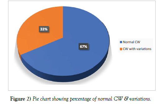
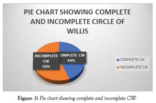
Of the total 100 circles which form the basis for this study, 33 (33%) were found to be normal in anatomical configuration and 67 (67%) associated with some variations. A circle is considered complete if all the component vessels are present, whether hypoplastic or duplicated but not absent. Among the 100 circle, 44 (44%) were found to be complete and 56 (56%) incomplete circles (Figure 2-3) (Table 1-2).
| No. of cases | Percentage | |
|---|---|---|
| Circle of Willis | 100 | |
| Normal circle of Willis | 33 | 33% |
| Circle of Willis with variations | 67 | 67% |
Table 1. Distribution of Circle of Willis with or without variations.
| Type | No. of cases | Percentage |
|---|---|---|
| Complete CW | 44 | 44% |
| Incomplete CW | 56 | 56% |
Table 2. Complete & incomplete Circle of Willis.
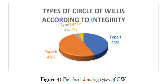
Among the 100 circles, 44 (44%) were found to be intact (Type I), out of which 33 CW were typical, complete, and arterial components with normal calibre. 11 circles (11%) were intact but have one or more components hypoplastic. 48 (48%) CW had complete anterior circulation but incomplete posterior circulation (Type II) whereas 4 (4%) CW had posterior complete circulation with incomplete anterior circulation (Type III). However anteriorly as well as posteriorly incomplete circles (Type IV) were found in 4 cases (4%) (Figure 4) (Table 3).
| Type of integrity | No. of cases | Percentage |
|---|---|---|
| Type I | 44 | 44% |
| Type II | 48 | 48% |
| Type III | 4 | 4% |
| Type IV | 4 | 4% |
Table 3. Classification of CW according to integrity of circle.
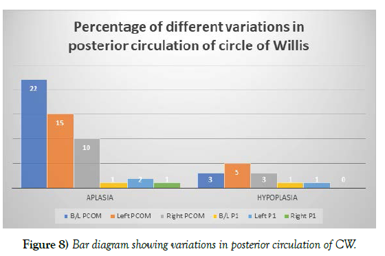
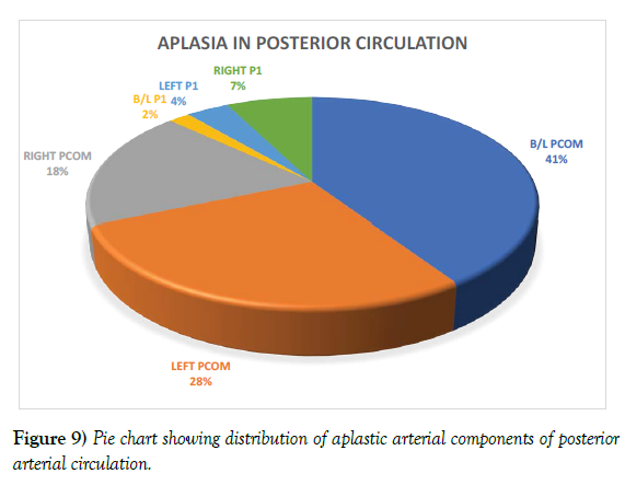
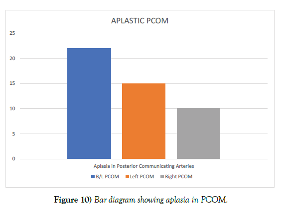
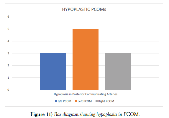
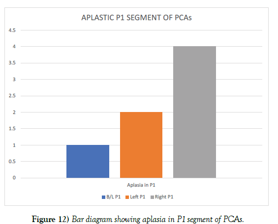
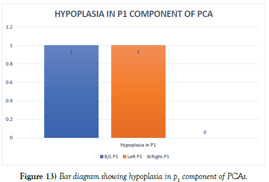
There are 25 different types of variations with or without association found in this study. 58% variations seen in PCOM, 3% in A1 & 9% in P1. In posterior communication circulation we have seen different types of variations with or without association namely B/L PCOM aplasia, Right PCOM aplasia, Left PCOM aplasia, B/L PCOM hypoplasia, Right PCOM hypoplasia and Left PCOM hypoplasia in 22%, 10%, 15%, 3%, 3% and 5% cases respectively. Among posterior Circle of Willis configurations, unilateral posterior communicating artery (PCOM) aplasia (25%) is the most common type of variation we have found and bilaterally PCOM aplasia (22%) is the second most commonly observed variant. Normal morphology of posterior arterial circulation is seen in (35%) of the circles. In pre-communicating posterior cerebral artery (P1 Segment), we have Right & Left P1 aplasia, Left P1 hypoplasia, B/L P1 hypoplasia in each 1% of cases. In P1 segment there are two cases of foetal origin in posterior cerebral arteries one in left side and other bilaterally.
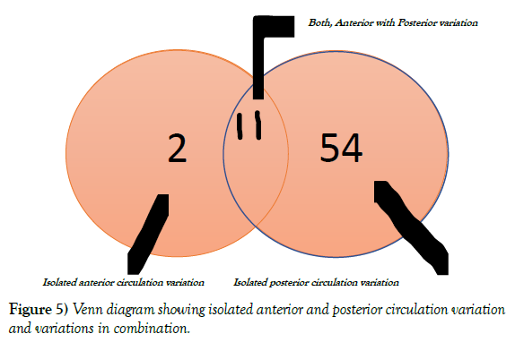
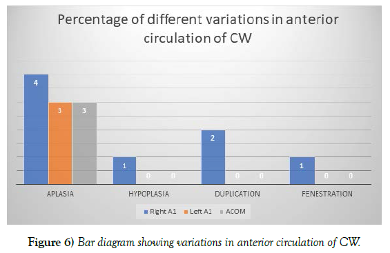
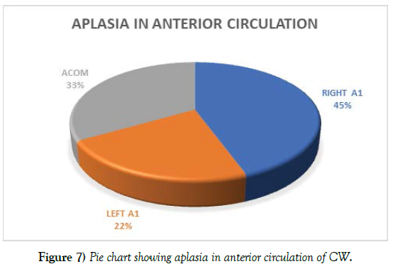
There are 14 anterior arterial variations in 13 circles. Two anterior variations in same circle are left A1 aplasia associated with ACOM aplasia. There are 65 posterior arterial variations out of which 54 variations are found only in posterior circulation and 11 variations are associated with anterior circulation. Among posterior circulation of Circle of Willis unilateral posterior communicating artery (PCOM) aplasia (25%) is the most common type of variation we have found and bilaterally aplastic PCOM (22%) is the second most commonly observed variant. Normal morphology of posterior arterial circulation is seen in (35%) of the circles (Figure 5-13) (Table 4-8).
| Vessels | Variations | Their association | No. of cases | Total No. of Cases | Total No. of Cases |
|---|---|---|---|---|---|
| PCOM | B/L PCOM Aplasia | Without association | 18 (18%) | 22 | 58 (58%) |
| With left A1 Aplasia | 1 (1%) | ||||
| With right A1 Hypoplasia | 1 (1%) | ||||
| With right A1 Fenestration | 1 (1%) | ||||
| With right A1 Duplication | 1 (1%) | ||||
| Right PCOM Aplasia | Without association | 8 (8%) | 10 | ||
| With right A1 Aplasia | 1 (1%) | ||||
| With left A1 Aplasia | 1 (1%) | ||||
| Left PCOM Aplasia | Without association | 12 (12%) | 15 | ||
| With right P1 Aplasia | 1 (1%) | ||||
| With right A1 duplication | 1 (1%) | ||||
| With right A1 and right P1 Aplasia | 1 (1%) | ||||
| B/L PCOM Hypoplasia | Without association | 3 (3%) | 3 | ||
| Right PCOM Hypoplasia | Without association | 2 (1%) | 3 | ||
| With ACOM Aplasia | 1 (1%) | ||||
| Left PCOM Hypoplasia | Without association | 4 (4%) | 5 | ||
| With ACOM Aplasia | 1 (1%) | ||||
| A1 | Right A1 Aplasia | Without Aplasia | 1 (1%) | 2 | 3 (3%) |
| With right P1 Aplasia and right persistent trigeminal | 1 (1%) | ||||
| Left A1 Aplasia | With ACOM Aplasia | 1 (1%) | 1 | ||
| P1 | Right P1 Aplasia (pcom as PTA) | Without association | 1 (1%) | 1 | 6 (6%) |
| Left P1 Aplasia (left foetal origin of pca-1) | Without association | 2 (2%) | 2 | ||
| B/L P1 Aplasia (b/l foetal origin of pca-1) | Without association | 1 (1%) | 1 | ||
| Left P1 Hypoplasia | Without association | 1 (1%) | 1 | ||
| B/L P1 Hypoplasia | Without association | 1 (1%) | 1 | ||
| TOTAL | 67 | 67 | |||
Table 4. Showing different variations and their association.
| Right A1 | Left A1 | ACOM | ||||||
|---|---|---|---|---|---|---|---|---|
| Variations | No. of cases | % | No. of cases | % | No. of cases | % | Total | % |
| Aplasia | 4 | 4 | 3 | 3 | 3 | 3 | 10 | 10 |
| Hypoplasia | 1 | 1 | 0 | 0 | 0 | 0 | 1 | 1 |
| Duplication | 2 | 2 | 0 | 0 | 0 | 0 | 2 | 2 |
| Fenestration | 1 | 1 | 0 | 0 | 0 | 0 | 1 | 1 |
| 8 | 8% | 3 | 3% | 3 | 3% | 14 | 14% | |
Table 5. Variations in anterior arterial circulation of CW.
| Vessels | Side | Variation | No. of cases | Total no. of cases |
|---|---|---|---|---|
| PCOM | Right | Aplasia | 10 | 58 |
| Hypoplasia | 3 | |||
| Left | Aplasia | 15 | ||
| Hypoplasia | 5 | |||
| B/L | Aplasia | 22 | ||
| Hypoplasia | 3 | |||
| P1 | Right | Aplasia | 4 | 9 |
| Hypoplasia | 0 | |||
| Left | Aplasia | 2 | ||
| Hypoplasia | 1 | |||
| B/L | Aplasia | 1 | ||
| Hypoplasia | 1 | |||
| PCOM | PCOM as a persistent trigeminal A. | 1 | 1 | |
| PCOM | PCOM as part of PCA (foetal origin) | 2 | 2 | |
| PCA | Bilateral PCA from right ICA | 1 | 1 | |
| PCOM | Fusion of left PCOM and P1 absent | 1 | 1 | |
Table 6. Variations in posterior circulation of CW.
| Types of arteries | Total variations | Percentage % |
|---|---|---|
| PCOM | 62 | 62 |
| A1 | 11 | 11 |
| P1 | 9 | 9 |
| ACOM | 3 | 3 |
Table 7. Variations In different components of CW.
| Artery | Aplasia | Hypoplasia |
|---|---|---|
| A1 | 7 (7%) | 1 (1%) |
| ACOM | 3 (3%) | 0 |
| PCOM | 47 (47%) | 11 (11%) |
| P1 | 7 (7%) | 2 (2%) |
Table 8. Showing Aplasia & Hypoplasia in different segment of circle.
DISCUSSION
Circle of Willis is formed at the base of brain in order to preserve the cerebral perfusion and to avoid the symptoms of ischemia in case of blockage of part of the cerebral arterial system. The benefit of arterial circle is to equalize the pressure and under normal condition little interchange of blood takes place along the anastomotic channel due to equality of blood pressure.
The normal function of brain is usually not affected by anatomical variation in arteries of circle of Willis. This happens due to the collateral circulation and compensation from the arteries of the other side.
The knowledge of the segmental anatomy and the prevalence of the arterial variants is important while evaluating multidetector CTA images. Analysis and establishing the pattern of circle of Willis is one of the most important prerequisites of various diagnostic & therapeutic neurovascular procedures [38]. We evaluated anatomical variations of the cerebral arteries in 100 patients and found that those anatomical variations are frequent and could be evaluated correctly by CTA.
A thorough knowledge of the variations in cerebral arterial circle has become essential with the increasing number of procedures like aortic arch surgery, microsurgical clipping of anterior communicating artery aneurysm etc. Its variations are common and the textbook picture of symmetrical, large, approximately equal sized vessels were present only in 30% subjects [34].The study looked at the completeness of the circle and also the calibre of vessels in the posterior communicating arteries with external diameters less than 0.5mm as hypoplasia.
The Circle of Willis and its branches are found to be associated with numerous variations. The variations not only differ from person to person but also on the right and left side of the same individual [39] as well as in anterior and posterior circulation. In this study we found that right side of the circles showed similar variation (25%) as compared with left side of circles (26%).
Ranil [24] (2009) defined a circle as typical if all the component vessels of the CW were present, origin of the vessels forming the CW was from its typical source and the size of a component vessel was more than 1 mm in diameter. In this study typical Circle of Willis without any variation was found in 33 Circle (33%) and atypical circle was found in rest 67 CW (67%). This result was very close to study done by Klimek Piotrowska [32] and Hina Siddqi [29] in which typical CW was 27% and 29.4% respectively and atypical CW was 73% and 76.4%. The present observation is at great variation with those of Fisher et al [40-42] study who recorded typical or classic configuration in 4.8% and atypical in 95.2% of the circle (Table 9-15).
| Study | Typical CW | Atypical CW |
|---|---|---|
| Kapoor et al9 | 45.2% | 54.8% |
| Fisher40 | 4.8% | 95.2% |
| Ramanuj34 | 73.3% | 26.6% |
| Alpers10 | 52% | 48% |
| Klimek Piotrowska32 | 27% | 73% |
| Hina29 | 29.4% | 76.4% |
| S. Iqbal11 | 48% | 52% |
| Present study | 33% | 67% |
Table 9. Comparison of typical and atypical CW.
| Study | Complete CW | Incomplete CW |
|---|---|---|
| Gunnal31 | 60 | 10.66 |
| Alpers10 | 52.30 | 0.60 |
| Klimek Piotrwska32 | 57 | 16 |
| Krabbe et al22 | 42 | |
| Hafez KA41 | 45 | |
| Present study | 44 | 56 |
Table 10. Comparison of complete and incomplete CW.
| Study | Aplasia | Hypoplasia | Duplication | Fenestration |
|---|---|---|---|---|
| Sahin H.36 | 2.53 | 14.6 | 1.06 | |
| Dimmick26 | 1-2 | 10 | 0.058 | |
| Syed yaseen33 | 3.22 | |||
| S. Iqbal11 | 4 | |||
| Dr. K.S. Kaviyavarshini35 | 10.9 | 9.7 | ||
| Alpers10 | 0 | 2 | ||
| Puchades42 | 0.8 | 6.4 | ||
| Kanchan kapoor9 | 0.4 | 1.7 | 2.6 | |
| Present study | 7 (7%) | 1 (1%) | 2 (2%) | 1 (1%) |
Table 11. Comparison of different variations of anterior cerebral artery (A1).
| Study | Aplasia | Hypoplasia | Duplication |
|---|---|---|---|
| Alpers10 | 2 | ||
| Fawcett19 | 0.14 | 7.2 | |
| Windle43 | 3 | ||
| Syed yaseen32 | 3.22 | 6.45 | |
| S. Iqbal11 | 8 | ||
| Sahin H.36 | 3.86 | ||
| Ramanuj34 | 4 | ||
| Kanchan Kapoor9 | 1.8 | 10 | |
| Present study | 3 | - |
Table 12. Comparison of different variations of ACOM.
| Study | Aplasia | Hypoplasia |
|---|---|---|
| Alpers et al.10 | 0.6 | 22 |
| Fawcett19 | 3.6 | 22.2 |
| Windle 43 | 12.5 | 25 |
| Riggs & Rupp13 | 51 | |
| Ramanuj 34 | 12 | 21 |
| Hina siddique29 | 23.4 | 33.3 |
| Hilal Sahin.36 | 47.3 | |
| Present study | 47 | 11 |
Table 13. Comparison of Aplasia & Hypoplasia of PCOM.
| Type | Chen chi12 | Present study |
|---|---|---|
| Type I | 34.64 % | 44 % |
| Type II | 47.71 % | 48 % |
| Type III | 5.23 % | 4 % |
| Type IV | 12.42 % | 4 % |
Table 14. Comparison of type of CW.
| Name of study | Incidence of embryonic origin of PCA from ICA% |
|---|---|
| Alpers10 | 15 |
| Riggs& Rupp13 | 22 |
| Milenkovic45 | 21 |
| S. Iqbal11 | 10 |
| Our study | 2 |
Table 15. Showing comparison of % of embryonic origin of PCA.
Atypical CW in all the studies ranged between 40% to 75%. Though the percentage of atypical CW in our study (67%) was nearly same to that of Klimek Piotrowska [32] (73%), they varied with study of Gunnal [31] (64%). Alpers [10] (1963, USA), Kapoor9 (2008, India) and Macchi21 (2002, Italy) studied the CW in their respective region and found that about 50% of the circles are of typical configuration.
In the present study, a circle was considered complete if all the component vessels were present, whether hypoplastic or duplicated but not absent. The incidence of incomplete circle of Willis is extremely varies in the literature. Several authors did not find any incomplete CW29. Incomplete CW is mostly because of absence of PCOM. The incidence of an incomplete CW in this study was 56%.
Fenestration and duplication of vessel were more commonly seen in anterior circulation as compared with the posterior circulation. In this study, 2 circles (2%) having duplication of A1 segment on the right side and 1 circle (1%) with fenestration of A1 segment on the same side in ACA. Simon J. Dimmick et al [26] observed that fenestration of the anterior cerebral artery is a rare finding. The prevalence of fenestration of the A1 segment is between 0% and 4% in anatomic imaging studies and 0.058% in angiographic studies, while fenestration of anterior communicating artery was more common in their study. In cadaveric study, fenestration of anterior communicating artery was 12% -21% whereas in computerised tomographic angiographic it was only 5.3%. Hilal Sahin [36] observed that fenestration/duplication were more common in anterior circulation as compared with posterior one. Fenestration of the posterior cerebral artery, an extremely rare occurrence, has been found in both the P1 and the P2 segments. Duplication of the posterior communicating artery was not found in this study as well as previous study.
In this study, aplasia was found to be the most common type of variation. Total 64 cases of aplasia were found, out of which 10 in anterior circulation and 54 in posterior arterial circulation. Unilateral aplasia of the posterior communicating arteries was the most common (25%). In this study, Aplasia of posterior communicating artery was found in 47% of circles and this result is very similar to study done by Hilal Sahin et al [36] which was 47.3%. Bilateral PCOM Aplasia were seen in 22% in this study, which is nearly similar to the finding of Hilal Sahin et al36in which bilateral PCOM aplasia was 27.56%.
Thin string like vessel was found in 14% of the circles and was present in every type of vessel except anterior communicating artery and left anterior cerebral pre-communicating segment (left A1). However, hypoplastic artery was seen only in pre-communicating segment of anterior cerebral artery (A1) in Hilal Sahin et al [36] study. In this study PCOM hypoplasia was seen in 11% of circle. In Ramanuj et al [34] study, hypoplastic PCOM was seen in 21%. Bilateral hypoplasia was the most common type of variation in most of the studies, but in present study it was only 3%. Posterior communicating artery aplasia was not seen in Lazorthes [16] (1979), DeSilva [25] (2011) and Klimek-Piotrowska [32] (2016) study. Alpers et al10, Riggs and Rupp [13], Raja Reddy et al [14], and Fetterman and Moran15 have reported that hypoplastic vessels were more commonly seen in posterior communicating arteries followed by the posterior cerebral arteries in contrast to study done by Kamath18 and Fawcett et al [19], hypoplasia was more common in the posterior cerebral arteries than in the posterior communicating arteries.
In this study, type II (complete anterior and incomplete posterior) was the most common type seen in 48% of circle which is similar to study done by Chen Chi [12] (47.71%).
In the foetal origin of posterior cerebral artery the posterior communicating artery (PCOM) is larger than the P1 segment of the posterior cerebral artery (PCA) and supplies the bulk of the blood to the PCA. The term is typically used to refer to the situation where the PCOM is larger than the P1 [43- 45]. The embryonic origin of the posterior cerebral artery from the internal carotid persisted as a fairly common anomaly and was found in 10% of the circles in Iqbal (2013) study. In this study foetal origin of posterior cerebral artery was found in 2% of circle.
Persistent primitive trigeminal artery (PPTA): is the most common of the remaining persistent foetal arteries. Zampakis et al [46] stated that persistent trigeminal artery was present in 0.1–1 % of the angiograms and autopsies. In their series the percentage of patients having this uncommon variation was 0.057 %, lower than the reported angiographic percentage in other published CTA studies, as well as with our finding i.e 1%.
In 1% of circle both side of posterior cerebral arteries arise from the right side of internal carotid artery.
CONCLUSION
Among the 100 cases, normal CW were seen in 33% and variations in 67% of cases. We observed 25 different types of variations some of which were isolated variation and some in combination with others. A wide variability in the posterior arterial component of the CW was observed. The variability was mainly hypoplasia or absent vessels in the PCOM. Most common variations were seen in PCOM (62%) followed by A1 (11%) respectively. On comparing the result with other studies, it has been found that the results were variable in different studies as well as in the present study. These variations may be due to racial factor, ethnicity and genetic make-up or due to small sample size in different studies. But the posterior circulation was the commonest site for variation in most of the study. As it confirms high percentage of variations in the formation of Circle of Willis, all surgical interventions should be preceded by angiography. Awareness of these anatomical variations is important in the neurovascular procedures.
REFERENCES
- Tihomir Z. Encyclopædia Britannica Online. Studia lexicographica. 2020; 27(14):109-123
- Pai SB, Kulkarni RN, Varma RG. Microsurgical anatomy of the anterior cerebral artery-anterior communicating artery complex. Neurology Asia 2005; 10(6):21-28.
- de Gast AN, van Rooij WJ, Sluzewski M. Fenestrations of the anterior communicating artery: incidence on 3D angiography and relation with aneurysms. AJNR Am J Neuroradiol 2008; 29(2):296-298.
- Haines DE. Fundamental Neuroscience for Basic and Clinical Applications. (Fifth Edition). 2018:496-516.
- Betsy B. Love, José Biller. Textbook of Clinical Neurology. (Third Edition). 2007.
- Gaillard F, Hacking C. Circle of Willis. Radiopaedia.org. 2020.
- Edwin Ocran Mbchb. Circle of Willis. Last Reviewed: September 11, 2021.
- Satheesha Nayak B, Somayaji S, Soumya K. Variant arteries at the base of the brain. Ijav. 2009; 2(1):60-61.
- Kapoor K, Singh B, Dewan Lij. Variations in the configuration of the circle of willis. Anat Sci Int. 2008; 83(2):96-106.
- Alpers Bj, Berry Rg. Circle Of Willis in Cerebral Vascular Disorders. The Anatomical Structure. Arch Neurol. 1963 4(8):398-402.
- Iqbal S. A comprehensive study of the anatomical variations of the circle of Willis in adult human brains. J Clin Diagn Res. 2013; 7(11): 2423-2427.
- He J, Liu H, Huang B. Investigation of morphology and anatomic variations of circle of Willis and measurement of diameter of cerebral arteries by 3D-TOF angiography. Sheng Wu Yi Xue Gong Cheng Xue Za Zhi. 2007; 24(1):39-44. Chinese. PMID: 17333889.
- Riggs HE, Rupp C. Variation in form of circle of Willis. The relation of the variations to collateral circulation: anatomic analysis. Arch Neurol. 1963; 8(8):24-30.
- Reddy DR, Prabhakar V, Rao BD. Anatomical study of circle of Willis. Neurol India. 1972; 20(1):8-12.
- Fetterman GH, Moran TJ. Anomalies of the circle of Willis in relation to cerebral softening. Arch Pathol. 1941; 32: 251-57.
- Lazorthes G, Gouaze A, Santini JJ. The arterial circle of the brain (circulus arteriosus cerebri). Anatomia Clinica. 1979, 1:241-257.
- El Khamlichi A, Azouzi M, Bellakhdar F. Anatomic configuration of the circle of Willis in the adult studied by injection technics. Apropos of 100 brains. Neurochirurgie. 1985; 31(4):287-293.
- Kamath S. Observations on the length and diameter of vessels forming the circle of Willis. J Anat. 1980; 133(3):419-23.
- Fawcett E, Blachford JV. The Circle of Willis: an Examination of 700 Specimens. J Anat. 1905; 40(1):63.2-70.
- Hillen B. The variability of the circulus arteriosus (Willisii): Order or anarchy. Acta Anat (Basel). 1987; 129(1):74-80.
- Macchi C, Catini C, Federico C. Magnetic resonance angiographic evaluation of circulus arteriosus cerebri (circle of Willis): a morphologic study in 100 human healthy subjects. Ital J Anat Embryol. 1996; 101(2):115-23.
- Krabbe- Hartkamp MJ, van der GJ, de LFE. Circle of Willis: Morphologic variation on three-dimensional time-of-flight MR angiograms. Radiology, 1998; 207(1):103-111.
- Eftekhar B, Dadmehr M, Ansari S. Are the distributions of variations of circle of Willis different in different populations? Results of an anatomical study and review of literature. BMC Neurol. 2006; 24:6-22.
- Hashemi SM, Mahmoodi R, Amir Jamshidi A. Variations in the Anatomy of the Willis' circle: A 3-year cross-sectional study from Iran (2006-2009). Are the distributions of variations of circle of Willis different in different populations? Result of an anatomical study and review of literature. Surg Neurol Int. 2013 May 17:4:65.
- De Silva KRD, Silva R, Gunasekera WSL. Prevalence of typical circle of Willis and the variation in the anterior communicating artery: A study of a Sri Lankan population. Ann Indian Acad Neurol. 2009; 12(3):157-61.
- Dimmick SJ. Normal variations of the cerebral circulation at multidetector CT angiography. Radiographics. 2009; 29(4):1027-1043.
- Zhou HW, Fu J, Lu ZL. Fenestration of the anterior cerebral artery detected by magnetic resonance angiography. Chin Med J Engl. 2009; 122:1139-1142.
- Makowicz G, Poniatowska R, Lusawa M. Variants of cerebral arteries - anterior circulation. Pol J Radiol. 2013; 78(3):42-47.
- Hina Siddiqi, Mohammad Tahir, Khalid Parvez. Lone3Variations in Cerebral Arterial Circle of Willis in Adult Pakistani Population. J Coll Physicians Surg Pak. 2013; 23(9):615-619.
- Cooke DL, Stout CE, Kim WT. Cerebral arterial fenestrations. Int Neuro. 2014; 20(3):261-274.
- Gunnal SA, Farooqui MS, Wabale RN. Anatomical variations of the circulus arteriosus in cadaveric human brains. Neuro Res IN. 2014:1:6872-6881.
- Wiesława Klimek-Piotrowska. A multitude of variations in the configuration of the circle of Willis: an autopsy study. Anat Sci Int. 2016; 91(4):325-33.
- Yaseen S, Riyaz ZH, Siddiqui AA. Variations in the brain circulation-the circle of Willis. IP Indian J Neurosci. 2016; 2(2):37-42
- Singh R.Anatomical variations of circle of Willis a cadaveric study. Int Surg J. 2017; 4(4):1249-1258
- Kaviyavarshini KS, Madhumita C, Prashant Moorthy SMVMCH. Puducherry Normal Variants of Circle of Willis in MR Angiography among Puducherry Population. JMSCR 2018; 6(4).
- Şahin H, Pekçevik Y. Anatomical variations of the circle of Willis:evaluation with CT angiography. Anatomy. 2018; 20-26.
- Lars Hindenes B, Asta K, Håberg. Vangberg Variations in the Circle of Willis in a large population sample using 3D TOF angiography. PLoS One. 2020; 15(11):e0241373.
- Milo MacBain Persistent primitive trigeminal artery. 2021.
- Raghavendra Shirol Vs, Daksha Dixit, Desai S. Circle of Willis and Its Variations; Morphometric Study in Adult Human Cadavers. Int J Med Res Health Sci. 2014; 3(2):394-400.
- Fisher CM. The circle of Willis: anatomical variations. Vasc Dis. 1965; 2:99-105.
- Hafez KA, Amm NM, Saudi FZ. Anatomical variations of the circle of Willis in males and females on 3D MR angiograms. Egypt J Hosp Med. 2007; 26:106-121.
- Puchades-Orts A, Nombela-Gomez M, Ortuno-Pacheco G. Variation in form of circle of Willis: Some anatomical and embryological considerations. Anat Rec. 1976; 185:119-123.
- Windle BCA. On the arteries forming circle of Willis. J Anat and Phy. 1888; 22:289.
- Gaillard F, Glick Y. Fetal posterior cerebral artery. J Investig Med High Impact Case Rep. 2016; 4(3).
- Milenkovic Z, Vucetic R, Puzic M. Asymmetry and anomalies of the circle of Willis in fetal brain. Microsurgical study and functional remarks. Surg Neurol. 1985; 24: 563-570.
- Zampakis P, Panagiotopoulos V, Petsas T. Common and uncommon intracranial arterial anatomic variations in multi-detector computed tomography angiography (MDCTA). What radiologists should be aware of Insights. Imaging. 2015; (6):33-42.
Indexed at, Google Scholar, Crossref
Indexed at, Google Scholar, Crossref
Indexed at, Google Scholar, Crossref
Indexed at, Google Scholar, Crossref
Indexed at, Google Scholar, Crossref
Indexed at, Google Scholar, Crossref
Indexed at, Google Scholar, Crossref
Indexed at, Google Scholar, Crossref
Indexed at, Google Scholar, Crossref
Indexed at, Google Scholar, Crossref
Indexed at, Google Scholar, Crossref
Indexed at, Google Scholar, Crossref
Indexed at, Google Scholar, Crossref
Indexed at Google Scholar, Crossref
Indexed at, Google Scholar, Crossref
Indexed at, Google Scholar, Crossref
Indexed at, Google Scholar, Crossref
Indexed at, Google Scholar, Crossref
Indexed at, Google Scholar, Crossref
Indexed at, Google Scholar, Crossref
Indexed at, Google Scholar, Crossref
Indexed at, Google Scholar, Crossref


