Anatomical variations of the clavicle and main vascular structures in two pediatric patients: subclavicular vein cannulation with supraclavicular approach
Hafize Oksuz*, Nimet Senoglu, Huseyin Yildiz and Hilmi Demirkiran
Kahramanmaras Sutcu Imam University, Faculty of Medicine, Department of Anesthesiology and Reanimation, Kahramanmaras, Turkey
- *Corresponding Author:
- Hafize Oksuz, MD
Assistant Professor of Anesthesiology Kahramanmaras Sutcu Imam University Faculty of Medicine Department of Anesthesiology and Reanimation 46050 Kahramanmaras, Turkey
Tel: +90 344 2212337
Fax: +90 344 2212371
E-mail: drhoksuz@hotmail.com
Date of Received: December 25th, 2008
Date of Accepted: April 6th, 2009
Published Online: May 26th, 2009
© IJAV. 2009; 2: 51–53.
[ft_below_content] =>Keywords
supraclavicular approach, subclavicular vein cannulation, ultrasound guidance, cerebral palsy, anatomical variations
Introduction
Central venous catheterization (CVC) is implemented for volume resuscitation, hemodynamic monitoring, vasopressor administration, frequent blood sampling, parenteral nutritional support and the administration of long-term chemotherapy [1].
Generally, it is attempted using the internal jugular, subclavian or femoral veins. The subclavian vein access has been the recommended approach for CVC both for short and long term catheterization. The advantages of this approach can be attributed to the fact that it is a large vein [2,3].
The standard technique for placement of central venous catheters includes the use of anatomical landmarks which may not correlate with vessel location [3]. Although ultrasound guidance improved the success rate, reduced the number of needle passes and decreased complications associated with the internal jugular vein and CVC, it is currently uncommon for central venous catheter placement [1].
In this report, we present the use of a supraclavicular approach for subclavian cannulation with ultrasound guidance in two pediatric patients who had anatomical variations of the clavicles and main vascular structures due to cerebral palsy.
Case Reports
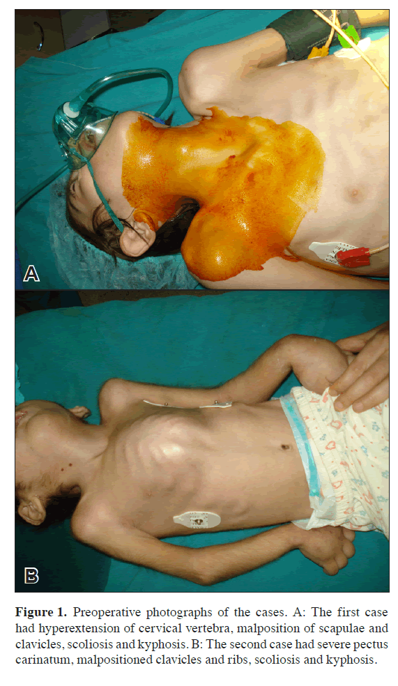
After informed consents were taken from their families, a seven year old male (first case) and four year old female (second case) were scheduled for a central venous catheterization under general anesthesia to gain access to the venous system for fluid resuscitation. Both cases had a history of cerebral palsy and treatment with mechanical ventilation due to pneumonia. They presented with hyperextension of the cervical vertebra and malpositioning of the clavicles and ribs. A physical examination revealed a pectus carinatum in the second case. Scoliosis and kyphosis were present in both cases (Figure 1). The mediastinum and cardiorespiratory shadows were enlarged in chest radiographs. Due to anatomical variations, catheterizations were planned under ultrasound guidance.
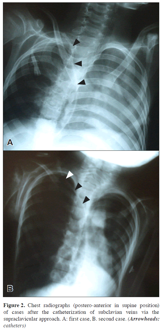
After the induction of anesthesia, the subclavian vein was located by positioning the transducer superiorly until the vessel was clearly visualized. The needle was introduced under ultrasound guidance from the supraclavicular region. The wall of the right subclavian vein was verified prior to needle insertion which was achieved on the first attempt in both cases. The Seldinger technique was used and the wire placement confirmed with ultrasound. The catheter and port were then introduced as per standard practice. Image intensification was used to confirm the position of the catheters in the superior vena cava. After the procedures were completed, a radiological examination was performed. Direct chest radiographs were used to confirm the positions of the catheters in the superior vena cava (Figure 2).
Discussion
Central venous catheters are used daily in clinical practice for the management of critical patients. They are usually placed with a blind, external landmark guided technique [2]. Mechanical complications are reported to occur during or after the process in 5% to 19% of patients. These complications increase in association with several characteristics including patient anatomy (e.g., morbid obesity, cachexia or local scarring from surgery or radiation therapy), patient setting (e.g., patients under mechanical ventilation or in emergency conditions such as cardiac arrest), comorbidities and operator’s experience [4]. Complications resulting from patient factors like anatomical variations play an important role in the difficulty of central venous access. Malpositioning of central venous catheters may lead to serious complications including intravascular knotting, rupture of the heart and great vessels, incorrect central venous pressure readings and thrombosis formation due to delivery of hyperosmolar solutions [5].
Patients with hemiplegic cerebral palsy typically have multiple upper extremity impairments such as impaired forearm rotation and movement patterns of the upper arm and trunk. It disregards rotations of the scapula and clavicle [6].
In our patients, the puncture was made into the subclavian vein using the supraclavicular approach under ultrasound guidance due to the anatomical variations including the impaired rotation and movement patterns of the upper arm and trunk. Because of these variations in the presented cases, the external landmark-guided technique was not useful for the subclavian vein cannulation. It could be used for the supraclavicular approach although this is a less popular technique.
The ultrasound-guided technique is recommended in high-risk patients such as those with coagulation defects, obesity or unusual anatomy. This technique has advantages for visualization of the desired vein and the surrounding anatomic structures prior to and during the procedure and is associated with a reduced number of attempts, higher success rates and a reduced incidence of arterial puncture [4].
Kusminsky suggested that the supraclavicular approach is an appropriate and safe method for subclavian vein cannulation [7].
Traditional anterior or posterior triangle approaches for subclavian vein cannulation for central venous access may be unsuccessful, especially in patients whose anatomy renders cannulation difficult. The supraclavicular approach is an easy cannulation technique for inexperienced physicians and a useful alternative to traditional approaches for the experienced physicians [8]. Pirotte and Veyckemans showed that when using an ultrasound guide for internal jugular vein cannulation, the subclavian vein could be easily visualized in the supraclavicular area [9].
Ultrasound cannot be used to show the subclavian vein as, by definition, this vessel is under the clavicle. It may be used for the supraclavicular approach although this is a less popular technique. Ultrasound helps with accurate cannulation of many central veins thus reducing complications associated with cannulation [10].
In conclusion, this ultrasound guided approach for subclavian vein catheterization offers a successful procedure to anesthesiologists for central venous access in patients having anatomical variations. The best way to prevent complications during central venous catheterization is to use an ultrasound guide which helps to identify the needle within the lumen of the vein in addition to underlying anatomy, anatomical variations and possible pathologies (thrombosis). We conclude that in patients having altered anatomy due to cerebral palsy, central vein catheterization should be performed under ultrasound guidance in order to provide greater safety and an increased success rate.
References
- Randolph AG, Cook DJ, Gonzales CA, Pribble CG. Ultrasound guidance for placement of central venous catheters: a meta- analysis of the literature. Crit Care Med. 1996; 24: 2053–2058.
- Paoletti F, Ripani U, Antonelli M, Nicoletta G. Central venous catheters. Observations on the implantation technique and its complications. Minerva Anestesiol. 2005; 71: 555–560.
- Jensen MO. Anatomical basis of central venous catheter fracture. Clin Anat. 2008; 21: 106–110.
- Karakitsos D, Labropoulos N, De Groot E, Patrianakos AP, Kouraklis G, Poularas J, Samonis G, Tsoutsos DA, Konstadoulakis MM, Karabinis A. Real-time ultrasound-guided catheterisation of the internal jugular vein: a prospective comparison with the landmark technique in critical care patients. Crit Care. 2006; 10: R162.
- Yerdel MA, Karayalcin K, Anadol E. Malpositioning of subclavian vein catheters in left and right sided attempts: a prospective study. Neth J Surg. 1991; 43: 178–180.
- Kreulen M, Smeulders MJ, Veeger HE, Hage JJ. Movement patterns of the upper extremity and trunk associated with impaired forearm rotation in patients with hemiplegic cerebral palsy compared to healthy controls. Gait Posture. 2007; 25: 485–492.
- Kusminsky RE. Complications of central venous catheterization. J Am Coll Surg. 2007; 204: 681–696.
- Conroy JM, Rajagopalan PR, Baker JD 3rd, Bailey MK. A modification of the supraclavicular approach to the central circulation. South Med J. 1990; 83: 1178–1181.
- Pirotte T, Veyckemans F. Ultrasound-guided subclavian vein cannulation in infants and children: a novel approach. Br J Anaesth. 2007; 98: 509–514.
- Galloway S, Bodenham A. Ultrasound imaging of the axillary vein-anatomical basis for central venous access. Br J Anaesth. 2003; 90(5): 589–595.
Hafize Oksuz*, Nimet Senoglu, Huseyin Yildiz and Hilmi Demirkiran
Kahramanmaras Sutcu Imam University, Faculty of Medicine, Department of Anesthesiology and Reanimation, Kahramanmaras, Turkey
- *Corresponding Author:
- Hafize Oksuz, MD
Assistant Professor of Anesthesiology Kahramanmaras Sutcu Imam University Faculty of Medicine Department of Anesthesiology and Reanimation 46050 Kahramanmaras, Turkey
Tel: +90 344 2212337
Fax: +90 344 2212371
E-mail: drhoksuz@hotmail.com
Date of Received: December 25th, 2008
Date of Accepted: April 6th, 2009
Published Online: May 26th, 2009
© IJAV. 2009; 2: 51–53.
Abstract
Central venous catheterization is a routine application in the management of patients in critical condition. However, the placement of central venous catheters is not without risk. The standard technique for central venous cannulation includes the use of anatomical landmarks. However, an ultrasound-guided method is recommended for catheterization in high-risk patients. In this report, we present two pediatric cases which had anatomical variations of the clavicles and main vascular structures due to cerebral palsy and were treated with mechanical ventilation because of pneumonia. The subclavian vein cannulation was performed using a supraclavicular approach under ultrasound guidance in both cases. We conclude that central venous catheterization of critical patients who have anatomical variations must be performed under ultrasound guidance as it provides greater safety and a higher success rate.
-Keywords
supraclavicular approach, subclavicular vein cannulation, ultrasound guidance, cerebral palsy, anatomical variations
Introduction
Central venous catheterization (CVC) is implemented for volume resuscitation, hemodynamic monitoring, vasopressor administration, frequent blood sampling, parenteral nutritional support and the administration of long-term chemotherapy [1].
Generally, it is attempted using the internal jugular, subclavian or femoral veins. The subclavian vein access has been the recommended approach for CVC both for short and long term catheterization. The advantages of this approach can be attributed to the fact that it is a large vein [2,3].
The standard technique for placement of central venous catheters includes the use of anatomical landmarks which may not correlate with vessel location [3]. Although ultrasound guidance improved the success rate, reduced the number of needle passes and decreased complications associated with the internal jugular vein and CVC, it is currently uncommon for central venous catheter placement [1].
In this report, we present the use of a supraclavicular approach for subclavian cannulation with ultrasound guidance in two pediatric patients who had anatomical variations of the clavicles and main vascular structures due to cerebral palsy.
Case Reports
After informed consents were taken from their families, a seven year old male (first case) and four year old female (second case) were scheduled for a central venous catheterization under general anesthesia to gain access to the venous system for fluid resuscitation. Both cases had a history of cerebral palsy and treatment with mechanical ventilation due to pneumonia. They presented with hyperextension of the cervical vertebra and malpositioning of the clavicles and ribs. A physical examination revealed a pectus carinatum in the second case. Scoliosis and kyphosis were present in both cases (Figure 1). The mediastinum and cardiorespiratory shadows were enlarged in chest radiographs. Due to anatomical variations, catheterizations were planned under ultrasound guidance.
After the induction of anesthesia, the subclavian vein was located by positioning the transducer superiorly until the vessel was clearly visualized. The needle was introduced under ultrasound guidance from the supraclavicular region. The wall of the right subclavian vein was verified prior to needle insertion which was achieved on the first attempt in both cases. The Seldinger technique was used and the wire placement confirmed with ultrasound. The catheter and port were then introduced as per standard practice. Image intensification was used to confirm the position of the catheters in the superior vena cava. After the procedures were completed, a radiological examination was performed. Direct chest radiographs were used to confirm the positions of the catheters in the superior vena cava (Figure 2).
Discussion
Central venous catheters are used daily in clinical practice for the management of critical patients. They are usually placed with a blind, external landmark guided technique [2]. Mechanical complications are reported to occur during or after the process in 5% to 19% of patients. These complications increase in association with several characteristics including patient anatomy (e.g., morbid obesity, cachexia or local scarring from surgery or radiation therapy), patient setting (e.g., patients under mechanical ventilation or in emergency conditions such as cardiac arrest), comorbidities and operator’s experience [4]. Complications resulting from patient factors like anatomical variations play an important role in the difficulty of central venous access. Malpositioning of central venous catheters may lead to serious complications including intravascular knotting, rupture of the heart and great vessels, incorrect central venous pressure readings and thrombosis formation due to delivery of hyperosmolar solutions [5].
Patients with hemiplegic cerebral palsy typically have multiple upper extremity impairments such as impaired forearm rotation and movement patterns of the upper arm and trunk. It disregards rotations of the scapula and clavicle [6].
In our patients, the puncture was made into the subclavian vein using the supraclavicular approach under ultrasound guidance due to the anatomical variations including the impaired rotation and movement patterns of the upper arm and trunk. Because of these variations in the presented cases, the external landmark-guided technique was not useful for the subclavian vein cannulation. It could be used for the supraclavicular approach although this is a less popular technique.
The ultrasound-guided technique is recommended in high-risk patients such as those with coagulation defects, obesity or unusual anatomy. This technique has advantages for visualization of the desired vein and the surrounding anatomic structures prior to and during the procedure and is associated with a reduced number of attempts, higher success rates and a reduced incidence of arterial puncture [4].
Kusminsky suggested that the supraclavicular approach is an appropriate and safe method for subclavian vein cannulation [7].
Traditional anterior or posterior triangle approaches for subclavian vein cannulation for central venous access may be unsuccessful, especially in patients whose anatomy renders cannulation difficult. The supraclavicular approach is an easy cannulation technique for inexperienced physicians and a useful alternative to traditional approaches for the experienced physicians [8]. Pirotte and Veyckemans showed that when using an ultrasound guide for internal jugular vein cannulation, the subclavian vein could be easily visualized in the supraclavicular area [9].
Ultrasound cannot be used to show the subclavian vein as, by definition, this vessel is under the clavicle. It may be used for the supraclavicular approach although this is a less popular technique. Ultrasound helps with accurate cannulation of many central veins thus reducing complications associated with cannulation [10].
In conclusion, this ultrasound guided approach for subclavian vein catheterization offers a successful procedure to anesthesiologists for central venous access in patients having anatomical variations. The best way to prevent complications during central venous catheterization is to use an ultrasound guide which helps to identify the needle within the lumen of the vein in addition to underlying anatomy, anatomical variations and possible pathologies (thrombosis). We conclude that in patients having altered anatomy due to cerebral palsy, central vein catheterization should be performed under ultrasound guidance in order to provide greater safety and an increased success rate.
References
- Randolph AG, Cook DJ, Gonzales CA, Pribble CG. Ultrasound guidance for placement of central venous catheters: a meta- analysis of the literature. Crit Care Med. 1996; 24: 2053–2058.
- Paoletti F, Ripani U, Antonelli M, Nicoletta G. Central venous catheters. Observations on the implantation technique and its complications. Minerva Anestesiol. 2005; 71: 555–560.
- Jensen MO. Anatomical basis of central venous catheter fracture. Clin Anat. 2008; 21: 106–110.
- Karakitsos D, Labropoulos N, De Groot E, Patrianakos AP, Kouraklis G, Poularas J, Samonis G, Tsoutsos DA, Konstadoulakis MM, Karabinis A. Real-time ultrasound-guided catheterisation of the internal jugular vein: a prospective comparison with the landmark technique in critical care patients. Crit Care. 2006; 10: R162.
- Yerdel MA, Karayalcin K, Anadol E. Malpositioning of subclavian vein catheters in left and right sided attempts: a prospective study. Neth J Surg. 1991; 43: 178–180.
- Kreulen M, Smeulders MJ, Veeger HE, Hage JJ. Movement patterns of the upper extremity and trunk associated with impaired forearm rotation in patients with hemiplegic cerebral palsy compared to healthy controls. Gait Posture. 2007; 25: 485–492.
- Kusminsky RE. Complications of central venous catheterization. J Am Coll Surg. 2007; 204: 681–696.
- Conroy JM, Rajagopalan PR, Baker JD 3rd, Bailey MK. A modification of the supraclavicular approach to the central circulation. South Med J. 1990; 83: 1178–1181.
- Pirotte T, Veyckemans F. Ultrasound-guided subclavian vein cannulation in infants and children: a novel approach. Br J Anaesth. 2007; 98: 509–514.
- Galloway S, Bodenham A. Ultrasound imaging of the axillary vein-anatomical basis for central venous access. Br J Anaesth. 2003; 90(5): 589–595.


