Anomalous right coronary artery from the pulmonary artery (ARCAPA) in a 63-year-old male patient with a stable angina pectoris and positive stress test
Received: 14-Dec-2020 Accepted Date: Feb 15, 2021; Published: 22-Feb-2021, DOI: 10.37532/1308-4038.14(2).165-166
Citation: Sasani H, Gur DO. Anomalous right coronary artery from the pulmonary artery (ARCAPA) in a 63-year-old male patient with a stable angina pectoris and positive stress test. Int J Anat Var. 2021;14(2):53-54.
This open-access article is distributed under the terms of the Creative Commons Attribution Non-Commercial License (CC BY-NC) (http://creativecommons.org/licenses/by-nc/4.0/), which permits reuse, distribution and reproduction of the article, provided that the original work is properly cited and the reuse is restricted to noncommercial purposes. For commercial reuse, contact reprints@pulsus.com
Abstract
Anomalous origin of the right coronary artery from the pulmonary artery (ARCAPA) is a very rare congenital cardiac anomaly. Herein, computed tomography and conventional coronary angiography imaging findings of the patient with ARCAPA are presented.
Keywords
Coronary angiography; Computed tomography angiography; Coronary veins
Introduction
Anomalous origin of the coronary arteries from the pulmonary artery (ACAPA) is a rare congenital anomaly which has two subtypes including right ACAPA (ARCAPA) and left ACAPA (ALCAPA) coronary arteries. The left sided anomalous origin of coronary artery (ALCAPA) is more commonly seen variation compared to ARCAPA with an incidence of one per 300.000 live births and 0.25% - 0.5% of all congenital cardiac diseases. However, ARCAPA is a rare congenital coronary anomaly with reported incidence of 0.002%, which constitutes 0.12% of all coronary artery anomalies [1–3].
Although it is more commonly encountered in the young population, some patients may remain undiagnosed until middle age. The timing of onset of symptoms in ARCAPA patients is variable and is related to the presence of collateral circulation [4]. Early symptoms may be secondary to heart failure, valve insufficiency or myocardial infarction [5]. Surgical treatment is recommended even if patients are asymptomatic due to their high risk profile [6].
The pathophysiology of ARCAPA depends on the direction of blood flow in the coronary artery and its ability to deliver oxygen to the myocardium. Since patients with right dominant blood circulation cannot tolerate ARCAPA well compared to those with left dominant system [2] Coronary steal phenomenon in ARCAPA may cause diastolic pressure differences between systemic and pulmonary artery beds and increased myocardial oxygen demand and risk of myocardial ischemia [7]. Although the symptoms of ARCAPA can range from effort angina to dyspnea or fatigue, it can also manifest itself in the form of ischemic myocardial damage sometimes causing sudden cardiac death [8,9].
In this article, the imaging findings of, ARCAPA with conventional coronary angiography (CCA) and coronary computed tomographic angiography (CCTA) of a 68-year-old male patient are presented.
Case Report
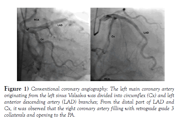
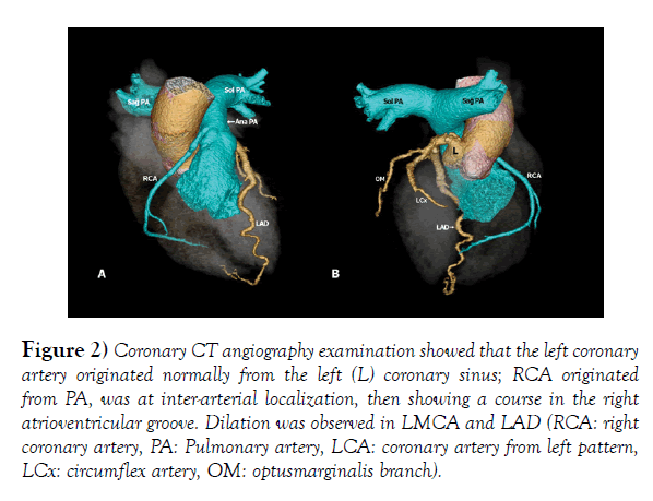
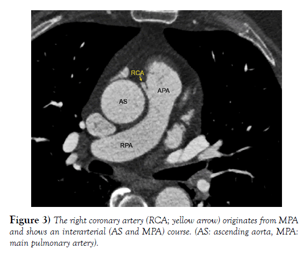
A 68-year-old male patient applied to the hospital with a complaint of stable angina pectoris. There was no referred pain present. No remarkable finding was found on physical examination and electrocardiography. However due to the positive stress test of the patient, CCA examination was done, revealing that no coronary artery originated from the right sinus Valsalva. The left main coronary artery (LMCA) originated from the left coronary sinus, divided into the branches of the circumflex (Cx) and the left anterior descending artery (LAD). From the distal part of LAD and Cx, the right coronary artery (RCA) was filled with retrograde grade 3 collaterals and opening to the pulmonary artery (PA) (Figure 1). Based on these findings, CCTA examination was performed showing that LMCA originated from the left coronary sinus; however, RCA originated from PA, was at inter-arterial localization, then showing a course in the right atrioventricular groove. Dilation was observed in LMCA and LAD (Figures 2 & 3). These findings were evaluated as compatible with ARCAPA.
Figure 1) Conventional coronary angiography: The left main coronary artery originating from the left sinus Valsalva was divided into circumflex (Cx) and left anterior descending artery (LAD) branches; From the distal part of LAD and Cx, it was observed that the right coronary artery filling with retrograde grade 3 collaterals and opening to the PA.
(RCA: right coronary artery, PA: Pulmonary artery, LCA: coronary artery from left pattern, LCx: circumflex artery, OM: optusmarginalis branch)
Figure 2) Coronary CT angiography examination showed that the left coronary artery originated normally from the left (L) coronary sinus; RCA originated from PA, was at inter-arterial localization, then showing a course in the right atrioventricular groove. Dilation was observed in LMCA and LAD.
(AS: ascending aorta, MPA: main pulmonary artery)
Figure 3) The right coronary artery (RCA; yellow arrow) originates from MPA and shows an interarterial (AS and MPA) course.
Discussion
Abnormal origin of the coronary arteries, typically from the contralateral side of the sinus Valsalva, is a relatively common finding during routine CCA and is associated with sudden cardiac death in athletes [10]. Coronary arteries originating anomalously from the PA, however, is a rare incidence.
Compared to ARCAPA, patients with abnormal origin of the left coronary artery from the pulmonary artery (ALCAPA) are diagnosed early in life as they are usually symptomatic [1]. The diagnosis of ARCAPA, which is even less common than ALCAPA, can be delayed due to presence of collaterals, as was the case with our patient. ARCAPA can be seen alone or can be accompanied with other congenital cardiac defects such as tetralogy of Fallot, bicuspid aortic valve, aortic stenosis, septal defects or aortic coarctation [1,2]. We could not demonstrate any other cardiac defect in this specific patient.
As imaging modalities, transthoracic echocardiography (TTE), magnetic resonance angiography (MRA) and CCTA are used in the diagnosis of ARCAPA, but, MRA and CCTA are more reliable. TTE is a non-invasive method used to for evaluate cardiac defects. Although Doppler US may show the abnormally located ostium and proximal intramural course of coronary artery in children, this cannot be imaged in adults. Intracoronary collaterals within the ventricular septum, thought to be an indicative of ARCAPA, can, however, be shown using color Doppler sonography [11]. In this patient, CCA could demonstrate the anatomy of right coronary circulation due to presence of excellent collaterals from left coronary system. This seems to delay the symptoms of the patient.
By using quantitative blood flow parameters and phase-contrast imaging, cardiac magnetic resonance imaging (CMR) can demonstrate pulmonary to systemic blood flow ratio, can be used in quantification of the flow difference in assessment of the shunt, in evaluation of right ventricular size and function and myocardial viability assessment [12]. Due to a high spatial resolution, CTA can provide 3D volume rendering reconstructions images for better clarifying the variation and also reveal the possible inter-arterial course of the anomalous coronary artery [13].
Conclusion
Although CCTA, CCA and MRA are among the most useful imaging methods in the diagnosis of ARCAPA; as in the current case, the origin and course of the anomalous coronary artery which cannot always be visualized in CCA, was shown on CCTA. Therefore, in order to confirm the diagnosis, the most appropriate imaging method can be selected individually based on the specific patient’s clinical findings.
Acknowledgement
None.
REFERENCES
- Rawal H, Mehta S. Anomalous Right Coronary Artery Originating from the pulmonary artery. (ARCAPA): A systematic review of literature. J Lung Health Dis. 2018;2:7-9.
- Williams IA, Gersony WM, Hellenbrand WE. Anomalous right coronary artery arising from the pulmonary artery: a report of 7 cases and a review of the literature. Am Heart J. 2006;152:1004.e9-17.
- Pena E, Nguyen ET, Merchant N, et al. ALCAPA syndrome: not just a pediatric disease. Radiographics. 2009;29:553-65.
- Balakrishna P, Illovsky M, Al-Saghir YM, et al. Anomalous Origin of Right Coronary Artery Originating from the Pulmonary Trunk (ARCAPA): an Incidental Finding in a Patient Presenting with Chest Pain. Cureus. 2017;9:e1172.
- Rodriguez-Gonzalez M, Tirado AM, Hosseinpour R, et al. Anomalous Origin of the Left Coronary Artery from the Pulmonary Artery: Diagnoses and Surgical Results in 12 Pediatric Patients. Tex Heart Inst J. 2015;42:350-6.
- Radke PW, Messmer BJ, Haager PK, et al. Anomalous origin of the right coronary artery: preoperative and postoperative hemodynamics. Ann Thorac Surg. 1998;66:1444-9.
- Mehta SS, Sattiraju S. Multimodality Imaging of Anomalous Origin of Right Coronary Artery From Pulmonary Artery (ARCAPA). J Invasive Cardiol. 2017;29:E104.
- McAlindon E, Johnson TW, Strange J, et al. Isolated anomalous right coronary artery from the pulmonary artery in adulthood: anatomical features and ischemic burden. Circulation. 2012;125:1183-5.
- Rajbanshi BG, Burkhart HM, Schaff HV, et al. Surgical strategies for anomalous origin of coronary artery from pulmonary artery in adults. J ThoracCardiovasc Surg. 2014;148:220–224.
- Maron BJ, Thompson PD, Puffer JC, et al. Cardiovascular preparticipation screening of competitive athletes. A statement for health professionals from the Sudden Death Committee (clinical cardiology) and Congenital Cardiac Defects Committee (cardiovascular disease in the young), American Heart Association. Circulation. 1996;94:850-6.
- Kühn A, Kasnar-Samprec J, Schreiber C, et al. Anomalous origin of the right coronary artery from the pulmonary artery (ARCAPA). Int J Cardiol. 2010;139:e27-28.
- Shariat M, Grosse-Wortmann L, Seed M, et al. Isolated anomalous origin of the right coronary artery from the pulmonary artery in an asymptomatic 12-year-old girl: role of MRI in depicting the anatomy, detecting the ischemic burden, and quantifying the amount of left-to-right shunt. World J Pediatr Congenit Heart Surg. 2013;4:201-5.
- Svensson A, Themudo R, Cederlund K. Anomalous origin of right coronary artery from the pulmonary artery. Eur Heart J. 2017;38:3069.


