Anthropometric study on the anatomical variation of the external ear amongst Port Harcourt students, Nigeria
2 Department of Anatomy, College of Medicine and Health Sciences, Gregory University, Uturu, Nigeria
Received: 05-Oct-2018 Accepted Date: Dec 11, 2018; Published: 19-Dec-2018
Citation: Osunwoke EA, Vidona WB, Atulegwu GC. Anthropometric study on the anatomical variation of the external ear amongst Port Harcourt students, Nigeria. Int J Anat Var. Dec 2018;11(4):143-146.
This open-access article is distributed under the terms of the Creative Commons Attribution Non-Commercial License (CC BY-NC) (http://creativecommons.org/licenses/by-nc/4.0/), which permits reuse, distribution and reproduction of the article, provided that the original work is properly cited and the reuse is restricted to noncommercial purposes. For commercial reuse, contact reprints@pulsus.com
Abstract
Background: The morphology of an individual‘s external ear and its dimensions vary amongst persons to the point that the right and left ears differ.
Aims: This study is aimed at establishing the anatomical variation in the auricular structure of the students of University of Port Harcourt, Nigeria.
Methods: The study utilizes a total of 200 students, 101 male and 99 female subjects within the age range of 16-30 years. Total ear length and breadth of both ears were measured using a digital vernier caliper (in mm).
Results: Result obtained showed the total ear length for the right ear to be 54.3 ± 4.12 while that of left ear is 54.2 ± 4.10. The total ear breadth for the right ear of the subject was 31.4 ± 2.51 while that of left ear was 31.3 ± 2.33. Of the t otal population, oval ear shape was commonly noted (58%), followed by round (25.5%), rectangular (10%) and triangular (6.5%) respectively. 100% symmetry was noted with the shape of the right and left ears. Pearson’s correlation between shape of auricle and gender was not significant (p=0.798). Inference statistics shows the right and left ears of each parameter to be significant (p<0.05).
Conclusion: The results obtained from this study will serve useful purposes in ear morphology and anthropometric considerations.
Keywords
Anthropometric; Ear morphology; Auricular structure; External ear; Ear dimensions; Ear shape
Introduction
Anthropometric data is needed in the design of products as it varies between individuals and nations [1]. As products are designed for specific types of consumers, anthropometric data provides a valuable source of information to ergonomist and designers who attempt to consider a range of body sizes and abilities in the design of occupational environments and products [2]. Reliable anthropometric data of the ear for a particular population is necessary when designing for that population otherwise the product may not be suitable for the user, [1]. Direct measurement of a subject’s ear can be difficult because of distortion of soft tissues of the natural ear during measurements and the problem of the subjects in maintaining the orientation of the head for several minutes, which may alter the operators’ perspectives, [3].
Humans show a wide range of biological variation; this variation makes us unique and distinguishes one individual from another. People vary in shape, size, skin colour and many numbers of other characteristics. A characteristic that is often overlooked is the structure of the human external ear. The external ear is highly variable to the point that even two ears of a single individual may be notably different [4]. The length and breadth of the ear varies from person to person and anthropometric points have been described to investigate the dimensions of the external ear [5].
The ear is an important and under-recognized defining feature of the face whose shape and size conveys information about age and sex, [6]. Although the primary function of the pinna is to collect sound waves that are transmitted to ear drum through the external auditory meatus, the ear is also recognized as a cosmetic organ and its importance is more related to the aesthetics and physiognomy of the face, [7]. People having abnormal set of ears through congenital malformations or loss of the auricle through trauma usually feel depressed and uncomfortable, [8]. Any auricular defect in form of inappropriate size, abnormal elongation of the auricular lobe, or missing part is corrected by surgery, [9]. For rectifying such abnormalities plastic surgeons require information about normal auricular dimension, the auricles bilateral position on the face, the general conformations and its variation. But these auricular data vary in different ethnic groups. So the morphometric measurements given in western literatures are less likely to be of much use, [7]. Ear biometrics can positively identify an individual using comparative analysis of the human ear and its morphology. The dimensions of the pinna have been found to vary among different individuals who which can be utilized in forensics for personal identification in the absence of valid finger print.
Anatomy of the External Ear
The ear is desperately divided into the external, middle and internal ear. The external ear consists of the auricle or pinna and the external acoustics meatus, at the medial end of which lies the tympanic membrane, separating the ear from the middle ear [10].
The auricle or pinna has a skeleton of resilient yellow elastic cartilage which is thrown into folds. The folds give the auricle its characteristic shape. The cartilage is covered on both surfaces with adherent skin; it does not extend into the lobule of the ear. The lobule of the tag of the skin containing soft fibro fatty tissue, it is easily pierced for earrings. The cartilage of the auricle is prolonged inwards tubular fashion as the cartilaginous part of the external acoustic meatus, whose attachment to bone establishes the auricle in position. Small anterior, superior and posterior auricular muscles attach the auricle to the scalp and skull [10].
The main nerves of the skin of the auricles are great auricular and auriculotemporal nerves. The great auriculotemporal nerve supplies the cranial surface commonly called the back of the ear and the posterior part of the lateral surface. The auriculotemporal nerve, a branch of CN V3, supplies the skin of the auricle anterior to the external acoustic meatus [11].
The arterial supply to the auricle is derived mainly from the posterior auricular and superficial temporal arteries [11].
The lymphatics of the auricle are as follows: the lateral surface of the superior half of the auricle drains to the superficial parotid lymph nodes. The cranial surface of the posterior half of the auricle drains to the mastoid lymph nodes and deep cervical lymph nodes and the reminder of the auricle, including the lobule, drains into the cervical lymph node [11].
Embryologically, the auricle develops from six mesenchymal proliferations at the dorsal ends of the first and second pharyngeal cleft. These swellings (auricular hillocks), three on each side of the external meatus, later fuse and form the definitive auricles. As fusion of the auricular hillock is completed, developmental abnormalities of the auricle are common. Initially, the external ears are in the lower neck region but with development of the mandible, they ascend to the side of the head at the level of the eyes [8].
Anatomical Landmarks of the Ear
Anti-helix
A Y- shaped curved cartilaginous ridge arising from the anti-tragus and separating the concha, triangular fossa and scapha. The anti-helix presents a folding of the conchal cartilage and it usually has similar prominence to a well-developed helix. The stem of the normal helix is gently curved and branches about two thirds of the way along its course to form the broad fold of the superior (posterior) anti-helical crus, and the more sharply folded inferior (anterior crus). The inferior and superior crura of the anti-helical can vary both in volume and degree of folding [12].
Anti-helix, inferior crus
The lower cartilaginous ridge arising at the bifurcation of the anti-helix that ends beneath the fold of the ascending helix, and separates the concha from the triangular fossa.
Anti-helix super crus
The upper cartilaginous ridge arising at the bifurcation of the anti-helix that separates the scapha from the triangular fossa.
Anti-tragus
The anterior superior cartilaginous protrusions lying between the incisura and the origin of the anti-helix. The anterio-superior margin of the antitragus forms the posterior wall of the incisura.
Helix
The outer rim of the ear that extend from the superior insertion of the ear on the scalp to the termination of the cartilage at the earlobe. The helix can be divided into three approximate parts: the ascending helix, which extends vertically from the root: superior helix, which begins at the top of the ascending portion, extends horizontally and curves posterior to the site of Darwin tubercle, the descending helix which begins inferior to Darwin tubercle and extends to the superior border of the earlobe. The lower portion of the posterior part is often non-cartilaginous. The border of the helix usually forms a rolled rim but the helix is highly variable in shape [12].
Lobe
The soft fleshly inferior part of the pinna. It is bounded on it postero superior border by the end of the descending helix, on the anterio superior border by the inferior border of the antitragus and superiorly by the incisura. The ear lobe is highly variable in size, and in the degree of attachment of the anterio-inferior portion of the face.
Scapha
The groove between the helix and the anti-helix.
Tragus
A posterior, slightly inferior protrusion of the skin covered cartilage, anterior to the auditory meatus.
Triangular fossa
The concavity bounded by the superior and inferior crura of the anti-helix and the ascending portion of the helix.
Frankfort Horizontal
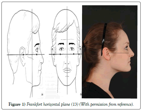
This is a plane connecting the lowest point on the lower margin of each orbit and highest point on the upper margin of the external auditory meatus. The Frankfort horizontal plane or Frankfurt plane is used as the general horizontal plane of the head and as reference point for other planes and structures [5].
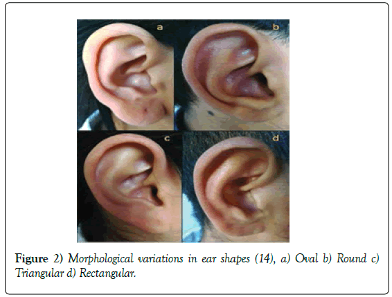
This is a plane connecting the lowest point on the lower margin of each orbit and highest point on the upper margin of the external auditory meatus. The Frankfort horizontal plane or Frankfurt plane is used as the general horizontal plane of the head and as reference point for other planes and structures (Figures 1 and 2) [5].
Variation in Ear Shape
The four basic ear morphological shapes are oval, rectangular, triangular and round. Also, the ear lobule attachments are either attached or free [14].
Aim and Objectives
The aim of this study is to establish the anatomical variation in the auricular structure of students in University Of Port Harcourt, Nigeria; with the specific objectives to establish the total ear length, total ear breadth and auricular shape; to investigate the difference between the right and left ear and to determine the association between gender and auricular shape.
Thus the significance of this study is that Statistical data about the variation of the ear in the population are useful for optimizing products, in diagnosis of congenital malformation, planning plastic surgery, and in designing hearing instruments.
Materials and Methods
The research was carried on a total of 200 students, 101 male and 99 female subjects between the ages of 16-30 years were collected randomly.
Exclusion criteria included subjects that are not from the university. Also Subjects whose normal ear external morphology has been altered by trauma, accidents or surgery. The present study was conducted after taking approval for human population study. Before carrying out the experiments, the subjects were informed of requirements and procedures of the measurements with assurance for confidentiality of information for openness in line with standard protocol.
Methods
With the subjects head in Frankfurt horizontal plane, measurements were taken using vernier calliper with a resolution of 0.01 mm.
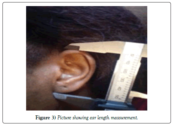
The ear length of the subjects were gotten by measuring the distance between the superior most point of the auricle and the inferior most point of the earlobe with a vernier caliper (Figure 3).
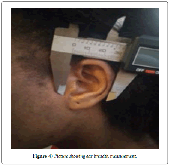
The ear breadth was gotten by measuring the distance between the maximum convexity of the helix and the root of a vernier caliper (Figure 4).
The ear morphological shape of each subject was noted by the anatomical positions of the helix and lobes when viewed using the Frankfort horizontal plane. Methods of data analysis include Chi square test used to test for sex differences in the auricular shape while independent T-test was used to test for the variability in the right and left ear, using the SPSS statistical analysis program. Statistical significance was considered at p<0.05.
Results
The results of this study are presented in the tables as shown. Table 1 shows the descriptive estimate of the tool car length and total ear breadth of the students. The table shows that the mean ± S.D. of the total ear breadth of the right ear was 54.3 ± 4.12 while the total ear length of the left ear was 54.2 ± 4.10. The total ear breadth of the right ear was 31.4 ± 2.5, while the total ear breadth of the left ear was 31.3 ± 2.3. This shows that all the parameters of the right ear are larger than that of the left ear.
| Parameters | Right | Left |
|---|---|---|
| Total ear breadth | 54.3 ± 4.120 | 54.2 ± 4.10 |
| Total ear length | 31.4 ± 2.451 | 31.3 ± 2.33 |
Table 1: Descriptive estimate of the total ear length and total ear breadth of the students.
Table 2 shows the descriptive estimate of the auricular shape of the student’s and the variability of the right and the left ear. The table shows the classification of ear shape into oval which was 116 (58%) of the total population, rectangular shape 20 (10%), round 51 (25%) and triangular mass which was 13 (6.5) respectively. Complete 100% symmetry was noted regarding shapes of the right and left ears among the students.
| Variable | Frequency | Percentage (%) |
|---|---|---|
| Oval | 116 | 58.0 |
| Round | 51 | 25.5 |
| Triangular | 13 | 6.5 |
| Rectangular | 20 | 10 |
Table 2: Descriptive estimate of the auricular shape of the students.
Analysis of students T-test on the variability of the right and left ear was highly significant (P<0.05). This is shown in Tables 3a and 3b.
| Variable | F | T | Df | p-value | Variable |
|---|---|---|---|---|---|
| Right | Right | ||||
| Equal of variances assumed | 1.441 | 7.851 | 198 | 0.000 | Equal of variances assumed |
| Equal of variances not assumed | 7.868 | 190.776 | 0.000 | Equal of variances not assumed | |
| Left | Left | ||||
| Equal of variances assumed | 7.944 | 8.505 | 198 | 0.000 | Equal of variances assumed |
| Equal of variances not assumed | 8.539 | 173.267 | 0.000 | Equal of variances not assumed | |
| Variable | F | T | Df | p-value | Variable |
| Right | Right | ||||
| Equal of variances assumed | 1.441 | 7.851 | 198 | 0.000 | Equal of variances assumed |
Table 3a: Analysis of students T-test on the variability of the right and left ear for total ear length.
| Variable | F | T | Df | p-value | Variable |
|---|---|---|---|---|---|
| Right | Right | ||||
| Equal of variances assumed | 0.112 | 5.909 | 198 | 0.000 | Equal of variances assumed |
| Equal of variances not assumed | 5.086 | 196.376 | 0.000 | Equal of variances not assumed | |
| Left | Left | ||||
| Equal of variances assumed | 0.450 | 6.779 | 198 | 0.000 | Equal of variances assumed |
| Equal of variances not assumed | 6.774 | 196.014 | 0.000 | Equal of variances not assumed | |
| Variable | F | T | Df | p-value | Variable |
| Right | Right | ||||
| Equal of variances assumed | 0.112 | 5.909 | 198 | 0.000 | Equal of variances assumed |
Table 3b: Analysis of students T-test on the variability of the right and left ear for total ear breadth.
The study was able to identify that among the 101 male students, 56 had oval ear shape, 12 had rectangular shape dear, 26 had round shaped ear and 7 had triangular shaped ear, as shown in Table 4.
| Variable | Oval | Rectangular | Round | Triangular | Total |
|---|---|---|---|---|---|
| Male Count | 56 | 12 | 26 | 7 | 101 |
| % within gender | 55.4% | 11.9% | 25.7% | 6.9% | 100.0% |
| % within the ear shape | 48.3% | 60.0% | 51.0% | 53.8% | 50.5% |
| Female Count | 60 | 8 | 25 | 6 | 99 |
| % within gender | 60.6% | 8.1% | 25.3% | 6.1% | 100.0% |
| % within the ear shape | 51.7% | 40.0% | 49.0% | 46.2% | 49.5% |
| Total Count | 116 | 20 | 51 | 13 | 200 |
| % within gender | 58.0% | 10.0% | 25.5 | 6.5% | 100.0% |
| % within the ear shape | 100% | 100.0% | 100.0% | 100.0% | 100.0% |
Table 4: The association between gender and auricular shape of the students.
Table 5 shows that there is no significant difference between ear shape and gender, P- value >0.05 (0.798). This means that ears are the same in shape in both male and female students.
| Value | Df | p-value | |
|---|---|---|---|
| Person Chi-Square | 1.015 | 3 | 0.798 |
| Likelihood ratio | 1.020 | 3 | 0.798 |
| Linear-by-Linear association | 0.257 | 1 | 0.612 |
| N of Valid Cases | 200 |
Table 5: Analysis of students’ Chi-square test on the association of gender and ear shape.
Discussion
Human ear shapes and variations can be useful for identification in the absence of fingerprints and facial recognition software. The variation in external ear shape and its dimensions are important in the diagnosis of congenital malformations, treatment planning and surgeries as the ear reaches it mature height at 13 years in males and 12 years in females as according to Verma et al., [14].
In the present study attempt was made to establish the anatomical variation of the external ear amongst university of Port Harcourt students which can be useful for identification, prosthetics and in manufacturing personalised ear products.
The mean total ear length obtained in this study was lower than that on their morphometric study of the external ear, age and sex related differences as their total mean length was 63.0 mm.
The right and left ears of most individuals are totally different in its length and breadth. In this study, it was observed that all parameters were significantly larger on the right side than on the left. This was similar to the findings of Talisumak et al., [15], their study addressed the symmetry, handedness and auricle morphometry. The same result of the right ear parameters being larger than the left ear parameters was also obtained by Taura et al., [16] on their study on external ear morphometry, the search for sexual dimorphism and correlations among Nigerians and also in a study carried out by Shiren and Karad [17] on the morphometric measurement of human external measurement of human external ear on 147 medical students of BRIMS, India. The result was not in line with Akpa et al., [7] on their study on anthropometry of the pinna carried out on 148 male and female Nigerian Igbo’s of the south east zone between the ages of 6 and 70 years. They found out that there was no statistical difference between right and left ears. Barut and Aktune [18] on their anthropometric measurement of the external ear of 153 primary schools in Turkey found the left ear indices for all the subjects. In this study, bilateral symmetry was observed in the shape of the right and left ears.
Most studies in literature tends to furnish data for the length, breadth and other parameters of the external ear among male and female, only few research body has been done on the correlation between gender and auricular shape of individuals. Like most parts of the body, the human external ear has a fascinating shape. The ear also conveys age and gender although not easily defined.
In this study, an attempt was made to determine the association between gender and auricular shape which showed no significance. Of the total population, oval ear shape was commonly noted followed by round, rectangular and triangular. This was not in the line with Verna et al., [14] on their study on morphological variation and biometrics of the ear, an aid to personal identification carried out on 80 students from an Indian population. They discovered that oval ear shape was commonly noted followed by triangular, rectangular and round. Other variables that affect ear morphology and dimension apart from six includes: ethnicity and age. In the data which was analysed by Brucker et al., [6] in regards to sex of volunteers, it was observed that total ear height was larger in males than females. They explained it to be a result of the release of growth hormones in females. But in this study, the total ear length was not studied in a sexual dimorphic way.
Auricular size increased significantly wit age in both men and women and these structural changes were associated with changes in the elastic fibres after adulthood [19].
Conclusion
In conclusion, the data obtained in this present study would serve some very useful purposes in ear morphology and for anthropometric considerations for groups with shared anthropometric identities with our subjects and also for supportive evidence in forensic field for the identification of individuals.
Recommendation
In recommendation, we suggest that further study on other parameters which includes ear length above tragus, ear length below tragus, tragus length, concha length, concha breadth, lobule height, lobule width and ear print which are not included in this study should be done for individualization and recognition of the students and also for supportive evidence in forensic field for the identification of individuals.
REFERENCES
- Ismaila O. Anthropometric data of hand, foot and ear of university students in Nigeria. Leonardo J of Sci. 2009;15-20
- Feathers J, Pageut V, Drury C. Measurements consistency and three dimensional electromechanical anthropometry. Int J of Industrial ergonomics. 2004;33:181-90.
- Alexander K, Scott J, Sivakumar B, et al. A morphometric study of the human ear. J Plast Reconstr Aesthet Surg. 2010;10:1016.
- Keith A. The Significance of certain features and types of the External Ear. Nature. 1901;65:16-21.
- Farkas G. Anthropometric of the Normal and Defective ear. Clin Plast Surg. 1990;173:213-21.
- Bruker J, Pater J, Sullivan P. A morphometric study of the external ear-age and sex related differences. Plast Reconstr Surg. 2003;112:647-52.
- Akpa A, Ibiam A, Ugwu C. Anthropometric study of the pinna among south East Nigerians. J of expt Res. 2013;1.
- Sadler T. Langman’s Medical Embryology. Lippincott Williams and Wilkins Publish House, USA. 2012.
- Kumar B, Selvi G. Morphometry of the ear pinna in sex determination. Int J of Anatomy and Res. 2016;4:2480-84.
- Sinnatamby C. Last’s Anatomy. Elservier Limited, USA. 2011; 137-154.
- Moore K, Dalley A, Augur A. Clinically oriented anatomy. Lippincott Williams and Wilkins, USA. 2010;pp:789-813.
- https://elementsofmorphology.nih.gov/anatomy-ear.shtml
- Thomas C. Standardized anatomical alignment of the head in a clinical photography studio. The Royal Photographic society. 2015;2.
- Verma P, Sandhu K, Verma G, et al. Morphological variations and biometrics of ear-an aid to personal identification. J Clin Diag Res. 2016;10.
- Tatlisumak E, Yavuz S, Kutulu N, et al. Asymmetry, handedness and auricle morphometry. Int J Morpho. 2015;33:1542-8.
- Taura MG, Adamu LH, Modibbo MH. Exetrnal ear anthropometry among hausas of Nigeria; the search of sexual dimorphism and correlations. WSRJ. 2013;1:91-5.
- Shireen S, Karad V. Anthropometric measurements of Human External Ear. J of Evolution of Medical and Dental Sciences. 2015;4 :10333-8.
- Barut C, Aktune A. Anthropometric Measurement of the external ear in a group of Turkish Primary School Students. Aesth Plast Surg. 2006;30:225-59.
- Ekanem A, Garba S, Musa S, et al. Anthropometric study of the Pinna (Auricle) among adult Nigerians resident in Maiduguri metropolis. J of Med Sci. 2010;10:176-80.


