Arteria lusoria, truncus bicaroticus, variant left vertebral artery
Francis LSB*, Gardner MT, Lodenquai PB
Department of Basic Medical Sciences, University of the West Indies Medical School, Mona, West Indies.
- *Corresponding Author:
- Francis LSB
Section of Anatomy, Department of Basic Medical Science
University of the West Indies Medical School
Mona, Kingston 7, Jamaica, West Indies
Telephone (876)830 4838
E-mail: landkfrancis@gmail.com
Published Online: 2 June 2017
Citation: Francis LSB, Gardner MT, Lodenquai PB. Arteria lusoria, truncus bicaroticus, variant left vertebral artery. Int J Anat Var. 2017;10(2):024-25.
© This open-access article is distributed under the terms of the Creative Commons Attribution Non-Commercial License (CC BY-NC) (http:// creativecommons.org/licenses/by-nc/4.0/), which permits reuse, distribution and reproduction of the article, provided that the original work is properly cited and the reuse is restricted to noncommercial purposes. For commercial reuse, contact reprints@pulsus.com
[ft_below_content] =>Keywords
Multiple aortic arch variants, Clinical significance, Embryology
Introduction
The Variations of the branching patterns of arteries have always received considerable attention in anatomical literature. Although these variants generally are asymptomatic, some are associated with significant symptomatology. It is also very important to recognize these variants when procedures are being performed as failure to do so may lead to harm for the patient. A right retrooesophageal subclavian artery can cause dysphagia lusoria [1,2] and an aneurysmal dilatation of this vessel (Kommerell’s diverticulum/aneurysm) [2] can cause thrombosis with resulting arterial embolization to the right upper extremity [3]. Failure to recognize a truncus bicaroticus may lead to complications during tracheostomy or thyroidectomy. Also injury to the vertebral artery in cervical spine surgery is increased in a case with a variant vertebral artery [4] and preoperative imaging studies such as a MRI should be carefully reviewed to prevent this complication. We will also be looking at the embryological origin of these variants.
Case Report
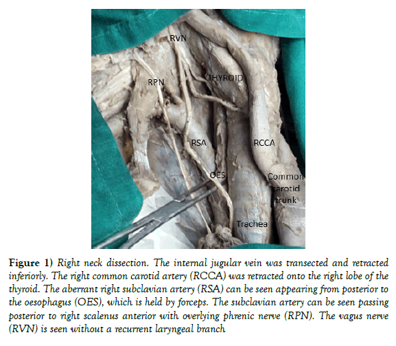
It is the routine in our Anatomy Department at the University of the West Indies Medical School to dissect the anterior neck before dissecting the superior mediastinum. During dissection of the right side of the neck of the cadaver (Figure 1) of an elderly male it was noted that the right common carotid artery (9.5 mm in diameter) did not branch from a brachiocephalic trunk and the right subclavian artery (10 mm in diameter) entered the right neck by passing behind the oesophagus. The right recurrent laryngeal nerve was not seen and the nerves to the larynx and oesophagus branched directly from the right vagus as it descended in the neck to run on the right side of the trachea in the superior mediastinum. Dissection of the left side of the neck revealed a left common carotid artery (10 mm in diameter) and a left subclavian artery (10 mm in diameter) (Figure 1).
Figure 1: Right neck dissection. The internal jugular vein was transected and retracted inferiorly. The right common carotid artery (RCCA) was retracted onto the right lobe of the thyroid. The aberrant right subclavian artery (RSA) can be seen appearing from posterior to the oesophagus (OES), which is held by forceps. The subclavian artery can be seen passing posterior to right scalenus anterior with overlying phrenic nerve (RPN). The vagus nerve (RVN) is seen without a recurrent laryngeal branch
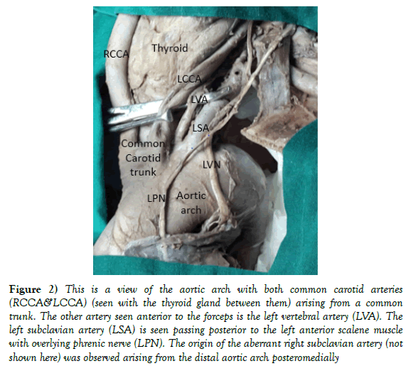
On removing the anterior chest wall and dissecting the superior mediastinum (Figure 2), it was noted that there was a short truncus bicaroticus (22 mm in transverse diameter and 20 mm in length) with two common carotid arteries straddling the trachea behind the upper manubrium sterni. The next branch arising from the arch was the left vertebral artery (4 mm in diameter), followed by the left subclavian artery. There was 1.5 mm of the superior surface of the aortic arch between the left vertebral artery and the origin of the left subclavian artery. The origin of the aberrant right subclavian artery was 11 mm in diameter and it arose posteromedially from the distal aortic arch 25 mm after the origin of the left subclavian artery.
Figure 2: This is a view of the aortic arch with both common carotid arteries (RCCA&LCCA) (seen with the thyroid gland between them) arising from a common trunk. The other artery seen anterior to the forceps is the left vertebral artery (LVA). The left subclavian artery (LSA) is seen passing posterior to the left anterior scalene muscle with overlying phrenic nerve (LPN). The origin of the aberrant right subclavian artery (not shown here) was observed arising from the distal aortic arch posteromedially
Discussion
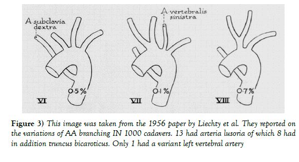
Variations in the branching pattern of the aortic arch are not rare. We reviewed four large studies looking for the incidence of the variants seen in our case and the incidence of these variants occurring together. Lale et al. [5] reported on 881 CT angiograms of the aortic arch (AA). 2.8% had left vertebral artery arising from the arch and 1.9% had an aberrant right subclavian artery. Berko et al. [6] reported on 1000 CT angiograms of AA. 6.6% had direct origin of left vertebral artery from the arch and 1.2% had an aberrant right subclavian artery. Klinkhamer [7] reported that in 295 patients with an aberrant right subclavian artery, 85 patients (29%) had truncus bicaroticus. This is not surprising as the most common variant seen in aortic arh branching is the ‘bovine arch’ where there is a common origin of the brachiocephalic trunk and the left common carotid artery. However it was Liechty et al. [1] who looked at 1000 cadavers and drew diagrams (Figure 3). 1.3% of the cadavers had an aberrant right subclavian artery and 0.1% of all cadavers had all the variants seen in our case.
A full discussion of the embryology of the aortic arch is beyond the scope of this article. The left fourth aortic arch and the left dorsal aorta caudal to it gives rise to the aortic arch, with the left 7th cervical intersegmental artery becoming the left subclavian artery [8]. The right fourth aortic arch and the right 7th cervical intersegmental artery give rise to the right subclavian artery; associated with this is regression of the right dorsal aorta rostrally up to entry of the right 3rd aortic arch and caudally to the descending aorta. In arteria lusoria, what happens is that the right 4th aortic arch regresses and it is the right dorsal aorta caudal to it that persists, joining with the right 7th cervical intersegmental artery to form the right subclavian artery. This will therefore arise from the distal aortic arch and thus passes posterior to the oesophagus and occasionally posterior to the trachea.
The remaining cervical dorsal intersegmental arteries disappear but not before giving rise to several longitudinal ateries [9] by longitudinal anastomoses between them. The vertebral artery is one of these; usually its first part is a longitudinal contribution from the 7th dorsal cervical intersegmental artery; hence its origin from the subclavian artery. The vertebral artery represents a postcostal or pretransverse anastomosis of these arteries. Then the costal element of the cervical spine fuses with the transverse process to form the foramen transversarium. When the left vertebral artery arises from the aortic arch, it appears that the origin of the first part of the vertebral artery is from the origin of the left 6th cervical intersegmental artery which persists. There are other similar longitudinal anastomoses of branches of these dorsal cervical intersegmental arteries in the neck. Thus the ascending cervical represents a precostal longitudinal anastomosis and the deep cervical artery represents a posttransverse longitudinal anastomosis.
Again we wish to point out that these variants generally are asymptomatic but some are associated with significant symptomatology as mentioned in the introduction. It is also very important to recognize these variants [10] when procedures are being performed as failure to do so may lead to harm for the patient.
References
- Liechty JD, Shields TW, Anson BJ. Variations pertaining to the aortic arches and their branches. Q Bull Northwest Univ Med Sch. 1957;31:136-43.
- Brown DL, Chapman WC, Edwards WH, et al. Dysphagia lusoria: aberrant right subclavian artery with a Kommerell’s diverticulum. Am Surg. 1993;59:582-6
- Akers DL, Fowl RJ, Plettner J, et al. “Complications of anomalous origin of the right subclavian artery: case report and review of the literature,” Annals of Vascular Surgery. 1991;5:385-8.
- Singh R. Two Rare Variants of Left Vertebral Artery. J Craniofac Surg. 2017.
- Lale P, Toprak U, Yagız G, et al. Variations in the Branching Pattern of the Aortic Arch Detected with Computerized Tomography Angiography, Advances in Radiology. 2014.
- Berko NS, Jain VR, Godelman A, et al. Variants and anomalies of thoracic vasculature on computed tomographic angiography in adults. J Comput Assist Tomogr. 2009;33:523-8.
- Klinkhamer AC. Aberrant right subclavian artery. Clinical and roentgenologic aspects. Am J Roent-genol Radium Ther Nucl Med. 1966;97:438-46.
- Sadler TW. Langman’s Medical Embryology, Lippincott Williams & Wilkins, 12th Edn., 2012;185.
- Brookes M, Anthony Zietman, Clinical embryology, a color atlas and text. Chapter 33. 1998;136.
- Suresh R, Ovchinnikov N, McRae A. “Variations in the branching pattern of the aortic arch in three Trinidadians”. West Indian Medical Journal. 2006;55:351-3.
Francis LSB*, Gardner MT, Lodenquai PB
Department of Basic Medical Sciences, University of the West Indies Medical School, Mona, West Indies.
- *Corresponding Author:
- Francis LSB
Section of Anatomy, Department of Basic Medical Science
University of the West Indies Medical School
Mona, Kingston 7, Jamaica, West Indies
Telephone (876)830 4838
E-mail: landkfrancis@gmail.com
Published Online: 2 June 2017
Citation: Francis LSB, Gardner MT, Lodenquai PB. Arteria lusoria, truncus bicaroticus, variant left vertebral artery. Int J Anat Var. 2017;10(2):024-25.
© This open-access article is distributed under the terms of the Creative Commons Attribution Non-Commercial License (CC BY-NC) (http:// creativecommons.org/licenses/by-nc/4.0/), which permits reuse, distribution and reproduction of the article, provided that the original work is properly cited and the reuse is restricted to noncommercial purposes. For commercial reuse, contact reprints@pulsus.com
Abstract
The normal aorta has three branches from its arch, the brachiocephalic trunk, the left common carotid and the left subclavian artery. Variations in the branching pattern of the aortic arch are not uncommon. We report a case of aberrant branching of the aortic arch involving 3 variants. This case was observed during cadaveric dissection and a review of the literature indicates that this pattern of branching is uncommon. There was a right retro-oesophageal subclavian artery (arteria lusoria). The right and left common carotid arteries arose from the arch by a short common trunk (truncus bicaroticus). Also, the left vertebral artery arose directly from the aortic arch between the common trunk of the right and left common carotid arteries and the left subclavian artery. In a seminal study done by Liechty et al. they reviewed the branching pattern of the aortic arch in 1000 cadavers, finding thirteen cases of arteria lusoria; eight of these had associated truncus bicaroticus and only one had all three variants. We will be discussing the embryology of these variants and their clinical importance.
-Keywords
Multiple aortic arch variants, Clinical significance, Embryology
Introduction
The Variations of the branching patterns of arteries have always received considerable attention in anatomical literature. Although these variants generally are asymptomatic, some are associated with significant symptomatology. It is also very important to recognize these variants when procedures are being performed as failure to do so may lead to harm for the patient. A right retrooesophageal subclavian artery can cause dysphagia lusoria [1,2] and an aneurysmal dilatation of this vessel (Kommerell’s diverticulum/aneurysm) [2] can cause thrombosis with resulting arterial embolization to the right upper extremity [3]. Failure to recognize a truncus bicaroticus may lead to complications during tracheostomy or thyroidectomy. Also injury to the vertebral artery in cervical spine surgery is increased in a case with a variant vertebral artery [4] and preoperative imaging studies such as a MRI should be carefully reviewed to prevent this complication. We will also be looking at the embryological origin of these variants.
Case Report
It is the routine in our Anatomy Department at the University of the West Indies Medical School to dissect the anterior neck before dissecting the superior mediastinum. During dissection of the right side of the neck of the cadaver (Figure 1) of an elderly male it was noted that the right common carotid artery (9.5 mm in diameter) did not branch from a brachiocephalic trunk and the right subclavian artery (10 mm in diameter) entered the right neck by passing behind the oesophagus. The right recurrent laryngeal nerve was not seen and the nerves to the larynx and oesophagus branched directly from the right vagus as it descended in the neck to run on the right side of the trachea in the superior mediastinum. Dissection of the left side of the neck revealed a left common carotid artery (10 mm in diameter) and a left subclavian artery (10 mm in diameter) (Figure 1).
Figure 1: Right neck dissection. The internal jugular vein was transected and retracted inferiorly. The right common carotid artery (RCCA) was retracted onto the right lobe of the thyroid. The aberrant right subclavian artery (RSA) can be seen appearing from posterior to the oesophagus (OES), which is held by forceps. The subclavian artery can be seen passing posterior to right scalenus anterior with overlying phrenic nerve (RPN). The vagus nerve (RVN) is seen without a recurrent laryngeal branch
On removing the anterior chest wall and dissecting the superior mediastinum (Figure 2), it was noted that there was a short truncus bicaroticus (22 mm in transverse diameter and 20 mm in length) with two common carotid arteries straddling the trachea behind the upper manubrium sterni. The next branch arising from the arch was the left vertebral artery (4 mm in diameter), followed by the left subclavian artery. There was 1.5 mm of the superior surface of the aortic arch between the left vertebral artery and the origin of the left subclavian artery. The origin of the aberrant right subclavian artery was 11 mm in diameter and it arose posteromedially from the distal aortic arch 25 mm after the origin of the left subclavian artery.
Figure 2: This is a view of the aortic arch with both common carotid arteries (RCCA&LCCA) (seen with the thyroid gland between them) arising from a common trunk. The other artery seen anterior to the forceps is the left vertebral artery (LVA). The left subclavian artery (LSA) is seen passing posterior to the left anterior scalene muscle with overlying phrenic nerve (LPN). The origin of the aberrant right subclavian artery (not shown here) was observed arising from the distal aortic arch posteromedially
Discussion
Variations in the branching pattern of the aortic arch are not rare. We reviewed four large studies looking for the incidence of the variants seen in our case and the incidence of these variants occurring together. Lale et al. [5] reported on 881 CT angiograms of the aortic arch (AA). 2.8% had left vertebral artery arising from the arch and 1.9% had an aberrant right subclavian artery. Berko et al. [6] reported on 1000 CT angiograms of AA. 6.6% had direct origin of left vertebral artery from the arch and 1.2% had an aberrant right subclavian artery. Klinkhamer [7] reported that in 295 patients with an aberrant right subclavian artery, 85 patients (29%) had truncus bicaroticus. This is not surprising as the most common variant seen in aortic arh branching is the ‘bovine arch’ where there is a common origin of the brachiocephalic trunk and the left common carotid artery. However it was Liechty et al. [1] who looked at 1000 cadavers and drew diagrams (Figure 3). 1.3% of the cadavers had an aberrant right subclavian artery and 0.1% of all cadavers had all the variants seen in our case.
A full discussion of the embryology of the aortic arch is beyond the scope of this article. The left fourth aortic arch and the left dorsal aorta caudal to it gives rise to the aortic arch, with the left 7th cervical intersegmental artery becoming the left subclavian artery [8]. The right fourth aortic arch and the right 7th cervical intersegmental artery give rise to the right subclavian artery; associated with this is regression of the right dorsal aorta rostrally up to entry of the right 3rd aortic arch and caudally to the descending aorta. In arteria lusoria, what happens is that the right 4th aortic arch regresses and it is the right dorsal aorta caudal to it that persists, joining with the right 7th cervical intersegmental artery to form the right subclavian artery. This will therefore arise from the distal aortic arch and thus passes posterior to the oesophagus and occasionally posterior to the trachea.
The remaining cervical dorsal intersegmental arteries disappear but not before giving rise to several longitudinal ateries [9] by longitudinal anastomoses between them. The vertebral artery is one of these; usually its first part is a longitudinal contribution from the 7th dorsal cervical intersegmental artery; hence its origin from the subclavian artery. The vertebral artery represents a postcostal or pretransverse anastomosis of these arteries. Then the costal element of the cervical spine fuses with the transverse process to form the foramen transversarium. When the left vertebral artery arises from the aortic arch, it appears that the origin of the first part of the vertebral artery is from the origin of the left 6th cervical intersegmental artery which persists. There are other similar longitudinal anastomoses of branches of these dorsal cervical intersegmental arteries in the neck. Thus the ascending cervical represents a precostal longitudinal anastomosis and the deep cervical artery represents a posttransverse longitudinal anastomosis.
Again we wish to point out that these variants generally are asymptomatic but some are associated with significant symptomatology as mentioned in the introduction. It is also very important to recognize these variants [10] when procedures are being performed as failure to do so may lead to harm for the patient.
References
- Liechty JD, Shields TW, Anson BJ. Variations pertaining to the aortic arches and their branches. Q Bull Northwest Univ Med Sch. 1957;31:136-43.
- Brown DL, Chapman WC, Edwards WH, et al. Dysphagia lusoria: aberrant right subclavian artery with a Kommerell’s diverticulum. Am Surg. 1993;59:582-6
- Akers DL, Fowl RJ, Plettner J, et al. “Complications of anomalous origin of the right subclavian artery: case report and review of the literature,” Annals of Vascular Surgery. 1991;5:385-8.
- Singh R. Two Rare Variants of Left Vertebral Artery. J Craniofac Surg. 2017.
- Lale P, Toprak U, Yagız G, et al. Variations in the Branching Pattern of the Aortic Arch Detected with Computerized Tomography Angiography, Advances in Radiology. 2014.
- Berko NS, Jain VR, Godelman A, et al. Variants and anomalies of thoracic vasculature on computed tomographic angiography in adults. J Comput Assist Tomogr. 2009;33:523-8.
- Klinkhamer AC. Aberrant right subclavian artery. Clinical and roentgenologic aspects. Am J Roent-genol Radium Ther Nucl Med. 1966;97:438-46.
- Sadler TW. Langman’s Medical Embryology, Lippincott Williams & Wilkins, 12th Edn., 2012;185.
- Brookes M, Anthony Zietman, Clinical embryology, a color atlas and text. Chapter 33. 1998;136.
- Suresh R, Ovchinnikov N, McRae A. “Variations in the branching pattern of the aortic arch in three Trinidadians”. West Indian Medical Journal. 2006;55:351-3.


