Assessment of anatomical variation of spleen in an adult human cadaver and its clinical implication: Ethiopian cadaveric study
Received: 22-Nov-2018 Accepted Date: Dec 05, 2018; Published: 12-Dec-2018
Citation: Tenaw B, Muche A. Assessment of anatomical variation of spleen in an adult human cadaver and its clinical implication: Ethiopian cadaveric study. Int J Anat Var. Dec 2018;11(4):139-142.
This open-access article is distributed under the terms of the Creative Commons Attribution Non-Commercial License (CC BY-NC) (http://creativecommons.org/licenses/by-nc/4.0/), which permits reuse, distribution and reproduction of the article, provided that the original work is properly cited and the reuse is restricted to noncommercial purposes. For commercial reuse, contact reprints@pulsus.com
Abstract
The spleen is vital lymphatic organ in the human body which located in the left hypochondrial region. Currently, its anatomical variation with immunological and hematological functions for its clinical significance gets attention. The aim of this study was to assess the anatomical variation of spleen in an adult human cadaver.
Methods: This study was conducted in 21 apparently normal adult male of unknown age human cadaveric spleens. institutional based observational descriptive study design was applied to assess the morphometric values of the spleen like its length, breadth, width and weight and the morphologic features like the shape, notches, borders, surfaces and impressions on the spleens were observed in all formalin fixed cadavers in selected university medical school of Ethiopian.
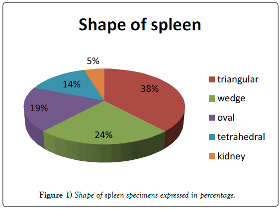
Result: Out of 21 spleen samples various shapes of the spleens with triangular shaped 8 (38.10%), wedge shaped 5 (23.81%), tetrahedral shaped 3 (14.29%), oval shaped 4 (19.04%) and kidney shaped 1 (4.76 %) of the spleens were observed .The lengths of the spleens varied between 9.7 cm to 13.7 cm, with an average of 11.2 cm. Their breadth was between 3.5 and 9.5 cm with an average breadth of 6.6 cm. Their widths varied between 2.3 and 7.3 cm with an average of 5.0 cm. The weights of the spleens showed great variations, ranging between 45.72 and 331.41 gm, with an average of 147.76 gm. Out of 21 spleen samples,18 (85.71%) of spleens had the hilar length varies from 4-8 cm with an average of 4.5 cm. The splenic notches were present on the superior, intermediate and inferior borders. However, accessory spleens were not found at the hilum of the spleen.
The knowledge on the anatomical variations of the spleen is of fundamental importance to the clinicians during the routine clinical examinations of the abdomen.
Keywords
Spleen; Anatomical variation; Morphology; Morphometry
Introduction
Spleen is the largest lymphoid organ in the body that to be found in the left hypochondrial region. It has various shapes and has anterior and posterior poles, diaphragmatic and visceral surfaces, and superior, inferior and intermediate margins. The mainly superior border of the spleen possesses characteristic notches. The normal adult human spleen is about 2.5 cm thick, 7.5 cm broad and 12.5 cm length and about 150 grams weight. Congenital anomalies of the spleen are believed to be rare and include absence of the spleen, spleen lobulation, wandering spleen, polysplenia and accessory spleens [1-3].
The spleen is enclosed by a capsule of uneven thickness that envaginates into the spleen parenchyma as a trabeculae. The splenic tissue between the capsule and trabeculae forms the cords or pulp. Histologically, the cords can be categorized as red or white pulp. Spleen plays an important role in regulating the number and quality of erythrocytes, eliminating cellular debris from the blood, and responding against antigens and/or virulent pathogens that may have entered the systemic circulation [4,5].
The embryonic development of the human spleen is not fully described. However, within the left dorsal mesogastrium around the 5th week of gestation multiple mesenchymal cells aggregate and give rise to lacunae of hematopoietic tissues. By the 8th week, the spleen has a segmented morphology based on arterial lobules, which gradually disappear around week 30, as the spleen develops its lymphoid structures. The immunological role of the spleen is mediated initially by the migration of B-lymphocytes which colonize these lacunae peripherally and then by T lymphocytes centrally around arterioles. As this tissue develops, a few nodules eventually fuse to form the spleen proper. The points of union of these nodules are believed to be the reason behind the splenic notches on its borders [6-8].
An enlarged spleen can be clinically detected in the left hypochondriac region of the abdomen through palpation. The notch on its superior border aids to identify the spleen and differentiate it from other abdominal organs.
Therefore, a variation in the number and location of notches may impede the clinical diagnosis of an enlarged spleen. Although traditional anatomical literature has invariably reported that the spleen has only one or two main notches. The number of notches may vary from one to six. More recently, a case where one spleen had seven notches [9,10].
Materials and Methods
This study was conducted in Department of Human Anatomy, Medical School of Ethiopia from January 15, 2017 to February 15, 2017. A total of 21 spleens obtained from embalmed cadavers found in dissection room were used for this study. All human spleens that have destructed surface and margins by any mechanical, pathological or other conditions were excluded from the study. The spleens were removed from adult human male unknown age cadavers during routine dissection sessions for undergraduate medical students and postgraduate human anatomy students. The spleens were inspected with naked eye in detail for sizes, shapes, surfaces, borders, hilum and presence or absence of accessory spleen.
The presence and number of notches on the superior, inferior and intermediate borders and the presence of fissures on the diaphragmatic or visceral surface were noted. The hilum of the spleen was observed for its presence and length. At the end of inspection, each spleen sample was photographed using 16.1 mega pixel camera.
For morphometric values, the length, width and breadth of the spleen were measured with the help of a measuring tape meter and sliding caliper and the weight of spleens were weighted by electronic digital weight balance. The maximum length was measured between the splenic anterior and posterior poles at the long axis of diaphragmatic surface of the spleen and maximum breadth was determined between the upper and lower border of the spleen at a plane perpendicular to the length. The thickness of the spleen was taken with the help of a slide caliper at maximum anterior- posterior dimension of the spleen that connects the diaphragmatic and visceral surfaces. The length of hilum was measured the longest distance between anterior and posterior poles and also from superior to inferior borders along visceral surface. The quality of data was assured by properly designed checklist, each spleen was examined in three different occasions by investigators and the mean of each value was taken. After Each data collection, the checklist was reviewed and checked for completeness and relevance and the necessary corrections were undertaken.
The entire checklist was checked visually, the data were analyzed by SPSS version 20 and MS office excels 2010. The result was presented in the form of tables, charts, figures and texts using frequencies and summary statistics to describe the relevant variables.
Results
Out of 21 spleens studied, triangular, wedge, tetrahedral, oval and kidney shaped spleens were identified in approximately 38%, 24%, 14%, 19% and 5% of the cases, respectively (Figure 1).
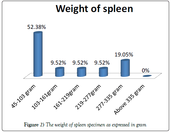
The weight of the spleens ranged from 45.72 to 331.61 grams and the average weight was found to be 147.68 grams (Figure 2).
In this study, the length of the spleens was between 9.7 cm and 13.7 cm with an average length of 11.23 cm. The width the spleen varied between 2.3 cm and 7.3 cm, with an average width of 5.0 cm. The breadth of the spleens was in the range of 3.2 cm to 8.2 cm, with an average breadth of 6.6 cm (Table 1).
| Parameters in cm | Interval of values | Number of spleen | Percent (%) | Mean value (cm) |
|---|---|---|---|---|
| Length of spleen | 9.7-10.7 | 10 | 47.62 | 11.23 |
| 10.7-11.7 | 4 | 19.05 | ||
| 11.7-12.7 | 2 | 9.52 | ||
| 12.7-13.7 | 4 | 19.05 | ||
| ≥ 13.7 | 1 | 4.76 | ||
| Width of spleen | 2.3-3.3 | 6 | 28.58 | 5 |
| 3.3-4.3 | 4 | 19.05 | ||
| 4.3-5.3 | 3 | 14.29 | ||
| 5.3-6.3 | 4 | 19.05 | ||
| 6.3-7.3 | 3 | 14.29 | ||
| ≥ 7.3 | 1 | 4.76 | ||
| Breadth of spleen | 3.2-4.2 | 4 | 19.05 | 6.6 |
| 4.2-5.2 | 0 | 0 | ||
| 5.2-6.2 | 3 | 14.29 | ||
| 6.2-7.2 | 5 | 23.81 | ||
| 7.2-8.2 | 2 | 9.52 | ||
| ≥ 8.2 | 7 | 33.33 |
Table 1: The morphometric values of length, width and breadth cadaveric spleen specimens expressed in centimeter (cm) and mean values of the parameters.
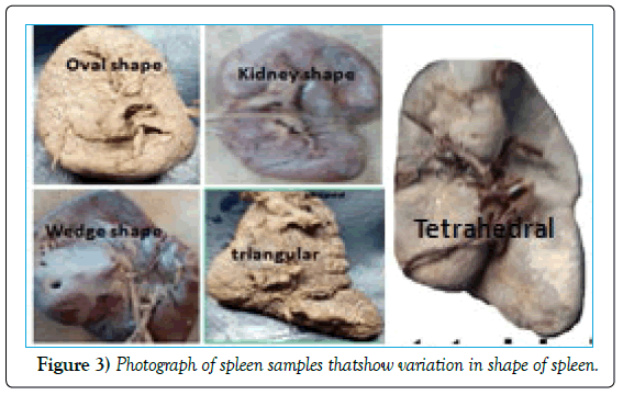
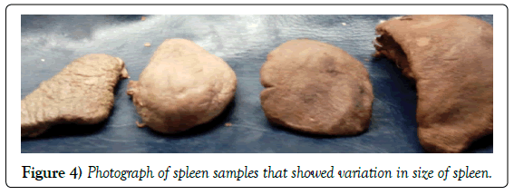
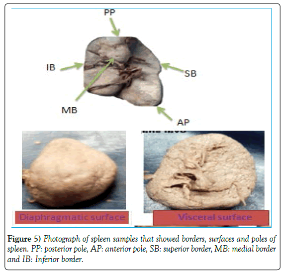
Different shapes and sizes, two poles, three borders and two surfaces of spleen have been observed. The diaphragmatic surface of spleen showed uniform morphology. However, its visceral surface had irregular morphology created for the pressure induced impression by neighboring structures such as stomach, kidney, colon pancreas (Figures 3-5).
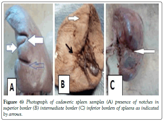
The variations in the number and depth of the splenic notches were observed. The number of spleens showing the notches on the superior and inferior borders were found to be 19 (90.48%) and 13 (61.90%), respectively.
Twelve specimens showed notches situated both on superior and inferior borders, and one specimen has notches on all three borders. Notches were totally absent on all three borders in one specimen (Table 2 and Figure 6).
| Border | Number of notches | Number of spleen | Percent |
|---|---|---|---|
| Superior border | 0 | 2 | 9.52 |
| 1 | 3 | 14.29 | |
| 2 | 9 | 42.86 | |
| 3 | 4 | 19.05 | |
| 4 | 3 | 14.29 | |
| Inferior border | 0 | 8 | 38.10 |
| 1 | 8 | 38.10 | |
| 2 | 5 | 23.81 | |
| Intermediate border | 0 | 20 | 95.24 |
| 1 | 1 | 4.76 | |
| Present in superior and inferior borders | 12 | 57.14 | |
| Present in all three borders | 1 | 4.76 | |
| Absent in all three borders | 1 | 4.76 | |
Table 2: Number of notches in each border of spleen of specimens
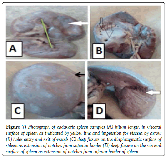
In two specimens, the deep notches were observed on the superior and inferior borders extended towards the diaphragmatic surface in the form of two fissures (Figure 7).
Figure 7) Photograph of cadaveric spleen samples (A) hilum length in visceral surface of spleen as indicated by yellow line and impression for viscera by arrow (B) holes entry and exit of vessels (C) deep fissure on the diaphragmatic surface of spleen as extension of notches from superior border (D) deep fissure on the visceral surface of spleen as extension of notches from inferior border of spleen.
In one specimen, the visceral surface of the spleen has been pierced by several irregularly spaced apertures for the entrance and exit of vessels and nerves in addition to hilum region (Figure 7).
The positions of the hilum in all specimens were more or less straight. The length of the splenic hilum measured ranged from 4.0-8.0 cm with average length of 4.5 cm. However, in 50% of the cases, the spleen has hilar length of greater than six centimeters (Table 3).
| Length of hilum(cm) | Number of spleen | Percent (%) | Mean value |
|---|---|---|---|
| 4.0-5.0 | 4 | 22.22 | |
| 5.0-6.0 | 5 | 27.78 | |
| 6.0-7.0 | 5 | 27.78 | |
| 7.0-8.0 | 4 | 22.22 | |
| Presence of hilum | 18 | 85.71 | |
| Absence of hilum | 3 | 14.29 |
Table 3: Length and presence of hilum of spleen specimens.
A clearly visible hilum was absent in 3 specimen, in which the splenic artery branches were seen to enter the splenic substance through separate perforations on the visceral surface (Table 3 and Figure 8). In all spleen samples that are included in this study, an accessory spleen was not observed.
By considering the length of hilum spleens were classified into three morphological types that are compact, intermediate and distributed types with 4 (22%), 5 (28%) and 9 (50%) respectively (Table 4).
| Type of morphology | Length of hilum (cm) | Number of spleen | Percent (%) |
|---|---|---|---|
| compacted | Less than 5 | 4 | 22 |
| intermediate | 5-6 | 5 | 28 |
| Distributed | Greater than 6 | 9 | 50 |
Table 4: Morphologic types of spleen based on Michels NA classification [23].
Discussion
The proportion of triangular, wedge, tetrahedral and oval shapes spleens of the present study not supported by previous studies. This difference may be due to body composition, sex and socioeconomic status differences across countries [11-22]. All spleen samples with clear hilum of the present study are congruent with the .morphological classification of spleen as distributed, intermediate compacted types by Michels NA [23]. The spleen usually presents a hilum on the visceral surface and the hilum is not covered by the peritoneum. Of the spleens, studied 14% did not have a classic hilum; instead, the blood vessels randomly pierced the visceral surface. Such cases have been reported in the previous study [10].
In the present study, there were variations in spleen length from 9.7 to 13.7 cm, with an average of 11.23 cm and breadth from 4.5 to 12.5 cm, with an average of 6.61.width from 2.3 to 7.3 with an average of 5.0 cm. The weight of the spleen varied from 45.72 to 331.41 gm, with an average of 147.40 gm. In the present study, the morphometry of spleen was compared with previous studies. The values for length, breadth and thickness were comparable with studies by Chaware et al. [16], Rao et al. [17] and Chaudari et al. [15]. The finding on the length, breadth, width and weight of the spleen was almost in line with recently reported studies [15-17].
This study indicates that the presence of notches on the superior, inferior and intermediate borders of spleen in approximately 90%, 62% and 5% of the cases, respectively. Recently reported studies support the notion that both superior and inferior borders of the spleen bear notches [11,14,18,23]. On the other hand, in (5%) spleens specimens notches were totally absent on all borders. Presence of notches on the superior margin is useful for the physician to palpate the spleen during enlargement of spleen [14].
Previous studies regarding the site of notches on the spleen have revealed the presence of notches on the superior order in 95% Girish et al. [18], 70% by Sivanageswara et al. [14], 50% by Chidambaram et al [11] and 70% by Rays et al. [23].
This variability in the presence and number of notches may be explained on developmental basis of the spleen. The spleen develops from the mesoderm during its development from different lobules, which fuse with each other later on. The indication of the lobulation in adult spleen is seen as notches on borders mostly seen on the superior as well as on the inferior borders [11,14,18,23].
Conclusion
This study provides the morphological and morphometric variations of spleen in Ethiopian cadavers similar with previous studies done in other countries. These cadaveric spleens were classified into three morphological types that are distributed, compact and intermediate types by considering its external features and the length of the hilum. It is interesting to note that the three types are present in spleens of all sizes and shapes.
Author’s Contribution
B.T. contributed to conception and design, acquisition of data collection, data entry, and data analysis, interpretation of data and preparation of manuscript. A.M was involved data collection, editing the work and revising the manuscript critically for important intellectual concepts and had given final approval of the version to be published
Conflict of Interest
The authors declare that there is no conflict of interest regarding the publication of this paper.
Acknowledgement
Authors sincerely acknowledge the Department of Human Anatomy Laboratory technicians for their support in sample preparation.
REFERENCES
- Hussein M, Hassan K, Yadav BDUU. Anatomical variations of spleen in north Indian population. Inn J Med Health Sci. 2013;3:190-2.
- Keith L. Moore. Essential clinical anatomy. 2007;160.
- Celiktas M, Ozandac S, Goker P, et al. Sonographic determination of normal spleen size in turkish adults. Int J Morphol. 2015;33:1401-5.
- Pathirana TI, Barton MJ, George M, et al. A rare anomaly of the human spleen with nine notches associated with multiple accessory spleens. A case study, hypothesis on origin and review of clinical significance. Italian Journal of Anatomy and Embryology. 2016;121:188-97.
- Dudek RW. Human Embryology, Development of dijestive system. 2011;pp:115-33 .
- Coetzee T. Clinical anatomy and physiology of the spleen. S Afr Med J. 1982;61:737-46.
- Ushakumari KD, Sushma M, Dash LS. Notches of the spleen. J Evi Based Med. 2014;1:1090-3.
- Kumar V, Kumar N, Jetti R. Unusual fissure on the diaphragmatic surface of the spleen-a case report. Int J Anat Var. 2012;5:96-8.
- Nayak BS, Somayaji S, Soumyank V. A study on the variations of size, shape and external features of the spleen in south indian population. Int J Morphol. 2011;29:675-7.
- Chidambaram RS, Sridhar SS. Morphological variations of spleen: A cadaveric study. J Evi Based Med Health. 2015;2:4248-54.
- Ka R, ASM N. Morphometric study of the postmortem human spleen. J Dhaka Med Col. 2011;20:32-6.
- Gandhi KR, Sao SKC. Spleen with multiple notches: a rare anatomical variant. Int J Students’ Res. 2013;3:24-5.
- Nayak SB. A lobulated spleen with multiple fissures and hila. J Cli Diag Res. 2014;8:8-9.
- Chaudari ML, Maheria PB, Lakhani C. Morphological variations of human spleen and its clinical significance. Int J Med Res Rev. 2014;2:16-9
- Chaware PN, The morphological variations of the human spleen. J Cli Diagn Res. 2012;6:159-62.
- Rao S, Setty S, Katikireddi RS. Morphometricstudy of human spleen. Int J Bio Med Res. 2013;4:3464-8.
- Patil GV, Apoorva D, Sharif J, et al. Study of splenic notches in a human cadaver. Int J Recent Adv Multidiciplinary. 2014;1:1-3.
- S AK, Dakshyani KR, Gangadhar MR. Multiple notches and fissures in the upper border of spleen-a case report. Ind J Med Case Reports. 2014;3:84-6.
- Moore K. The developing human clinically oriented embryology. 1997.
- Pushpalatha M, Sujana M. Accessory spleen: a case report. Int J Cur Res Rev. 2012;4:121-2.
- Sangeeta M, Varalakshmi KL, Ssahana BN. Cadaveric study of morphometry of spleen. J Med Sci. 2015;1:14-7.
- Nicholas MA. Anatomical variation of spleen and splenic artery . Ame J Anat. 1942:21-72.
- Ray SK, Shivhare PR, Mishra B, et al. Morphological study of human spleen. Int J Med Dental Res. 2016;2:89-92.


