Atypical Extensor Medii Proprius – A Case Report
2 Department of Anaesthesiology, All India Institute of Medical Sciences, Rishikesh, Uttarakhand, India
Received: 29-Apr-2022, Manuscript No. ijav-22-4873; Editor assigned: 02-May-2022, Pre QC No. ijav-22-4873 (PQ); Accepted Date: May 20, 2022; Reviewed: 16-May-2022 QC No. ijav-22-4873; Revised: 20-May-2022, Manuscript No. ijav-22-4873 (R); Published: 27-May-2022, DOI: 10.37532/1308-4038.15(5).196
Citation: Suyashi S. Anatomic Variant of Retrogressive Palmaris Longus: A Case Report. Int J Anat Var. 2022;15(4): 178-179.
This open-access article is distributed under the terms of the Creative Commons Attribution Non-Commercial License (CC BY-NC) (http://creativecommons.org/licenses/by-nc/4.0/), which permits reuse, distribution and reproduction of the article, provided that the original work is properly cited and the reuse is restricted to noncommercial purposes. For commercial reuse, contact reprints@pulsus.com
Abstract
The case presented here is of the presence of Extensor medii proprius (EMP) muscle. It is a variation in extensor group of muscles. Anatomic variations in musculotendinous structure of forearm and hand are very substantial in repairing or reconstructing hand injuries. EMP tendon originated from distal third of ulna. Muscle belly was nearly 1.7 cm in width. In the forearm EMP was lying deep to Extensor indicis. Phylogenetic comparisons advocate EMP as evolutionary remnant of normal developmental arrangement. Awareness of anatomy and variations of extensor tendons is important for health care practitioners for correct diagnosis and management of pain, disease and trauma of forearm and hand.
Keywords
Extensor digitorum; Intertendinous connection; Pain; Variation
Introduction
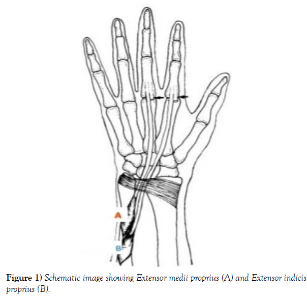
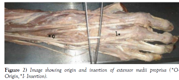
T endons of muscles of extensor compartment of forearm are assorted into six synovial tendon sheaths. They pass deep to extensor retinaculum. Extensor digitorum (ED) and extensor indicis proprius (EIP) lie in the fourth tendon sheath [1]. Variations of extensor muscles and flexor muscles have been reported in the past [2]. However, extensor medii proprius (EMP) has been reported in a very few studies. EMP is variant muscle which is akin to EIP; though EIP gets inserted on second digit of hand while EMP gets inserted on third digit [3] (Figure 1). As per the previous studies, EMP is found in the deep layer of extensor compartment of forearm. It originates from distal 1/3rd of ulna (distal to the origin of extensor indicis muscle) and inserts on dorsal digital expansion of the third digit [4,5] (dorsal aspect of the base of proximal phalanx of middle finger) (Figure 2). EMP has an incidence of nearly 0.8% [6] - 10.3% [3]. According to the systemic review in 2014, prevalence of EMP is remarkably higher in Japanese and North Americans compared to Indians and Europeans [7]. Extensor digiti medii proprius (EDMP) is the other name for EMP [8]. Intensive understanding of comparative myology is very crucial for understanding the functional morphology of muscles of human body. Anatomic variations in musculotendinous structure of forearm and hand are very substantial in repairing or reconstructing hand injuries. Inadequacy of knowledge of these variations can account for false diagnosis leading to incongruous treatment. Moreover, none of the studies till date has reported EMP in Indian population.
Case Report
We found a case of unusual bilateral variant of extensor muscle of forearm in male cadaver of Indian origin, during routine cadaveric dissection sessions of MBBS students in Department of Anatomy, All India Institute of Medical Sciences, and Jodhpur.
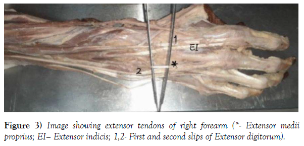
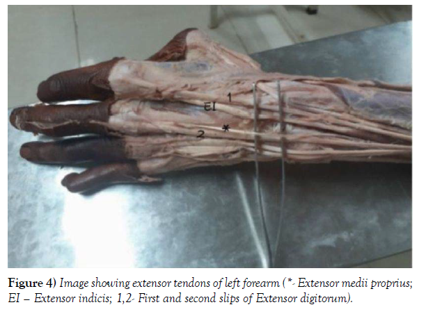
During dissection of extensor compartment of forearm and dorsum of hand, after removing skin and superficial fascia, extensor medii proprius (EMP) was seen bilaterally and was traced till its insertion. EMP tendon originated from distal third of ulna. Muscle belly was nearly 1.7 cm in width. In the forearm EMP was lying deep to Extensor indicis. Distal one-third portion of EMP tendon was very lean, and passed lateral to the second slip (in between extensor indicis and second slip of extensor digitorum), then it passed superficially on the dorsal surface of third dorsal interosseous muscle, and finally it got inserted on the dorsal surface of proximal phalanx of third digit (Figure 3 and 4).
Discussion
Important observations were done by us in our case report. EMP is found in the lower third of forearm and crosses radiocarpal and metacarpophalangeal (MCP) joints and then gets inserted on the third digit. EMP tendon passes between second and third slip of ED; hence the intertendinous connection is significant. Intertendinous connection will hinder bowstringing of EMP during contraction. As EMP crosses MCP, so it is possible for EMP to function as accessory MCP extensor. It is a tiny muscle; hence, its effect on MCP joint extension will be insignificant [1].
Two perspectives are there about EMP; according to some authors EMP is a variant of normal arrangement, whereas, according to others, it’s an evolutionary remnant. Embryologically, precursor extensor muscle of upper and lower limb migrates and differentiates into three segments: radial, superficial and deep. These segments lead to indivisualization of about 14 muscles in lower limb and 19 muscles in upper limb [1,5]. Extensor muscles of antebrachium form extensor digitorum profundus complex. They originate from the shaft of ulna and insertions depend on degree of differentiation of the muscle, and are supplied by posterior interosseous nerve. Extensor muscles of antebrachium crossing MCP joint evolve from superficial and deep categories of extensor muscle mass. As we observed EMP crossing MCP joint, hence EMP has been developed from superficial and deep portions of extensor or dorsal muscle mass. Depending on the topography of EMP, phylogenetic comparisons advocate EMP as evolutionary remnant of normal developmental arrangement [3]. It was found in Old World monkeys. While in chimpanzees and gorillas presence of EMP has been found to be variable as in humans [9].
Therefore, awareness of EMP is necessary for clinicians especially pain physicians, as this will give them wider spectrum if a patient complains of tenderness in forearm. Moreover it will also help surgeons to diagnose precisely and plan the operative measures accordingly. Presence of EMP can aggravate cross syndrome. Cross syndrome is the condition in which pressure gets elevated in the firm fibro-osseous compartment. It generally affects sportspersons like weight lifters and rowers. Repetitive wrist extension and flexion leads to this syndrome. The patient will complain of pain in dorsal forearm and wrist region; proximal to radial styloid process [10].
Conclusion
Knowledge of EMP is important for clinicians and surgeons, to treat functional defect of forearm extensor muscles, Though EMP muscle is rare and causes wrist pain rarely, but it is crucial to know about it before planning to repair or reconstruct via tendon transfer surgeries. Moreover, swelling or tenderness of EMP may be wrongly diagnosed as cyst or tumour on the dorsal part of forearm or hand. Hence, awareness of anatomy and variations of extensor tendons is important for health care practitioners for correct diagnosis and management of disease or trauma of forearm and hand. Understanding of presence of EMP is not only important for medical professionals but is also important for evolutionary biologists.
REFERENCES
- Standring S. Gray’s Anatomy: The Anatomical Basis of Clinical Practice. 41th ed. Edinburgh: Elsevier. 2016; 851-61.
- Ogura T, Inoue H, Tanabe G, et al. Anatomic and clinical studies of the extensor digitorum brevis manus. J Hand Surg Am. 1987; 12(1): 100-7.
- Von Schroeder HP, Botte MJ. The extensor medii proprius and anomalous extensor tendons to the long finger. J Hand Surg Am. 1991; 16(6):1141-5.
- Bergman RA, Thompson SA, Afifi AK. Compendium of human anatomic variation: Text, atlas, and world literature. Baltimore: Urb Schwar. 1988.
- Straus WI. Phylogeny of human forearm extensors. Ann Hum Biol. 1941; 13(5):203-38.
- Cauldwell EW, Anson BJ, Wright RR, et al. The extensor indicis proprius muscle: A study of 263 consequetive specimens. Q Bull Northwest Univ Med School. 1943; 17(4):267-9.
- Yammine K. The prevalence of the extensor indicis tendon and its variants: a systematic review and meta-analysis. Surg Radiol Anat. 2014; 37(3):247–254.
- Ross JA, Troy CA. The clinical significance of the extensor digitorum brevis manus. J Bone Joint Surg. 1969; 51(3):473–8.
- Ayer AA. The anatomy of semnopithecus entellus. Indian Publishing House.1948; 60-72.
- Vaida MA, Gug C, Jianu AM, et al. Bilateral anatomical variations in the extensor compartment of forearm and hand. Surg Radiol Anat. 2020; 43(5):697-702.
Indexed at, Google Scholar, Crossref
Indexed at, Google Scholar, Crossref
Indexed at, Google Scholar, Crossref
Indexed at, Google Scholar, Crossref
Indexed at, Google Scholar, Crossref
Indexed at, Google Scholar, Crossref


