Axillary origin of both radial and ulnar arteries: A cadaveric case report
2 Assistant Professor (Grade III) of the Departamento de Anatomia, Facultad de Medicina - Universidad de la Republica, Montevideo, Uruguay
3 Professor Aggregate (Grade IV) of the Department of Anatomy, Faculty of Medicine, Montevideo, Uruguay
Received: 28-Oct-2018 Accepted Date: Nov 08, 2018; Published: 19-Nov-2018
Citation: Joaquin C, Matilde L, Alejandra M, et al. Axillary origin of both radial and ulnar arteries: A cadaveric case report. Int J Anat Var. Dec 2018;11(4):131-133.
This open-access article is distributed under the terms of the Creative Commons Attribution Non-Commercial License (CC BY-NC) (http://creativecommons.org/licenses/by-nc/4.0/), which permits reuse, distribution and reproduction of the article, provided that the original work is properly cited and the reuse is restricted to noncommercial purposes. For commercial reuse, contact reprints@pulsus.com
Abstract
Variations of the arteries of the upper limb are frequent. Knowledge of these variants is crucial, not only from an anatomic stand point, but also clinically. We report a cadaveric case of proximal origin of both radial and ulnar arteries found during a routine dissection of and adult formalin fixed cadaver. The dis-section was carried out in the Department of Anatomy by the authors. Both arteries originated as terminal branches of the axillary artery. Radial artery fol-lowed a superficial path through the arm whereas the ulnar artery followed the path of the absent brachial artery. Anatomic and clinical implications of this variant are discussed in this paper.
Keywords
Radial artery; Ulnar artery; Axillary origin; Upper limb vessels; Anatomic variation
Introduction
Tariations of the arteries of the upper limb are frequent. They are interesting not only from the anatomical point of view but also surgical, for instance superficial positions of the ulnar and radial arteries make both arteries more vulnerable to trauma and bleeding, but at the same time, more accessible for arteriovenous fistula (AVF) creation [1]. The prevalence of anatomical variations in the arteries of the upper limb varies between 14% to 19.5% [2]. Its occurrence is reported in 1 out of 5 cases [3,4]. Aberrant dispositions do not develop randomly, but rather are a manifestation of more primitive conditions in embryological evolution. The aim of this manuscript is to report a cadaveric case of variant disposition of the upper limb arteries and to discuss its clinical and surgical implications.
Case Report
The variant was found during a routine dissection of an adult formalin fixed right upper limb. The procedure was performed in the Department of Anatomy of the Medical School of Montevideo, Uruguay by the authors.
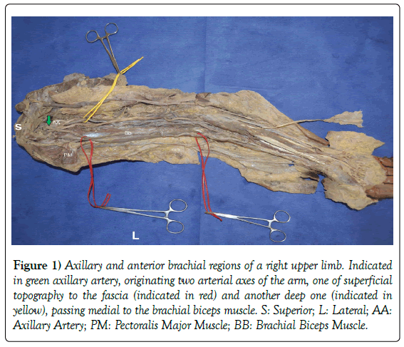
Two arterial axes were found at arm level that originated as terminal branches of the axillary artery (Figure 1). A lateral arterial axis originated from the anterior side of the Axillary Artery, and at arm level it passed anteriorly to the lateral edge of the biceps brachialis muscle, superficial to the fascia of the arm, remaining outside the brachial conduit, to reach the forearm superficially in front of the distal end of the radius, between the tendons of the brachioradialis and flexor carpi radialis, and then followed the typical path of the radial artery (RA) to finish in the hand as the deep palmar arch.
Figure 1) Axillary and anterior brachial regions of a right upper limb. Indicated in green axillary artery, originating two arterial axes of the arm, one of superficial topography to the fascia (indicated in red) and another deep one (indicated in yellow), passing medial to the brachial biceps muscle. S: Superior; L: Lateral; AA: Axillary Artery; PM: Pectoralis Major Muscle; BB: Brachial Biceps Muscle.
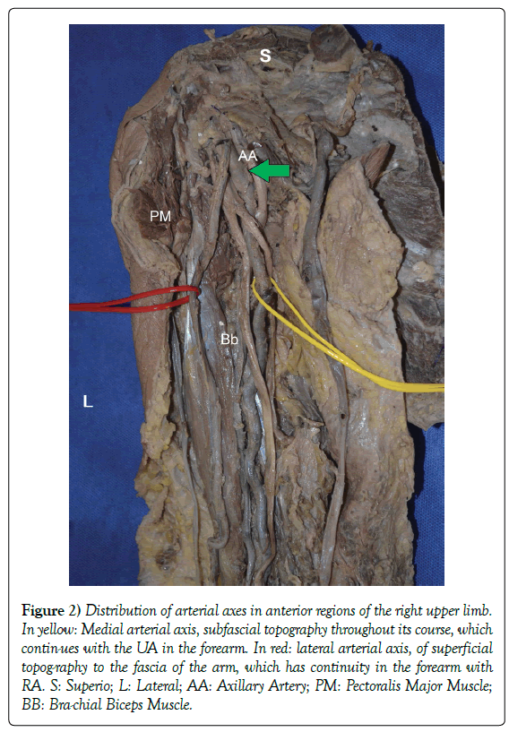
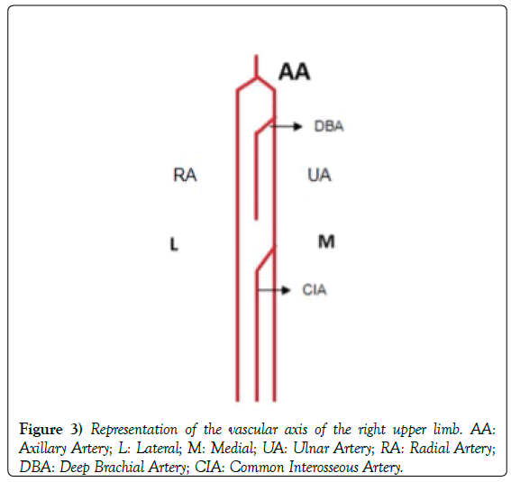
On the other hand, a medial arterial axis, which passed through the anteromedial aspect of the arm mimicking the brachial artery in the arm, to then continue as the usual path of the ulnar artery (Figures 2 and 3).
Figure 2) Distribution of arterial axes in anterior regions of the right upper limb. In yellow: Medial arterial axis, subfascial topography throughout its course, which contin-ues with the UA in the forearm. In red: lateral arterial axis, of superficial topog-raphy to the fascia of the arm, which has continuity in the forearm with RA. S: Superio; L: Lateral; AA: Axillary Artery; PM: Pectoralis Major Muscle; BB: Bra-chial Biceps Muscle.
Discussion
Variations in the origin and course of brachial, radial and ulnar arteries are not uncommon and have been described previously by various authors many times. For exapmle, Stanchev et al. [5] describes a case were a right axillary artery bifurcated into superficial brachial artery and deep brachial artery. Iliev et al. [6] found a posterior circumflex humeral arising as a branch of the brachial artery, and originating the deep brachial artery. But a superficial course of the radial artery is a rare variation that has many clinical implications, such as being more vulnerable to trauma.
Von Haller was the first to mention the variant here presented in the eighteenth century [7]. Contemporary authors mention that the high origin of the radial artery is the variation most frequently observed with a frequency of 14 to 19.5% [2,3]. The RA may not exist, or have its origin in the axillary artery, as in our case (2.13%) or in the brachial artery (12.4%). The high origin of the UA occurs in 2% [2,3]. Other authors such as Mehta et al. [4] and Frydman et al. [8] propose instead that this variant is a persistence of the superficial brachioradial artery, embryonic remnant of the superficial arterial system, which normally involutes.
In the case reported, we found two arterial axes that after originating from the axillary artery run through the arm (Figure 1). One of them taking the usual course of the brachial artery, originating the usual collateral branches of this artery, but that unlike the classic arrangement does not end bifurcating in two arteries at the level of the cubital fossa, but continues the path of the UA in the forearm. While the second arterial axis, has a superficial course, and continues as the RA in the forearm (Figures 2 and 3).
The classic anatomical texts (Testut, Latarjet- Ruiz Liard) [9,10] define the brachial artery as the continuation of the axillary artery, extended from the lower edge of the pectoralis major to the medial portion of the cubital fossa, where it divides into its two terminal branches: the RA and the UA. Following this definition, in the case here presented the lack of brachial artery seems clear.
The variations in the definitive arterial system are, for the most part, justified by the persistence or growth of the vessels that make up the primitive arterial sys-tem [7]. These variations represent a transient embryonic stage that persists in postna-tal life, which would usually suffer an involution [8].
Edward Singer [11] describes 5 stages in the arterial development of the upper limb that could explain the arterial variations discussed.
Initially, the subclavian artery extends to the wrist where it divides into two terminal branches for the fingers (stage I). The distal portions of this artery will become the interosseous of adults. The latter involutes and a median artery arises which anastomoses with the most distal portion of the interosseous artery and form the main channel for the digital branches, becoming the main artery of the forearm (Stage II). Then, the UA arises from the superficial brachial artery (continuation of the subclavian artery in the brachial region) and joins distally with the median artery to form the superficial palmar arch (stage III). This superficial brachial artery that arises from the main arterial trunk in the axillary region and crosses the middle surface of the arm, crosses the forearm from medial to lateral until it reaches the dorsal region of the wrist (Stage IV).
Finally, the median artery undergoes regression, becoming smaller and becoming the artery of the median nerve. The superficial brachial artery emits a distal branch that anastomoses with the superficial palmar arch. In the elbow, the brachial and brachial superficial arteries are anastomosed, forming the RA as the main forearm vessel from the distal portion of the latter. The proximal portion of the superficial brachial artery involutes (stage V) [8].
Following the exposed by Adachi [12], The superficial brachial artery owes this denomination for running superficial to the median nerve. The superficial brachial artery can, in a few cases, replace the main arterial trunk.
On the other hand, following Rodriguez-Baeza [13] the persistence of the superficial brachial artery with respect to Singer’s theory, is that the superficial brachial artery has hemodynamic predominance on the trunk of deep origin of the radial artery. In this way, the superficial brachial artery would not suffer regression and this explains its eventual persistence in the adult [14].
In the present case, it could be considered that the axillary artery emits the superficial brachial artery, which appears in the IV stage of Singer and continues to descend as the RA. There is an involution of the anastomotic branch be-tween the superficial brachial artery and the brachial artery at the cubital fossa, the superficial brachial artery persists and is continued as a RA occupying a superficial disposition in the arm and elbow.
Variations in blood vessels’ branching pattern, position or course can alter routine clinical procedures, like blood pressure monitoring or intravenous drug application. They also can create difficulties in complex interventions, such as cardiac catheterization, flap surgery or amputations [15].
From a surgical stand point, the presence of arterial variants in the upper limb may affect outcome in vascular access creation, including high failure rate and decreased functional patency of the AVF. This makes mandatory to investigate the presence of variants of the upper limb´s vascular tree prior to AVF creation and may influence access site selection [1]. The variation here presented is not an exception to this statement.
In conclusion, we present a cadaveric case of axillary origin of both RA and UA, this variant cannot be missed by both anatomists and clinicians, since it’s a quite frequent variation found both in cadavers and in the clinical scenario and may predispose to arterial injury and modify vascular access selection.
Acknowledgement
To the Anatomy Department of Medical School, UdelaR. Montevideo, Uruguay.
REFERENCES
- Kusztal M, Weyde W, Letachowicz K, et al. Anatomical vascular variations and practical implications for access creation on the upper limb. J Vas Access. 2014;15:S70-5.
- Medina Ruiz, Blás A, Mena Canata, et al. High origin of the radial artery-Case report and literature review. Rev Argent Anat Online. 2015;1:8-11.
- Medina RBA, Mena CCE, Romano S, et al. Clinical and surgical implications of the high origin of the radial artery. Int J Med Surg Sci. 2016;3:811-7.
- Mehta Vandana, Arora Jyoti, Suri RK, et al. Unilateral anomalous arterial pattern of human upper limb. Sultan Qaboos Univ Med J. 2008; 8:227-30.
- Stanchev AS, Iliev A, Georgiev GP, et al. А case of bilateral variations in the arterial branching in the upper limb and clinical implications. Chronicle J Clin Case Rep. 2017;1:006.
- Iliev BAA, Mitrov LG, Georgiev GP. A variation of the origin and course of the posterior circumflex humeral artery and the deep brachial artery. Clinical importance of the variation. J Biomed Clin Res. 2015;8:164-7.
- Rodríguez Niedenfuhr M, Vazquez T, Parkin I, et al. Arterial pat-terns of the human upper limb: update of anatomical variations and embryological development. Eur J Anat. 2003;7:21-8.
- Frydman J, Ostolaza M, Maroni MC, et al. Axillary origin of the radial artery. Rev Argent Anat Online. 2013;4:64-9.
- Testut L, Latarjet A. Human anatomy treaty. Angiology. 1984; 11:277-312.
- Latarjet M, Ruiz Liard A. Human anatomy. Medical Publishing Panamerican. 2004;pp:605-8.
- Singer E. Embryological pattern persisting in the arteries of the arm. The Anatomical Record. 1933;55:403-9.
- Adachi B. The arteries system of the Japanese. Kyoto. 1928;1:205-10.
- Rodríguez Baeza A, Nebot J, Ferreira B, et al. An anatomical study and ontogenic explanation of 23 cases with variations in main pattern of brachio antebraquial arteries. J Anat. 1995;187:473-9.
- Patnaik VVG, Kalsey G, Singla Rajan K. Bifurcation of Axillary Artery in its 3rd Part- A Case Report. J Anat Soc. 2001;50:166-9.
- Georgiev GP. Significance of anatomical variations for clinical practice. Inter J Anat Var. 2017;10:43-4.


