Bicipital origin of plantaris muscle – a case report
1Department of Anatomy, Government Medical College, Patiala, Punjab, India
2Department of Surgery, Government Medical College, Patiala, Punjab, India
- *Corresponding Author:
- Dr. Upasna
C2, Medical College Campus Govt. Medical College Patiala, Punjab, India
Tel: +91 987 2624818
E-mail: ashwaniupasna@yahoo.com
Date of Received: March 12th, 2011
Date of Accepted: October 6th, 2011
Published Online: October 12th, 2011
© Int J Anat Var (IJAV). 2011; 4: 177–179.
[ft_below_content] =>Keywords
plantaris, bicipital origin, variations, tennis leg
Introduction
Superficial flexors gastrocnemius, plantaris and soleus form the bulk of the calf. The small plantaris muscle is the lower limb equivalent of palmaris longus. It arises from the lower part of the lateral supracondylar line and the oblique popliteal ligament. Its fusiform belly is 7 to 10 cm long and ends in a long slender tendon which crosses obliquely between gastrocnemius and soleus, runs distally along the medial border of the calcaneal tendon and fuses or inserts with it [1]. Because of this long narrow tendon, it has been referred to as the freshman’s nerve. If it has origin from any two of the mentioned variations it is said to be bicipital [2].
Case Report
During dissection of lower limb of a middle aged female cadaver, bicipital origin of the plantaris muscle was observed on both sides. The muscle was displayed by careful dissection. Morphometric measurements were taken and the specimen was photographed.
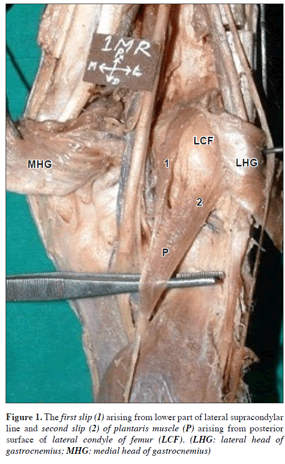
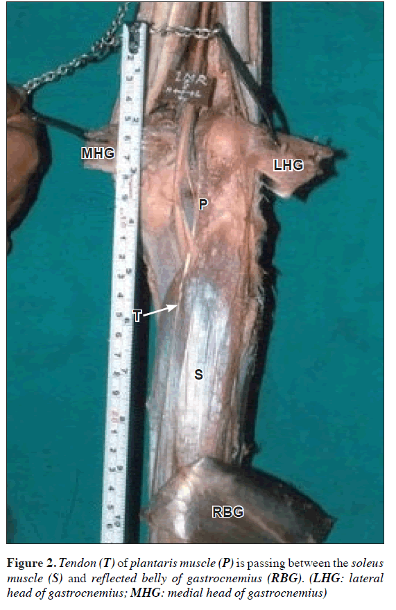
The first slip was arising from lower part of lateral supracondylar line and second slip was arising from posterior surface of lateral condyle of femur (LCF), some fibers of which were merging with the lateral head of gastrocnemius muscle (Figure 1). Tendon of plantaris muscle was passing between the soleus muscle and reflected belly of gastrocnemius. Tendon merged with medial border of calcaneal tendon (Figure 2). Nerve supply was from the tibial nerve (S1, S2).
Discussion
The phylogenetic trend in the foot is towards separation of musculature of the foot from that of the calf in the interest of efficiency in walking, the example being superficial flexors [3].
The plantaris is exceedingly variable in origin. It may take origin from the inferior extremity of the external limb of the linea aspera; the posterior ligament of the knee at the intercondylar space; the fascial covering of the popliteus; the fibula, between the flexor hallucis longus and the peroneus longus; the oblique line of the tibia, under cover of the soleus; the fascia of the leg; the lateral condyle of the femur above the origin of the lateral head of the gastrocnemius and when bicipital, any two of the above mentioned areas [2].
The small plantaris muscle, with its long slender tendon, is of interest both from anatomical and phylogenetic viewpoints. The muscle is regarded as vestigial in man, believing that with the assumption of an erect posture, the tendon lost its original insertion into the plantar aponeurosis and gained a secondary calcaneal attachment [2].
Embryological development in man supports the idea advocated by McMurrich that the plantaris is a derivative of the deeper portion of the lateral head of the gastrocnemius. When absent, it is likely that this separation has failed to take place during ontogeny. In many mammals, it is not differentiated (several edentates, carnivores, etc.); in others, especially in some rodents, it is highly developed [4].
The plantaris muscle itself can be a third head and may join gastrocnemius at a point where medial and lateral heads separate or may become continuous with the fascia beneath the muscle [5]. In our case, one of the slips (outer) was merging with few fibers of lateral head of gastrocnemius. Thus, it can be called the third head of gastrocnemius but the muscle fibers of this slip of plantaris muscle lower down merged with common belly of plantaris muscle. It is in consonance with the idea advocated by McMurrich that the plantaris is a derivative of the deeper portion of the lateral head of the gastrocnemius [6].
In terms of function the plantaris muscle acts with the gastrocnemius but is insignificant as either a flexor of the knee of a plantar flexor of the ankle. It has been considered to be an organ of proprioceptive function for the larger, more powerful plantar flexors as it contains a high density of muscle spindles [7]. Despite its small size injuries of plantaris muscle and tendon have been termed tennis leg. It has now been established through the use of MRI, sonography and surgical exploration that injuries to this muscle may in fact occur in isolation as well as in association with tears of gastrocnemius, soleus and anterior cruciate ligament. Proper diagnosis can be made by MRI scan or ultrasound [8].
Free plantaris tendon graft for reconstruction of anterior talofibular and calcaneofibular ligaments has been used [9]. It has been used successfully for flexor tendon replacement in hands and even for atrioventricular valve repair [10]. Thus, the existence and importance of bicipital origin of plantaris can be of academic, surgical and clinical interest.
References
- Standring S, ed. Gray’s Anatomy. The Anatomical Basis of Clinical Practice. 39th Ed., Philadelphia, Elsevier Churchill Livingstone. 2005; 1499.
- Daseler EH, Anson BJ. The plantaris muscle: an anatomical study of 750 specimens. J Bone Joint Surg Am. 1943; 25: 822-827.
- Barlow TE. The deep flexors of the foot. J Anat. 1953; 87: 308–310.
- Bardeen CR. Development and variation of the nerves and the musculature of the inferior extremity and of the neighboring regions of the trunk in man. Am J Anat. 1906; 6: 259–390.
- Bergman RA, Afifi AK, Miyauchi R. Illustrated Encyclopedia of Human Anatomic Variation: Opus I: Muscular System: Alphabetical Listing of Muscles: P: Plantaris. http://www.anatomyatlases.org/AnatomicVariants/MuscularSystem/Text/P/29Plantaris.shtml (accessed October 2011)
- McMurrich JP. The phylogeny of the crural flexors. Am J Anat. 1905; 4: 33–76.
- Moore KL, Dalley AF. Clinically Oriented Anatomy. 5. Ed., Philadelphia, Lipponcott Williams and Wilkins. 2006; 648–649.
- Spina AA. The plantaris muscle: anatomy, injury, imaging and treatment. J Can Chiropr Assoc. 2007; 51: 158–165.
- Pagenstert GI, Valderrabano V, Hintermann B. Lateral ankle ligament reconstruction with free plantaris tendon graft. Techniques in Foot & Ankle Surgery. 2005; 4: 104–112.
- Shuhaiber JH, Shuhaiber HH. Plantaris tendon graft for atrioventricular valve repair: a novel hypothetical technique. Tex Heart Inst J. 2003; 30: 42–44.
1Department of Anatomy, Government Medical College, Patiala, Punjab, India
2Department of Surgery, Government Medical College, Patiala, Punjab, India
- *Corresponding Author:
- Dr. Upasna
C2, Medical College Campus Govt. Medical College Patiala, Punjab, India
Tel: +91 987 2624818
E-mail: ashwaniupasna@yahoo.com
Date of Received: March 12th, 2011
Date of Accepted: October 6th, 2011
Published Online: October 12th, 2011
© Int J Anat Var (IJAV). 2011; 4: 177–179.
Abstract
The superficial flexors gastrocnemius, plantaris and soleus form the bulk of calf. The plantaris muscle is exceedingly variable in origin. Here, we present a case report where it originated by two slips, first slip from lower part of lateral supracondylar line (usual) and second slip arising from posterior surface of lateral condyle of femur (variant), some fibers of which were merging with the lateral head of gastrocnemius muscle. The existence and importance of bicipital origin of plantaris muscle can be of academic, surgical and clinical interest as it has been established by MRI scan and ultrasonography that injuries to this muscle can occur in isolation (tennis leg). Plantaris tendon grafts have been used successfully in reconstruction of anterior talofibular and calcaneofibular ligaments, for flexor tendon replacement in hand and for atrioventricular valve repair.
-Keywords
plantaris, bicipital origin, variations, tennis leg
Introduction
Superficial flexors gastrocnemius, plantaris and soleus form the bulk of the calf. The small plantaris muscle is the lower limb equivalent of palmaris longus. It arises from the lower part of the lateral supracondylar line and the oblique popliteal ligament. Its fusiform belly is 7 to 10 cm long and ends in a long slender tendon which crosses obliquely between gastrocnemius and soleus, runs distally along the medial border of the calcaneal tendon and fuses or inserts with it [1]. Because of this long narrow tendon, it has been referred to as the freshman’s nerve. If it has origin from any two of the mentioned variations it is said to be bicipital [2].
Case Report
During dissection of lower limb of a middle aged female cadaver, bicipital origin of the plantaris muscle was observed on both sides. The muscle was displayed by careful dissection. Morphometric measurements were taken and the specimen was photographed.
The first slip was arising from lower part of lateral supracondylar line and second slip was arising from posterior surface of lateral condyle of femur (LCF), some fibers of which were merging with the lateral head of gastrocnemius muscle (Figure 1). Tendon of plantaris muscle was passing between the soleus muscle and reflected belly of gastrocnemius. Tendon merged with medial border of calcaneal tendon (Figure 2). Nerve supply was from the tibial nerve (S1, S2).
Discussion
The phylogenetic trend in the foot is towards separation of musculature of the foot from that of the calf in the interest of efficiency in walking, the example being superficial flexors [3].
The plantaris is exceedingly variable in origin. It may take origin from the inferior extremity of the external limb of the linea aspera; the posterior ligament of the knee at the intercondylar space; the fascial covering of the popliteus; the fibula, between the flexor hallucis longus and the peroneus longus; the oblique line of the tibia, under cover of the soleus; the fascia of the leg; the lateral condyle of the femur above the origin of the lateral head of the gastrocnemius and when bicipital, any two of the above mentioned areas [2].
The small plantaris muscle, with its long slender tendon, is of interest both from anatomical and phylogenetic viewpoints. The muscle is regarded as vestigial in man, believing that with the assumption of an erect posture, the tendon lost its original insertion into the plantar aponeurosis and gained a secondary calcaneal attachment [2].
Embryological development in man supports the idea advocated by McMurrich that the plantaris is a derivative of the deeper portion of the lateral head of the gastrocnemius. When absent, it is likely that this separation has failed to take place during ontogeny. In many mammals, it is not differentiated (several edentates, carnivores, etc.); in others, especially in some rodents, it is highly developed [4].
The plantaris muscle itself can be a third head and may join gastrocnemius at a point where medial and lateral heads separate or may become continuous with the fascia beneath the muscle [5]. In our case, one of the slips (outer) was merging with few fibers of lateral head of gastrocnemius. Thus, it can be called the third head of gastrocnemius but the muscle fibers of this slip of plantaris muscle lower down merged with common belly of plantaris muscle. It is in consonance with the idea advocated by McMurrich that the plantaris is a derivative of the deeper portion of the lateral head of the gastrocnemius [6].
In terms of function the plantaris muscle acts with the gastrocnemius but is insignificant as either a flexor of the knee of a plantar flexor of the ankle. It has been considered to be an organ of proprioceptive function for the larger, more powerful plantar flexors as it contains a high density of muscle spindles [7]. Despite its small size injuries of plantaris muscle and tendon have been termed tennis leg. It has now been established through the use of MRI, sonography and surgical exploration that injuries to this muscle may in fact occur in isolation as well as in association with tears of gastrocnemius, soleus and anterior cruciate ligament. Proper diagnosis can be made by MRI scan or ultrasound [8].
Free plantaris tendon graft for reconstruction of anterior talofibular and calcaneofibular ligaments has been used [9]. It has been used successfully for flexor tendon replacement in hands and even for atrioventricular valve repair [10]. Thus, the existence and importance of bicipital origin of plantaris can be of academic, surgical and clinical interest.
References
- Standring S, ed. Gray’s Anatomy. The Anatomical Basis of Clinical Practice. 39th Ed., Philadelphia, Elsevier Churchill Livingstone. 2005; 1499.
- Daseler EH, Anson BJ. The plantaris muscle: an anatomical study of 750 specimens. J Bone Joint Surg Am. 1943; 25: 822-827.
- Barlow TE. The deep flexors of the foot. J Anat. 1953; 87: 308–310.
- Bardeen CR. Development and variation of the nerves and the musculature of the inferior extremity and of the neighboring regions of the trunk in man. Am J Anat. 1906; 6: 259–390.
- Bergman RA, Afifi AK, Miyauchi R. Illustrated Encyclopedia of Human Anatomic Variation: Opus I: Muscular System: Alphabetical Listing of Muscles: P: Plantaris. http://www.anatomyatlases.org/AnatomicVariants/MuscularSystem/Text/P/29Plantaris.shtml (accessed October 2011)
- McMurrich JP. The phylogeny of the crural flexors. Am J Anat. 1905; 4: 33–76.
- Moore KL, Dalley AF. Clinically Oriented Anatomy. 5. Ed., Philadelphia, Lipponcott Williams and Wilkins. 2006; 648–649.
- Spina AA. The plantaris muscle: anatomy, injury, imaging and treatment. J Can Chiropr Assoc. 2007; 51: 158–165.
- Pagenstert GI, Valderrabano V, Hintermann B. Lateral ankle ligament reconstruction with free plantaris tendon graft. Techniques in Foot & Ankle Surgery. 2005; 4: 104–112.
- Shuhaiber JH, Shuhaiber HH. Plantaris tendon graft for atrioventricular valve repair: a novel hypothetical technique. Tex Heart Inst J. 2003; 30: 42–44.


