Bifurcation of axillary artery – an unusual case
Received: 15-Oct-2012 Accepted Date: Jan 08, 2013; Published: 25-Sep-2013
Citation: © Int J Anat Var (IJAV). 2013; 6:161–164.
This open-access article is distributed under the terms of the Creative Commons Attribution Non-Commercial License (CC BY-NC) (http://creativecommons.org/licenses/by-nc/4.0/), which permits reuse, distribution and reproduction of the article, provided that the original work is properly cited and the reuse is restricted to noncommercial purposes. For commercial reuse, contact reprints@pulsus.com
Abstract
A male cadaver (approximately 60 years old) was seen to have a variant pattern of axillary artery branching.
On the right side, the axillary artery bifurcated behind the pectoralis minor into two trunks – superficial and deep. The median nerve was formed by the union of the two roots between these trunks. The deep trunk trifurcated in the upper arm into subscapular, a common trunk which gave origin to anterior and posteror circumflex humeral arteries, a branch that ran distally that gave rise to profunda brachii artery. The superficial trunk continued as the brachial artery and it bifurcated at the level of head of radius into radial and ulnar arteries.
On the left side, the axillary artery branching pattern was as usual.
The implication of this study occurs in various streams especially vascular surgery, general surgery, as well as in radiodiagnosis in Doppler and contrast imaging of vessels.
Keywords
axillary artery; bifurcation; variation; vascular
Introduction
Axillary artery is a continuation of the subclavian artery. It begins at the outer border of the first rib, and ends nominally at the inferior border of the teres major muscle, after which it is named the brachial artery. Pectoralis minor crosses the artery superficially and so divides it into three parts which are proximal (1st part), posterior (2nd part) and distal (3rd part). The branches of the artery are superior thoracic (from the 1st part); thoraco-acromial and lateral thoracic (from 2nd part) and subscapular and anterior and posterior circumflex humeral arteries from the third part [1].
The authors of the present paper intended to focus on the deviation from this classical pattern of vascular events at the axilla. This might have important clinical application especially in the management of vascular emergencies, where the accurate diagnosis and surgical repair of vessels is vital for satisfactory results. The necessary morphometric details found on dissection are also provided.
Case Report
The case was seen during routine undergraduate cadaveric dissection. It was a male cadaver of approximately 60 years of age. There were no obvious external malformations seen in the limbs. The axillae were dissected on both sides and the neurovascular structures were carefully traced. The following points were noted.
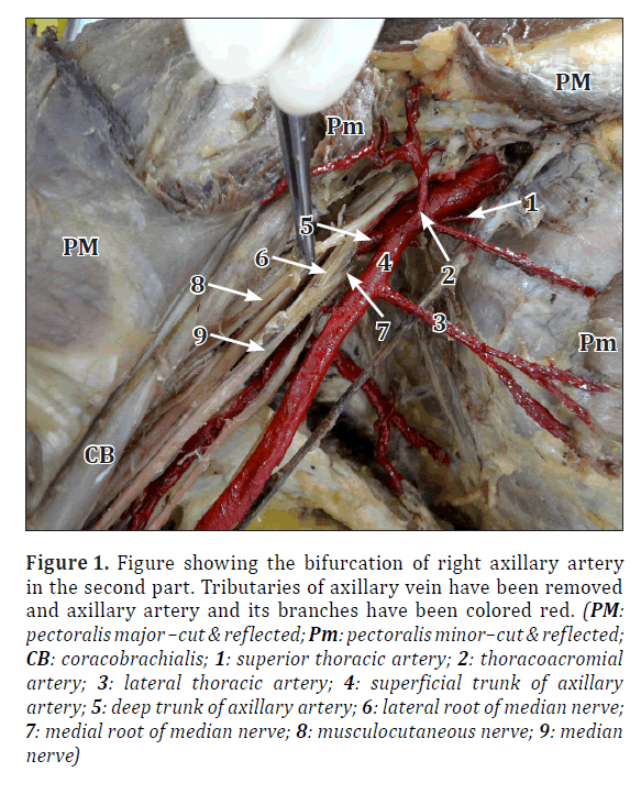
On the right side, the axillary artery was bifurcated into two almost similar caliber vessels, at the part behind the pectoralis minor, i.e., 2nd part (Figure 1, Table 1). The first part of axillary artery had an external circumference of 3.6 cm. The superficial axillary artery had an external circumference of 2 cm. The deep axillary artery had an external circumference of 2.5 cm.
Figure 1. Figure showing the bifurcation of right axillary artery in the second part. Tributaries of axillary vein have been removed and axillary artery and its branches have been colored red. (PM: pectoralis major –cut & reflected; Pm: pectoralis minor–cut & reflected; CB: coracobrachialis; 1: superior thoracic artery; 2: thoracoacromial artery; 3: lateral thoracic artery; 4: superficial trunk of axillary artery; 5: deep trunk of axillary artery; 6: lateral root of median nerve; 7: medial root of median nerve; 8: musculocutaneous nerve; 9: median nerve)
| Parameter | Left side | Right side | ||
|---|---|---|---|---|
| External circumference | 1st part of axillary a. | 3.3 cm | 3.6 cm | |
| 3rd part of axillary a. | 2.8 cm | Superficial axillary a. | 2 cm | |
| Deep axillary a. | 2.5 cm | |||
| Distance from outer border of first rib to bifurcation | N/A | 5 cm | ||
| Distance from bifurcation to trifurcation | N/A | 3.4 cm | ||
External circumferences were measured using a twine and measuring with a centimeter scale. Three readings were taken and average was obtained to avoid intraobserver variability.
Table 1. Morphometric details of vascular events.
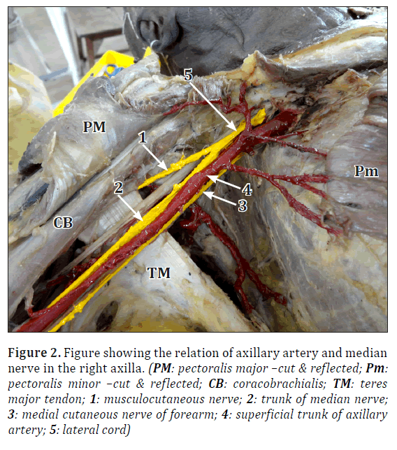
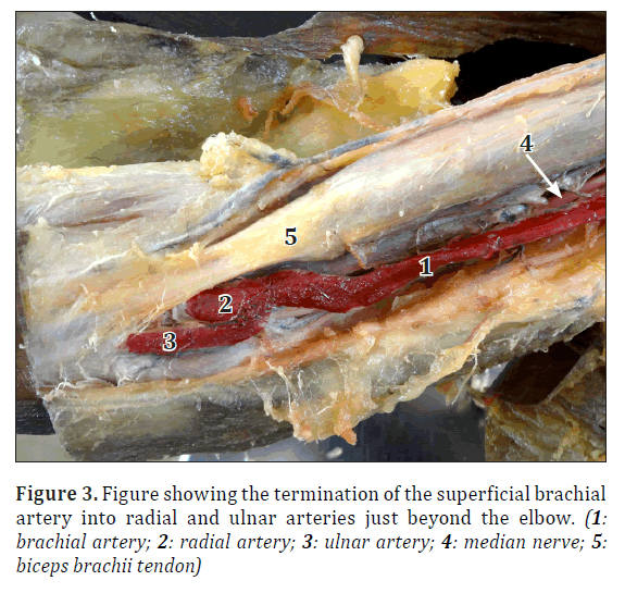
The superficial branch of the axillary artery continued distally as the brachial artery, but was seen superficial to the median nerve trunk (Figure 2). It bifurcated into radial and ulnar arteries below the elbow at the level of the head of radius (Figure 3). The rest of the course of the arteries was in accordance with the classic description.
Figure 2. Figure showing the relation of axillary artery and median nerve in the right axilla. (PM: pectoralis major –cut & reflected; Pm: pectoralis minor –cut & reflected; CB: coracobrachialis; TM: teres major tendon; 1: musculocutaneous nerve; 2: trunk of median nerve; 3: medial cutaneous nerve of forearm; 4: superficial trunk of axillary artery; 5: lateral cord)
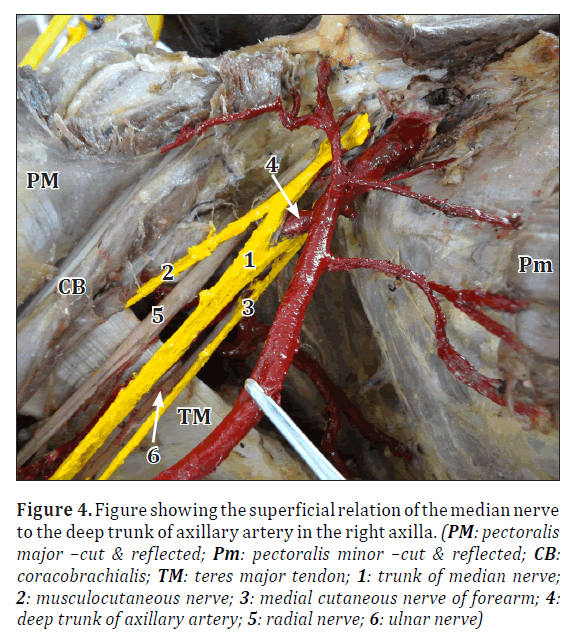
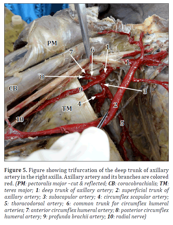
The deep axillary artery was seen to pass distally between and deep to the fork of the median nerve roots (Figure 4). The deep axillary artery was seen to trifurcate 3.4 cm distal to the axillary artery bifurcation (Figure 5). The branches were:
Figure 4. Figure showing the superficial relation of the median nerve to the deep trunk of axillary artery in the right axilla. (PM: pectoralis major –cut & reflected; Pm: pectoralis minor –cut & reflected; CB: coracobrachialis; TM: teres major tendon; 1: trunk of median nerve; 2: musculocutaneous nerve; 3: medial cutaneous nerve of forearm; 4: deep trunk of axillary artery; 5: radial nerve; 6: ulnar nerve)
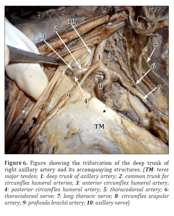
Subscapular artery, which gave rise to circumflex • scapular artery and thoracodorsal artery (accompanying thoracodorsal nerve) (Figures 5, 6).
Figure 5. Figure showing trifurcation of the deep trunk of axillary artery in the right axilla. Axillary artery and its branches are colored red. (PM: pectoralis major –cut & reflected; CB: coracobrachialis; TM: teres major; 1: deep trunk of axillary artery; 2: superficial trunk of axillary artery; 3: subscapular artery; 4: circumflex scapular artery; 5: thoracodorsal artery; 6: common trunk for circumflex humeral arteries; 7: anterior circumflex humeral artery; 8: posterior circumflex humeral artery; 9: profunda brachii artery; 10: radial nerve)
Figure 6. Figure showing the trifurcation of the deep trunk of right axillary artery and its accompanying structures. (TM: teres major tendon; 1: deep trunk of axillary artery; 2: common trunk for circumflex humeral arteries; 3: anterior circumflex humeral artery; 4: posterior circumflex humeral artery; 5: thoracodorsal artery; 6: thoracodorsal nerve; 7: long thoracic nerve; 8: circumflex scapular artery; 9: profunda brachii artery; 10: axillary nerve)
• A common trunk which gave rise to posterior (accompanying axillary nerve) and anterior circumflex humeral arteries (Figures 5, 6).
• A branch that ran distally from which arose profunda brachii artery (accompanying radial nerve) (Figures 5, 6).
Discussion
Variations in branching pattern of axillary artery have been reported in the past. Maraspin (1971) has reported the bifurcation of the second part of the axillary artery into superficial brachial and deep brachiothoracic branches [2].
Patnaik (2001) reported a case of bifurcation of axillary artery in its 3rd part [3]. Jurjus et al. (1999) observed bifurcation of the second part of the axillary artery into two medium sized arteries [4]. Compta (1991) noticed the division of third part of axillary artery into an anterior branch, which constituted the high origin of radial artery and a posterior branch which was the proper brachial artery [5]. Hollinshead’s textbook of anatomy describes the bifurcation of axillary artery into two branches that proceed down the arm, as a very rare, but striking anomaly. When this occurs, usually one of the arterial stems in the arm runs more superficially than does the other, and they are, therefore, sometimes distinguished as brachial artery and superficial brachial artery. This apparent doubling of the brachial artery is said to occur more commonly in the arm than in the axilla [6].The relationship of the bifurcated trunks with the median nerve is quite significant. Formation of median nerve behind the axillary artery has been commented as a rare occurrence [7].Gray’s Anatomy describes that occasionally the subscapular, circumflex humeral and profunda brachii arteries arises in common, in which case, branches of the brachial plexus surround this common vessel instead of the axillary artery [1].
The embryological background of this event has been hypothesized in similar case reports in the past. Jurjus et al. (1999) stated that the presence of a superficial brachial artery is based on the persistence of more than one intersegmental cervical artery, which does not deteriorate but instead remains and can even grow larger [8].
The variation described here is of great significance for vascular and cardiothoracic surgeons. Such a variant formation has to be confirmed in angiographic studies prior to coronary artery bypass grafting to avoid iatrogenic accidents. The variant anatomy is also significant during axillary dissection in breast cancer treatment. The different disposition of cords and branches of brachial plexus (especially the relation of median nerve, in this case) around the deep trunk of axillary artery is of much significance during arm surgeries, axillary dissections and brachial plexus blocks.
Acknowledgement
The authors thank the entire staff of the Department of Anatomy of Trivandrum Medical College. We also thank the students of the 2012 admission MBBS batch.
REFERENCES
- Standring S, ed. Gray’s Anatomy. The anatomic basis of clinical practice. 40th Ed., Elsevier.2008; 815–817.
- Maraspin LE. A report of an anomalous bifurcation of the right axillary artery in man. Vasc Surg. 1971;5: 133–136.
- Patnaik VVG, Kalsey G, Singla RK. Bifurcation of axillary artery in its 3rd part - a case report. J Anat Soc India. 2001; 50: 166–169.
- Jurjus A, Sfeir R, Bezirdjian R. Unusual variation of the arterial pattern the human upper limb. Anat Rec. 1986; 215: 82–83.
- Gonzalez-Compta X. Origin of the radial artery from the axillary artery and associated hand vascular anomalies. J Hand Surg Am. 1991; 16: 293–296.
- Hollinshead WH, Rosse C. Textbook of Anatomy. 4th Ed., Philadelphia Harper and Row Publishers.1985; 189.
- Chitra R. A rare variant formation of median nerve: a case report. Internet Journal of Neurology. 2007; 8(2).
- Jurjus AR, Correa-De-Aruaujo R, Bohn RC. Bilateral double axillary artery: Embryological basis and clinical implications. Clin Anat. 1999; 12:135–140.


