Bilateral absence of quadratus femoris and semimembranosus
Hao (Howe) Liu1*, James Fletcher2, M Kevin Garrison2 and Clayton Holmes1
1University of North Texas Health Science Center Physical Therapy Program, Forth Worth, TX, USA
2University of Central Arkansas Physical Therapy Department, Conway, AR, USA
- *Corresponding Author:
- Hao (Howe) Liu, PhD, PT
Physical Therapy Program, University of North Texas Health Science Center, 3500 Camp Bowie Blvd, CBH 461, Forth Worth, TX, 76107, USA
Tel: +1 817 735-2457
E-mail: hao.liu@unthsc.edu
Date of Received: November 9th, 2010
Date of Accepted: January 30th, 2011
Published Online: March 5th, 2011
© IJAV. 2011; 4: 40–42.
[ft_below_content] =>Keywords
variation, cadaver, hamstring muscle, lateral rotator of hip
Introduction
Usually, the origin of the quadratus femoris (QF) muscle is more anterolaterally located on the ischial tuberosity than the hamstrings origin [1]. In a medial-lateral direction, the QF muscle inserts onto the quadrate tubercle and the intertrochanteric crest of the femur [1]. The primary action of the QF muscle is to laterally rotate the hip joint.
The hamstrings are a muscle group formed by the biceps femoris, the semitendinosus, and the semimembranosus muscles. The long head of the biceps femoris, the semitendinosus and the semimembranosus all originate from the ischial tuberosity, with the origin of the semimembranosus (SM) being the most lateral [1,2]. The SM travels down with the semitendinosus inferomedially to become the superomedial border of the popliteal fossa. The SM tendon inserts onto the medial condyle of the tibia, the medial collateral ligament, popliteal fascia, and the oblique popliteal ligament (through its reflected tendon) [3,4]. The function of the SM muscle is flexion of the knee, medial rotation of a flexed knee, and as a stabilizer of the posteromedial aspect of the knee joint.
Variations of the QF have been reported before, including bilateral absence [5], unilateral absence [5,6], or double appearance of the muscle ipsilaterally [7]. Variation of the SM was not as common as the QF, but unilateral absence of the SM [8,9] or appearance of a variant SM [4] was described in the past literature. However, the bilateral absence of the SM or the coexistence of bilateral absence of both the QF and SM, as described in this manuscript, has not been previously reported.
Case Report
As shown in Figures 1 and 2, the absence of both QF and SM muscles was discovered on both the left and right lower extremities during a routine dissection of the posterior gluteal and thigh regions on a 73-year-old female cadaver in a gross anatomy laboratory in a university setting in 2009. The anal/perineal areas were covered with a folded white sheet for decency in Figures 1 and 2.
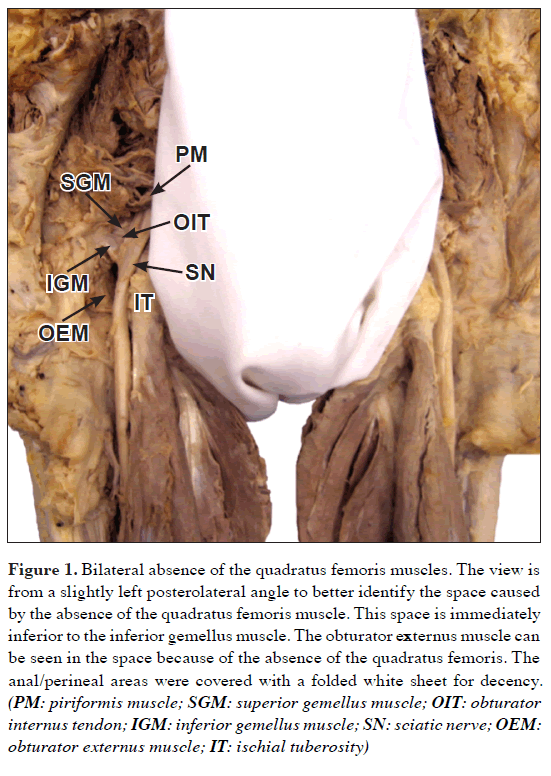
Figure 1: Bilateral absence of the quadratus femoris muscles. The view is from a slightly left posterolateral angle to better identify the space caused by the absence of the quadratus femoris muscle. This space is immediately inferior to the inferior gemellus muscle. The obturator externus muscle can be seen in the space because of the absence of the quadratus femoris. The anal/perineal areas were covered with a folded white sheet for decency. (PM: piriformis muscle; SGM: superior gemellus muscle; OIT: obturator internus tendon; IGM: inferior gemellus muscle; SN: sciatic nerve; OEM: obturator externus muscle; IT: ischial tuberosity)
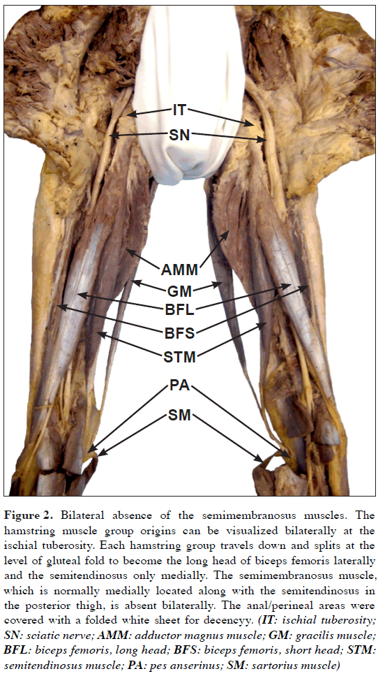
Figure 2: Bilateral absence of the semimembranosus muscles. The hamstring muscle group origins can be visualized bilaterally at the ischial tuberosity. Each hamstring group travels down and splits at the level of gluteal fold to become the long head of biceps femoris laterally and the semitendinosus only medially. The semimembranosus muscle, which is normally medially located along with the semitendinosus in the posterior thigh, is absent bilaterally. The anal/perineal areas were covered with a folded white sheet for decencyy. (IT: ischial tuberosity; SN: sciatic nerve; AMM: adductor magnus muscle; GM: gracilis muscle; BFL: biceps femoris, long head; BFS: biceps femoris, short head; STM: semitendinosus muscle; PA: pes anserinus; SM: sartorius muscle)
At both gluteal regions (Figure 1), the QF, which normally lies just inferior to the inferior gemellus muscle and attaches to the ischial tuberosity and the intertrochanteric crest, was completely absent bilaterally. The sciatic nerve was observed passing out of the piriformis foramen beneath the piriformis muscle and then crossing over the superior gemellus, tendon of obturator internus, and inferior gemellus to continue down over a shallow space where the QF muscle would typically be located. Over the area (Figure 1), the surrounding muscles (the superior and inferior gemelli, and tendon of obturator internus) are not visibly enlarged to compensate the missing QF. Due to the QF absence, the obturator externus muscle, which was anteriorly and superiorly located in the space, was exposed.
At both posterior thigh regions (Figure 2), the hamstring muscle origins were noted from the ischial tuberosities. As they passed down to the level of the gluteal folds, the bundles divided into the long head of the biceps femoris laterally and the semitendinousus medially, but the semimembranosus muscle belly was not present bilaterally. It could be observed (Figure 2) that the medially located semitendinosus tendon converged with tendons of the gracilis and sartorius muscles to become the pes anserinus as a conjoined tendon to insert on the anteromedial site of the proximal tibia. From the figure, the normally medially located semimembranosus muscle is absent bilaterally, which resulted in only the semitendinosus muscle as the superomedial border of the popliteal fossa. The oblique popliteal ligament, which is typically a reflected continuation of the semimembranosus tendon, could not be found over the popliteal fossa bilaterally. Dissection of the remaining areas of the gluteal and posterior thigh regions did not identify any additional muscular variants.
Discussion
A search of the literature revealed that variation of the QF has been reported more frequently than variation of the SM. The common QF variations reported include a double appearance unilaterally [7], absence bilaterally [5], and absence unilaterally [5,6]. In the cases of bilateral or unilateral QF absence, previous reports described compensatory development of the surrounding muscles such as well developed downward extension of gemelli muscles [6] or upward extension of the adductor magnus muscle [6]. However, in the present study, either gemelli muscles or the adductor magnus muscle was not extended to fill into the space resulted from the QF absence. Given that the QF is one of the muscles of lateral rotation at the hip, absence of the QF could cause weakness of hip lateral rotation to some extent.
Absence of the SM has been reported previously in both an infant [8] and an adult [9]. In both previous reports, the SM absence was reported as unilateral; while in the present study the absence is bilateral. Also, Cattrysse et al. [4] identified a muscle called the semimembranosus profundus. This muscle originated from the dorsolateral side of the femur between the origins of the adductor and short head of the biceps femoris, and inserted onto the medial condyle of the tibia deep to the normal insertion of the SM muscle at the posteromedial aspect of the knee joint capsule. After detailed observation and examination in our case, neither the semimembranosus profundus nor the typical semimembranosus was found.
Functionally, the SM is a substantial contributor to flexion of the knee, assists in hip extension, and medially rotates the knee when the joint is in a flexed position [1], Also, the SM plays an important role in stabilizing the posteromedial aspect of the knee joint [3,4] through distal attachments including two main expansions (the oblique popliteal ligament and thickening of the posteromedial knee joint capsule) [10], When the SM is absent bilaterally, an individual may experience weakness in performing the SM-related knee joint movements on both lower extremities and bilateral knee joint stability may be problematic as well.
In summary, this case presents a rare variation of bilateral absence of both the quadrates femoris and semimembranosus. This is of clinical relevance to clinicians who perform physical examinations or surgeries related to gluteal, posterior thigh, and knee regions.
References
- Moore KL, Dalley AF. Clinically Oriented Anatomy. 5th Ed., Philadelphia, Lippincott Williams & Wilkins. 2006; 592–594, 610–617.
- Miller SL, Webb GR. The proximal origin of the hamstrings and surrounding anatomy encountered during repair. Surgical technique. J Bone Joint Surg Am. 2008; 90 (Suppl 2 Pt 1): 108–116.
- Kim YC, Yoo WK, Chung IH, Seo JS, Tanaka S. Tendinous insertion of semimembranosus muscle into the lateral meniscus. Surg Radiol Anat. 1997; 19: 365–369.
- Cattrysse E, Barbaix E, Janssens V, Alewaeters K, Van Roy P, Clarijs JP. Observation of a supernumerary hamstring muscle: a state of the art on its incidence and clinical relevance. Morphologie. 2002; 86: 17–21.
- Gruber W. Absence of the quadratus femoris. Am J Medi Sci. 1879; 153: 238.
- Stibbe EP. Complete absence of the quadratus femoris. J Anat. 1929; 64 (pt 1): 97.
- Tanyeli E, Pestemalci T, Uzel M, Yildirim M. The double deep gluteal muscles. Saudi Med J. 2006; 27: 385–386.
- Peterson JE, Currarino G. Unilateral absence of thigh muscles confirmed by CT scan. Pediatric Radiol. 1981; 11: 157–159.
- Moncayo VM, Carpenter WA, Pierre-Jerome C, Smitson RD, Terk MR. Congenital absence of the semimembranosus muscle: case report. Surg Radiol Anat. 2010; 32: 519–523.
- LaPrade RF, Morgan PM, Wentorf FA, Johansen S, Engebretsen L. The anatomy of the posterior aspect of the knee. An anatomic study. J Bone Joint Surg Am. 2007; 89: 758–764.
Hao (Howe) Liu1*, James Fletcher2, M Kevin Garrison2 and Clayton Holmes1
1University of North Texas Health Science Center Physical Therapy Program, Forth Worth, TX, USA
2University of Central Arkansas Physical Therapy Department, Conway, AR, USA
- *Corresponding Author:
- Hao (Howe) Liu, PhD, PT
Physical Therapy Program, University of North Texas Health Science Center, 3500 Camp Bowie Blvd, CBH 461, Forth Worth, TX, 76107, USA
Tel: +1 817 735-2457
E-mail: hao.liu@unthsc.edu
Date of Received: November 9th, 2010
Date of Accepted: January 30th, 2011
Published Online: March 5th, 2011
© IJAV. 2011; 4: 40–42.
Abstract
Bilateral absence of both quadratus femoris and semimembranosus muscles was identified during a routine dissection of a 73-year-old Caucasian female cadaver. In the gluteal regions, the quadratus femoris was absent bilaterally from the area immediately inferior to the inferior gemellus muscle. The area presented as a shallow depression/space with the obturator externus muscle visible from a posterior view. In the posterior thigh regions, only the semitendinosus and biceps femoris were observed; no semimembranosus was found bilaterally. Only the semitendinosus traveled down to the superomedial border of the popliteal fossa. The absence of these muscles may present clinically as muscle weakness of hip lateral rotation (quadratus femoris action) and knee flexion (semimembranosus action) as well as hypermobility of the posteromedial knee joint (semimembranosus function). Surgeons, radiologists, and rehabilitation professionals should be aware of this anatomical variation.
-Keywords
variation, cadaver, hamstring muscle, lateral rotator of hip
Introduction
Usually, the origin of the quadratus femoris (QF) muscle is more anterolaterally located on the ischial tuberosity than the hamstrings origin [1]. In a medial-lateral direction, the QF muscle inserts onto the quadrate tubercle and the intertrochanteric crest of the femur [1]. The primary action of the QF muscle is to laterally rotate the hip joint.
The hamstrings are a muscle group formed by the biceps femoris, the semitendinosus, and the semimembranosus muscles. The long head of the biceps femoris, the semitendinosus and the semimembranosus all originate from the ischial tuberosity, with the origin of the semimembranosus (SM) being the most lateral [1,2]. The SM travels down with the semitendinosus inferomedially to become the superomedial border of the popliteal fossa. The SM tendon inserts onto the medial condyle of the tibia, the medial collateral ligament, popliteal fascia, and the oblique popliteal ligament (through its reflected tendon) [3,4]. The function of the SM muscle is flexion of the knee, medial rotation of a flexed knee, and as a stabilizer of the posteromedial aspect of the knee joint.
Variations of the QF have been reported before, including bilateral absence [5], unilateral absence [5,6], or double appearance of the muscle ipsilaterally [7]. Variation of the SM was not as common as the QF, but unilateral absence of the SM [8,9] or appearance of a variant SM [4] was described in the past literature. However, the bilateral absence of the SM or the coexistence of bilateral absence of both the QF and SM, as described in this manuscript, has not been previously reported.
Case Report
As shown in Figures 1 and 2, the absence of both QF and SM muscles was discovered on both the left and right lower extremities during a routine dissection of the posterior gluteal and thigh regions on a 73-year-old female cadaver in a gross anatomy laboratory in a university setting in 2009. The anal/perineal areas were covered with a folded white sheet for decency in Figures 1 and 2.
Figure 1: Bilateral absence of the quadratus femoris muscles. The view is from a slightly left posterolateral angle to better identify the space caused by the absence of the quadratus femoris muscle. This space is immediately inferior to the inferior gemellus muscle. The obturator externus muscle can be seen in the space because of the absence of the quadratus femoris. The anal/perineal areas were covered with a folded white sheet for decency. (PM: piriformis muscle; SGM: superior gemellus muscle; OIT: obturator internus tendon; IGM: inferior gemellus muscle; SN: sciatic nerve; OEM: obturator externus muscle; IT: ischial tuberosity)
Figure 2: Bilateral absence of the semimembranosus muscles. The hamstring muscle group origins can be visualized bilaterally at the ischial tuberosity. Each hamstring group travels down and splits at the level of gluteal fold to become the long head of biceps femoris laterally and the semitendinosus only medially. The semimembranosus muscle, which is normally medially located along with the semitendinosus in the posterior thigh, is absent bilaterally. The anal/perineal areas were covered with a folded white sheet for decencyy. (IT: ischial tuberosity; SN: sciatic nerve; AMM: adductor magnus muscle; GM: gracilis muscle; BFL: biceps femoris, long head; BFS: biceps femoris, short head; STM: semitendinosus muscle; PA: pes anserinus; SM: sartorius muscle)
At both gluteal regions (Figure 1), the QF, which normally lies just inferior to the inferior gemellus muscle and attaches to the ischial tuberosity and the intertrochanteric crest, was completely absent bilaterally. The sciatic nerve was observed passing out of the piriformis foramen beneath the piriformis muscle and then crossing over the superior gemellus, tendon of obturator internus, and inferior gemellus to continue down over a shallow space where the QF muscle would typically be located. Over the area (Figure 1), the surrounding muscles (the superior and inferior gemelli, and tendon of obturator internus) are not visibly enlarged to compensate the missing QF. Due to the QF absence, the obturator externus muscle, which was anteriorly and superiorly located in the space, was exposed.
At both posterior thigh regions (Figure 2), the hamstring muscle origins were noted from the ischial tuberosities. As they passed down to the level of the gluteal folds, the bundles divided into the long head of the biceps femoris laterally and the semitendinousus medially, but the semimembranosus muscle belly was not present bilaterally. It could be observed (Figure 2) that the medially located semitendinosus tendon converged with tendons of the gracilis and sartorius muscles to become the pes anserinus as a conjoined tendon to insert on the anteromedial site of the proximal tibia. From the figure, the normally medially located semimembranosus muscle is absent bilaterally, which resulted in only the semitendinosus muscle as the superomedial border of the popliteal fossa. The oblique popliteal ligament, which is typically a reflected continuation of the semimembranosus tendon, could not be found over the popliteal fossa bilaterally. Dissection of the remaining areas of the gluteal and posterior thigh regions did not identify any additional muscular variants.
Discussion
A search of the literature revealed that variation of the QF has been reported more frequently than variation of the SM. The common QF variations reported include a double appearance unilaterally [7], absence bilaterally [5], and absence unilaterally [5,6]. In the cases of bilateral or unilateral QF absence, previous reports described compensatory development of the surrounding muscles such as well developed downward extension of gemelli muscles [6] or upward extension of the adductor magnus muscle [6]. However, in the present study, either gemelli muscles or the adductor magnus muscle was not extended to fill into the space resulted from the QF absence. Given that the QF is one of the muscles of lateral rotation at the hip, absence of the QF could cause weakness of hip lateral rotation to some extent.
Absence of the SM has been reported previously in both an infant [8] and an adult [9]. In both previous reports, the SM absence was reported as unilateral; while in the present study the absence is bilateral. Also, Cattrysse et al. [4] identified a muscle called the semimembranosus profundus. This muscle originated from the dorsolateral side of the femur between the origins of the adductor and short head of the biceps femoris, and inserted onto the medial condyle of the tibia deep to the normal insertion of the SM muscle at the posteromedial aspect of the knee joint capsule. After detailed observation and examination in our case, neither the semimembranosus profundus nor the typical semimembranosus was found.
Functionally, the SM is a substantial contributor to flexion of the knee, assists in hip extension, and medially rotates the knee when the joint is in a flexed position [1], Also, the SM plays an important role in stabilizing the posteromedial aspect of the knee joint [3,4] through distal attachments including two main expansions (the oblique popliteal ligament and thickening of the posteromedial knee joint capsule) [10], When the SM is absent bilaterally, an individual may experience weakness in performing the SM-related knee joint movements on both lower extremities and bilateral knee joint stability may be problematic as well.
In summary, this case presents a rare variation of bilateral absence of both the quadrates femoris and semimembranosus. This is of clinical relevance to clinicians who perform physical examinations or surgeries related to gluteal, posterior thigh, and knee regions.
References
- Moore KL, Dalley AF. Clinically Oriented Anatomy. 5th Ed., Philadelphia, Lippincott Williams & Wilkins. 2006; 592–594, 610–617.
- Miller SL, Webb GR. The proximal origin of the hamstrings and surrounding anatomy encountered during repair. Surgical technique. J Bone Joint Surg Am. 2008; 90 (Suppl 2 Pt 1): 108–116.
- Kim YC, Yoo WK, Chung IH, Seo JS, Tanaka S. Tendinous insertion of semimembranosus muscle into the lateral meniscus. Surg Radiol Anat. 1997; 19: 365–369.
- Cattrysse E, Barbaix E, Janssens V, Alewaeters K, Van Roy P, Clarijs JP. Observation of a supernumerary hamstring muscle: a state of the art on its incidence and clinical relevance. Morphologie. 2002; 86: 17–21.
- Gruber W. Absence of the quadratus femoris. Am J Medi Sci. 1879; 153: 238.
- Stibbe EP. Complete absence of the quadratus femoris. J Anat. 1929; 64 (pt 1): 97.
- Tanyeli E, Pestemalci T, Uzel M, Yildirim M. The double deep gluteal muscles. Saudi Med J. 2006; 27: 385–386.
- Peterson JE, Currarino G. Unilateral absence of thigh muscles confirmed by CT scan. Pediatric Radiol. 1981; 11: 157–159.
- Moncayo VM, Carpenter WA, Pierre-Jerome C, Smitson RD, Terk MR. Congenital absence of the semimembranosus muscle: case report. Surg Radiol Anat. 2010; 32: 519–523.
- LaPrade RF, Morgan PM, Wentorf FA, Johansen S, Engebretsen L. The anatomy of the posterior aspect of the knee. An anatomic study. J Bone Joint Surg Am. 2007; 89: 758–764.


