Bilateral Accessory Piriformis Variant and its Implications – A Case Report
2 Texas College of Osteopathic Medicine, University of North Texas Health Science Center, Fort Worth, TX, USA
3 College of Natural Sciences, University of Texas at Austin, Austin, TX, USA
4 Department of Physiology and Anatomy, University of North Texas Health Science Center, TX, USA
Received: 01-Dec-2023, Manuscript No. ijav-23-6877; Editor assigned: 04-Dec-2023, Pre QC No. ijav-23-6877 (PQ); Reviewed: 21-Dec-2023 QC No. ijav-23-6877 ; Revised: 25-Dec-2023, Manuscript No. ijav-23-6877 (R); Published: 30-Dec-2023, DOI: 10.37532/1308-4038.16(12).330
Citation: Ballout B. Bilateral Accessory Piriformis Variant and its Implications: A Case Report. Int J Anat Var. 2023;16(12): 439-441.
This open-access article is distributed under the terms of the Creative Commons Attribution Non-Commercial License (CC BY-NC) (http://creativecommons.org/licenses/by-nc/4.0/), which permits reuse, distribution and reproduction of the article, provided that the original work is properly cited and the reuse is restricted to noncommercial purposes. For commercial reuse, contact reprints@pulsus.com
Abstract
The piriformis muscle is a major muscle of the gluteal region and can be a cause of sciatic pain known as piriformis syndrome. Routine dissection of a cadaver of unknown age presented with bilateral accessory piriformis muscles. The main piriformis muscle was noted bilaterally with a smaller, accessory muscle separate and distinct from the main piriformis muscles. These accessory muscles originated and attached at the same anatomical landmarks as the main piriformis. The tibial nerve exited from below both piriformis muscles and the common fibular nerve exited between the two muscle bellies. This presentation of accessory piriformis is unique and has seldom been presented in the literature. Anatomical accessory muscles such as this one has important clinical considerations including in surgery, radiology, and the diagnosis of sciatic pain caused by piriformis syndrome.
Keywords
Piriformis; Sciatic nerve; Sciatica; Piriformis syndrome; Accessory muscle
INTRODUCTION
The piriformis muscle is one of the major external rotators of the hip and is also responsible for hip abduction when the leg is flexed [1]. It originates from the anterior surface of the sacrum and traverses inferior and lateral through the greater sciatic foramen to attach on the greater trochanter of the femur [2]. The piriformis is innervated by the S1-S2 nerve roots and serves as a landmark for multiple other structures in the gluteal region including the sciatic nerve, and the superior and inferior gluteal neurovascular bundles [1]. The sciatic nerve innervates the posterior thigh and divides into the tibial nerve, innervating the posterior leg compartment and plantar foot, and the common fibular (peroneal) nerve, innervating the lateral and anterior leg compartments and dorsal foot [3]. The sciatic nerve has six well known variations of its course through the gluteal region, of which “Type A” exists in approximately 90% of patients and presents as the entire sciatic nerve passing inferior to the piriformis muscle [4]. The second most common variant presents as the sciatic nerve splitting superior to piriformis, with the common fibular nerve piercing through and the tibial nerve passing inferior to piriformis. This variation is referred to as “Type B” and has been reported in eight percent of patients [4].
The most common patient complaint regarding the piriformis muscle is piriformis syndrome. Piriformis syndrome is defined as entrapment of the sciatic nerve at the level of the ischial tuberosity and presents with back and/or gluteal pain with possible shooting pain down the leg [5]. Piriformis syndrome is the problem in up to six percent of low back pain and sciatica complaints [5]. Variations in the sciatic nerve may account for some cases of piriformis syndrome due to one or multiple parts of the nerve coursing through the muscle belly.
Accessory muscles are duplications of a muscle that may occur during development within the musculoskeletal system. These anatomic variants may or may not present with symptoms and often go unnoticed and untreated. Previous cases have been reported of an accessory piriformis muscle with merging of muscle fibers near the tendinous attachment [6]. These rare cases emerge from unknown causes. Accessory piriformis muscles are significant cases due to the possibility of causing piriformis syndrome and subsequent neuropathic pain. The presence of an accessory muscle also makes classifying sciatic nerve variations more difficult due to the exclusion of muscular anomalies in the classification system.
CASE REPORT
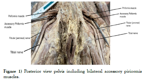
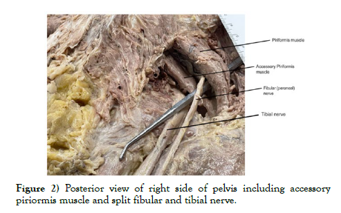
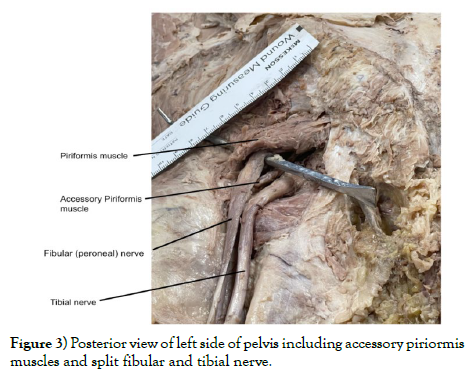
A case of bilateral accessory piriformis muscle presented during routine dissection of a middle-aged male cadaver. The cadaver was received through the HSC Willed Body Program. Upon dissection of the gluteal and perianal region, we noted an unusual appearance to the piriformis muscle. Further dissection revealed two distinct muscles comprising the piriformis muscle. Both of these piriformis muscles originate from the same attachment site on the anterior sacrum and insert on the greater trochanter of the femur. Dissection of the contralateral gluteal region showed a similar variation. The accessory piriformis muscles follow the same course as the primary piriformis muscles, but are distinctly separate from origin all the way to insertion with no visible merging of muscle fibers. In order to validate this finding, identification of the superior gemellus, obturator internus, inferior gemellus, obturator externus, quadratus femoris and the sacrotuberous ligament was performed bilaterally to assure this was indeed an accessory muscle of the piriformis. After completely clearing the region from subcutaneous tissue, we noted that the common fibular nerve exited between the primary and the accessory muscles bilaterally, while the tibial nerve exited inferior to the two piriformis muscles immediately inferior to the accessory piriformis muscle. As the two nerves were traced superiorly, we noted a unique feature: rather than being fused superiorly, they were separate and distinct from one another all the way to their exit from the greater sciatic foramen (Figures 1-3).
DISCUSSION
The piriformis muscle is a vital component to the overall mobility and stability of the gluteal, hip and proximal thigh regions. The piriformis is an external rotator of the hip alongside the other muscles neighboring it, such as the superior gemellus, quadratus femoris, inferior gemellus, obturator internus, and obturator externus [1]. The position of the piriformis also influences the anatomy and function of the sciatic nerve. The sciatic nerve, the largest nerve in the body, originates from spinal nerves L4 to S3, exits the pelvis inferior to the piriformis muscle, and courses inferiorly down the lower limb [3].
Piriformis muscle variants have several crucial implications, including its possible surgical relevance. An accessory piriformis muscle belly is a very rare case and can lead to piriformis syndrome [7]. Piriformis syndrome is generally classified as a rare entrapment neuropathy causing radicular pain down the affected limb, which may be caused by hypertrophy, injury, inflammation, or anatomical variation within the piriformis muscle affecting neighboring structures and nerves [8]. Piriformis syndrome is often misdiagnosed or completely missing as a differential due to its rarity and the difficulty in diagnosis. Not many cases of accessory piriformis muscles have been reported and this raises anatomical and surgical interest as a cause of undiagnosed pain in the deep gluteal region [6]. There are over 6 different classifications for piriformis and sciatic nerve anatomical variations within the Beaton and Anson’s classification system. Our cadaver most closely resembles the classification of Type B which states that the division of the sciatic nerve is between and below the undivided piriformis muscle [9]. However, our cadaver has two completely distinct muscle bellies, with the common fibular nerve passing between the two muscle bellies and the tibial nerve passing inferior to the accessory muscle. Therefore, our piriformis variant does not directly fall into any of the set classifications of the Beaton and Anson’s classification system making it even more important and challenging to properly diagnose. The importance of these variations is highlighted in a case report of a patient with 17 years of misdiagnosed low back pain who was eventually found to have Type-B piriformis variation causing piriformis syndrome and was treated accordingly [10]. The combination of the bilateral piriformis muscle with the unique anatomy of this cadaver’s sciatic nerve could have served as a source of pain and would have been difficult to diagnose. Variations in both muscles and nerves are important for clinicians to recognize a possibility of sciatic pain in patient’s refractory to usual treatments.
The common fibular nerve was found passing in between the two muscles,whereas the tibial nerve was found to be passing inferior to the accessory piriformis muscle. Therefore, there is no segment in which these two nerves combine with one another to form the sciatic nerve. This specific variant in which the common fibular nerve branches from L4-S2 spinal nerves and the tibial nerve branches from L4-S3 spinal nerves and remain as separate nerves has not previously been documented. A systematic review and meta-analysis assessing sciatic nerve variants found that Type-B of the Beaton and Anson’s classification system was only found in eight percent of sciatic variations, which is the variant that most closely resembles this cadaver’s findings [4].
However, given the anatomical positioning of the common fibular nerve and tibial nerve there is a high chance for nerve irritation and sciatica-like pain to have been present. This rare finding raises the need for further research to better understand the prevalence of similar variations in the general population.
Sciatic nerve variants and piriformis muscle variants are often difficult to distinguish in a clinical setting where there is lower limb and gluteal region pain. The differentiation is often done through techniques such as ultrasound or MRI aiding in proper diagnosis [11,12]. These anatomical variants of the piriformis muscle and peripheral nerves are vulnerable to surgical injury if not properly diagnosed by the surgeon ahead of time [13]. This further highlights how crucial conducting further research is to understand how these anatomical variants may impact the diagnosis of sciatica and piriformis syndrome. This added information will help clinicians treat these patients safely and effectively.
This cadaver had multiple muscular and peripheral nerve variations that were noted upon complete anatomical dissection of the musculoskeletal system. Noted for this cadaver were bilateral accessory piriformis muscle, bilateral sciatic nerve variation, bilateral accessory deltoid muscle which will have further research conducted on it. Multiple bilateral variations in one individual raise the question of a possible genetic predisposition. This elicits further research and genetic testing to see if there is any genetic explanation interconnecting these findings.
ACKNOWLEDGMENTS
The authors would like to thank the Willed Body Program at UNTHSC, the donors, and their families for their contribution to advancing medicine and anatomical knowledge.
REFERENCES
- Chang C, Jeno SH, Varacallo M. Anatomy Bony Pelvis and Lower Limb: Piriformis Muscle. In StatPearls Treasure Island (Fl), StatPearls Publishing. 2023.
- Shahid S. Piriformis Muscle. 2023.
- Giuffre BA, Black AC, Jeanmonod R. Anatomy Sciatic Nerve. In StatPearls Treasure Island (Fl) StatPearls Publishing. 2023.
- Poutoglidou F, Piagkou M, Totlis T, Tzika M et al. Sciatic Nerve Variants and the Piriformis Muscle: A Systematic Review and Meta-Analysis. Cureus.2020; 12(11):e11531.
- Hicks BL, Lam JC, Varacallo M. Piriformis Syndrome. In StatPearls Treasure Island (Fl), StatPearls Publishing. 2023.
- Ravindranath Y, Manjunath KY, Ravindranath R. Accessory Origin of the Piriformis Muscle. Singapore Med J. 2008; 49(8): e217.
- Kim HJ, Lee SY, Park HJ, Kim KW, Lee YT. Accessory Belly of the Piriformis Muscle as a Cause of Piriformis Syndrome: a Case Report with Magnetic Resonance Imaging and Magnetic Resonance Neurography Imaging Findings. iMRI. 2019;23(2):142-147.
- Gaillard F, Elhusseiny A, Knipe H. Piriformis syndrome. 2023.
- Jha AK, Baral P. Composite 0Anatomical Variations between the Sciatic Nerve and the Piriformis Muscle: A Nepalese Cadaveric Study. Case Rep Neurol Med. 2020; 2020(20):7165818.
- Polesello GC, Queiroz MC, Linhares JPT, Amaral DT et al. Anatomical variation of piriformis muscle as a cause of deep gluteal pain: diagnosis using MR Neurography and treatment. Rev Bras Ortop. 2013; 48(1):114-117.
- Ro TH, Edmonds L. Diagnosis and Management of Piriformis Syndrome A Rare Anatomic Variant Analyzed by Magnetic Resonance Imaging. J Clin Imaging Sci. 2018; 8(6):6.
- Gülec GG, Kurt Oktay KN, Aktas İ, Yılmaz B. Visualizing Anatomic Variants of the Sciatic Nerve Using Diagnostic Ultrasound During Piriformis Muscle Injection: An Example of 4 Cases. J Chiropr Med. 2022; 21(3):213-219.
- Honold S, Honis HR, Gruber H, Konschake M et al. Imaging of Anatomical Variants of the Lower Limb Nerves: Clinical and Preoperative Relevance. Semin Musculoskelet Radiol. 2023; 27(2):136-152.
Indexed at, Google Scholar, Crossref
Indexed at, Google Scholar, Crossref
Indexed at, Google Scholar, Crossref
Indexed at, Google Scholar, Crossref
Indexed at, Google Scholar, Crossref
Indexed at, Google Scholar, Crossref


