Bilateral axillary arch muscle with absence of posterior cord of brachial plexus
Dahiphale Varsha Prabhar*, Kulkarni Promod Raghunath and Sarda Swapnil Laxminarayan
Department of Anatomy, Government Medical College, Latur, Maharashtra, INDIA.
- *Corresponding Author:
- Dr. Dahiphale V.P.
Associate Professor Department of Anatomy Government Medical College Latur – 413512 Maharashtra, India
Tel: +91 965 7658650 .
E-mail: varsha.dahiphale@rediffmail.com
Date of Received: December 10th, 2012
Date of Accepted: May 19th, 2013
Published Online: January 30th, 2014
© Int J Anat Var (IJAV). 2014; 7: 1–3.
[ft_below_content] =>Keywords
axillary arch muscle,latissimus dorsi,brachial plexus,posterior cord
Introduction
Anatomical variations of axilla are of great importance due to the increasing surgical importance of this region during axillary surgery for breast cancer, reconstruction procedures and axillary by pass operations. These variations are well documented but the co-existence of double variant in the same area is very rare.
One of the rare variations reported has been the presence of a muscle extending from latissimus dorsi muscle to pectoralis major muscle called as Langer’s axillary arch, axillo-pectoral muscle, pecto-dorsal muscle or axillary arch muscle [1].
The axillary arch muscle is described as a thin muscular slip 7-10 cm in length and 2-5 cm in breadth extending from the latissimus dorsi to the pectoralis major muscle [2]. Variations of this muscular belly have been observed. Common cases include: the muscle adhering to the coracoid process of scapula, medial epicondyle of humerus, teres major, pectoralis minor, coracobrachialis, biceps brachii [3].
The muscular arch first identified by Ramsay in 1795 and described in 1812, was latter confirmed by Langer. These variations occur in 7-13 % of population. It is usually bilateral. It is commonly present anterior to axillary neurovascular bundle [1].
We report very unusual case of bilateral axillary arch muscle, posterior to axillary neurovascular bundle with absence of posterior cord formation of brachial plexus.
Case Report
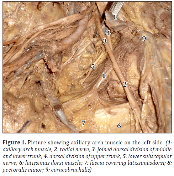
During the routine dissection of 60-year-old male cadaver in the Department of Anatomy, Government Medical College, Latur, we came across axillary arch muscles bilaterally. On both sides, the muscle took origin from fascia covering the latissimus dorsi muscle, then ran upwards and laterally in the axilla in between dorsal division of middle and lower trunk of brachial plexus anteriorly, and dorsal division of upper trunk posteriorly. Then the muscle split into two slips, medial slip inserted into coracoclavicular ligament behind the pectoralis minor, and lateral slip into coracobrachialis muscle. Length of muscle was 7 cm and breadth was 1.5 cm in the middle. The muscle was innervated by branch from lower subscapular nerve (Figure 1).
Figure 1: Picture showing axillary arch muscle on the left side. (1: axillary arch muscle; 2: radial nerve; 3: joined dorsal division of middle and lower trunk; 4: dorsal division of upper trunk; 5: lower subscapular nerve; 6: latissimus dorsi muscle; 7: fascia covering latissimusdorsi; 8: pectoralis minor; 9: coracobrachalis)
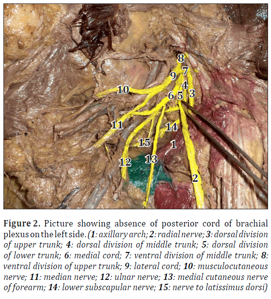
Also in the same cadaver, formation of posterior cord of brachial plexus was absent on both the sides. The dorsal division of upper trunk passed behind the axillary arch muscle, it gave axillary, upper subscapular and lower subscapular nerves; then joined with radial nerve. Dorsal division of middle trunk, after giving nerve to latissimus dorsi joined with dorsal division of lower trunk just above the axillary arch muscle,and then descended anterior to arch muscle and joined with dorsal division of upper trunk to form radial nerve just below axillary arch muscle (Figure 2).
Figure 2: Picture showing absence of posterior cord of brachial plexus on the left side. (1: axillary arch; 2: radial nerve; 3: dorsal division of upper trunk; 4: dorsal division of middle trunk; 5: dorsal division of lower trunk; 6: medial cord; 7: ventral division of middle trunk; 8: ventral division of upper trunk; 9: lateral cord; 10: musculocutaneous nerve; 11: median nerve; 12: ulnar nerve; 13: medial cutaneous nerve of forearm; 14: lower subscapular nerve; 15: nerve to latissimus dorsi)
Formation of lateral and medial cords and its branches was as usual. Similar findings were present on both sides.
Discussion
Usually, the muscular arch of axilla is described as extending from proximal border of latissimus dorsi to about the middle of posterior axillary fold, then arching across the axilla anterior the axillary vessels and nerves to join the under surface of tendon of pectoralis major muscle or coracobrachialis or fascia over the biceps brachii [3].
Ramsay initially mentioned muscular variations located in the axillary fossa in 1795 [4]. Carl Langer described the fibrous thickening of the medial edge of the axillary fascia between the borders of the pectoralis major and latissimus dorsi muscle as “Achselbogen” [5]. Later on, Testut called it as “axillary arch of Langer” [6]. Sochetella used the term “axillopectoral muscle in 1977 [7].
Arch shaped variations in the axilla could be considered in two groups, muscular form (type I) and tendinous form (type II). Clinical classification of axillary arches could be defined as superficial and deep arch. Superficial group arches cross in front of the vessels and nerves; and veins could be affected in these variations that may play role in intermittent obstruction of axillary vein. Deep group arches occurr deeply, these arches usually cross only parts of neurovascular bundle and axillary or radial nerves could possibly be affected [6].
Though the presence of an axillary arch muscle has been reported in 7-13% cadaver dissection [1], this is the first time that such a variation as described in this paper has been noticed in our dissection experience that goes back to the year 1997. Clinically the incidence of this variant muscle varies considerably from 0.25-25%. The highly variable incidence of axillary arch muscle may be related to differing sample size. Even racial variations could be responsible for this variation [2].
In the case reported here, the muscle passed behind the bulk of axillary neurovascular bundle, with absence of formation of posterior cord of brachial plexus with dorsal division of middle and lower trunk anterior and dorsal division of upper trunk posterior to the axillary arch muscle.
Though Pillay and Jacob made similar observation of axillary arch muscle posterior to axillary neurovascular bundle passing through posterior cord, in most of other studies reported, the axillary arch was observed to pass anterior to the axillary arch muscle [1].
Langer’s arch is usually seen as a single band, but it can divide into two or multiple slips across the axilla. In its complete and common form, it arises from the latissimus dorsi and inserts into pectoralis major; while in its incomplete form it present with varying insertions into pectoralis minor, coracobrachialis, biceps brachii, teres major, coracoid process, 1st rib and coracobrachial fascia [8].
In our case, the muscle was found attached to fascia covering latissimus dorsi and ended at the coracoclavicular ligament and coracobrachialis muscle. The bilateral presence, multiple attachments and absence of formation of posterior cord all put together, makes this case a very unique one.
According to Besana-Ciani and Greenall, the axillary arch muscle originates from panniculus carnosus, which is an embryological remnant of more extensive sheet of skin associated musculature lying at the junction between superficial fascia and subcutaneous fat. Panniculus carnosus is well developed in lower mammals, particularly in rodents, while in higher primates and humans it is only evident as muscle such as platysma and dartos, and its remaining part become vestigial [5]. In lower mammals, panniculus carnosus is highly developed to form pectoral group of muscle. However, in man it has regressed because its functional importance decreased during evolution in favor of wider upper limb mobility. In humans, therefore Langer’s arch is most common embryological remnant of panniculus carnosus in pectoral group of muscles [5,9].
Axillary arch muscle can be palpable during clinical examination and can be confused with enlarged lymph nodes and soft tissue tumors. Compression by the muscular arch should be considered in the differential diagnosis of patients with thoracic outlet and hyperabduction syndrome [10].
However, Langer’s arch is usually asymptomatic, its main importance is it can cause confusion during axillary surgery for breast cancer. The presence of muscular arch can give inadequate exposure of the axillary fat and may limit access to the lower lateral group of axillary lymph nodes resulting in an incomplete clearing of axilla [1].
References
- Pillay M, Jacob SM. Bilateral presence of axillary arch muscle passing through the posterior cord of brachial plexus. Int J Morphol. 2009; 27: 1047–1050.
- Babu ED, Khashaba A. Axillary arch and its implications in axillary dissection – review. Int J Clin Pract. 2000; 54: 524–525.
- Dharap A. An unusually medial axillary arch muscle. J Anat.1994; 184: 639–641.
- Georgiev GP, Jelev L, Surchev L. Axillary arch in Bulgarian population: clinical significance of the arches. Clin Anat. 2007; 20: 286–291.
- Besana-Ciani I, Greenall MJ. Langer’s axillary arch: anatomy, embryological features and surgical implications. Surgeon. 2005; 3: 325–327.
- Jelev L, Georgiev GP, Surchev L. Axillary arch in human: Common morphology and variety. Definition of “clinical” axillary arch and its classification. Ann Anat. 2007; 189: 473–481.
- Sachatello CR. Axillopectoral muscle (Langer’s axillary arch): A cause of axillary vein obstruction. Surgery. 1977; 81: 610–612.
- Loukas M, Noordeh N, Tubbs RS, Jordan R. Variation of the axillary arch muscle with multiple insertions. Singapore Med J. 2009; 50: 88–90.
- Sharma T, Singla RK, Agnihotri G, Gupta R. Axillary arch muscle. Kathmandu Univ Med J (KUMJ). 2009; 7: 432–434.
- Kanaka S, Phulipatil AK, Gaikwad MR. Axillary arch and its relations – a rare case report. Int J Biol Med Res. 2012; 3: 2277–2279.
Dahiphale Varsha Prabhar*, Kulkarni Promod Raghunath and Sarda Swapnil Laxminarayan
Department of Anatomy, Government Medical College, Latur, Maharashtra, INDIA.
- *Corresponding Author:
- Dr. Dahiphale V.P.
Associate Professor Department of Anatomy Government Medical College Latur – 413512 Maharashtra, India
Tel: +91 965 7658650 .
E-mail: varsha.dahiphale@rediffmail.com
Date of Received: December 10th, 2012
Date of Accepted: May 19th, 2013
Published Online: January 30th, 2014
© Int J Anat Var (IJAV). 2014; 7: 1–3.
Abstract
Axillary arch is an additional muscle slip usually joining the latissimus dorsi muscle to the pectoralis major or other neighboring muscles and bones. In this paper, rare case of bilateral axillary arch muscle is reported during routine dissection of the axillary region of 60-year-old male cadaver with absence of posterior cord formation of brachial plexus.
On both sides, the axillary arch muscle took origin from fascia covering latissimus dorsi muscle and passed upwards between dorsal divisions of middle and lower trunk anteriorly, and dorsal division of upper trunk posteriorly; but posterior to the bulk of axillary neurovascular bundle. Then it split into two slips, medial slip was inserted into coracoclavicular ligament and lateral slip was inserted into coracobrachialis muscle.
The presence of the arch muscle has important clinical implications and the bilateral presence, absence of posterior cord formation and multiple connective tissue attachments makes the case most unique.
Keywords
axillary arch muscle,latissimus dorsi,brachial plexus,posterior cord
Introduction
Anatomical variations of axilla are of great importance due to the increasing surgical importance of this region during axillary surgery for breast cancer, reconstruction procedures and axillary by pass operations. These variations are well documented but the co-existence of double variant in the same area is very rare.
One of the rare variations reported has been the presence of a muscle extending from latissimus dorsi muscle to pectoralis major muscle called as Langer’s axillary arch, axillo-pectoral muscle, pecto-dorsal muscle or axillary arch muscle [1].
The axillary arch muscle is described as a thin muscular slip 7-10 cm in length and 2-5 cm in breadth extending from the latissimus dorsi to the pectoralis major muscle [2]. Variations of this muscular belly have been observed. Common cases include: the muscle adhering to the coracoid process of scapula, medial epicondyle of humerus, teres major, pectoralis minor, coracobrachialis, biceps brachii [3].
The muscular arch first identified by Ramsay in 1795 and described in 1812, was latter confirmed by Langer. These variations occur in 7-13 % of population. It is usually bilateral. It is commonly present anterior to axillary neurovascular bundle [1].
We report very unusual case of bilateral axillary arch muscle, posterior to axillary neurovascular bundle with absence of posterior cord formation of brachial plexus.
Case Report
During the routine dissection of 60-year-old male cadaver in the Department of Anatomy, Government Medical College, Latur, we came across axillary arch muscles bilaterally. On both sides, the muscle took origin from fascia covering the latissimus dorsi muscle, then ran upwards and laterally in the axilla in between dorsal division of middle and lower trunk of brachial plexus anteriorly, and dorsal division of upper trunk posteriorly. Then the muscle split into two slips, medial slip inserted into coracoclavicular ligament behind the pectoralis minor, and lateral slip into coracobrachialis muscle. Length of muscle was 7 cm and breadth was 1.5 cm in the middle. The muscle was innervated by branch from lower subscapular nerve (Figure 1).
Figure 1: Picture showing axillary arch muscle on the left side. (1: axillary arch muscle; 2: radial nerve; 3: joined dorsal division of middle and lower trunk; 4: dorsal division of upper trunk; 5: lower subscapular nerve; 6: latissimus dorsi muscle; 7: fascia covering latissimusdorsi; 8: pectoralis minor; 9: coracobrachalis)
Also in the same cadaver, formation of posterior cord of brachial plexus was absent on both the sides. The dorsal division of upper trunk passed behind the axillary arch muscle, it gave axillary, upper subscapular and lower subscapular nerves; then joined with radial nerve. Dorsal division of middle trunk, after giving nerve to latissimus dorsi joined with dorsal division of lower trunk just above the axillary arch muscle,and then descended anterior to arch muscle and joined with dorsal division of upper trunk to form radial nerve just below axillary arch muscle (Figure 2).
Figure 2: Picture showing absence of posterior cord of brachial plexus on the left side. (1: axillary arch; 2: radial nerve; 3: dorsal division of upper trunk; 4: dorsal division of middle trunk; 5: dorsal division of lower trunk; 6: medial cord; 7: ventral division of middle trunk; 8: ventral division of upper trunk; 9: lateral cord; 10: musculocutaneous nerve; 11: median nerve; 12: ulnar nerve; 13: medial cutaneous nerve of forearm; 14: lower subscapular nerve; 15: nerve to latissimus dorsi)
Formation of lateral and medial cords and its branches was as usual. Similar findings were present on both sides.
Discussion
Usually, the muscular arch of axilla is described as extending from proximal border of latissimus dorsi to about the middle of posterior axillary fold, then arching across the axilla anterior the axillary vessels and nerves to join the under surface of tendon of pectoralis major muscle or coracobrachialis or fascia over the biceps brachii [3].
Ramsay initially mentioned muscular variations located in the axillary fossa in 1795 [4]. Carl Langer described the fibrous thickening of the medial edge of the axillary fascia between the borders of the pectoralis major and latissimus dorsi muscle as “Achselbogen” [5]. Later on, Testut called it as “axillary arch of Langer” [6]. Sochetella used the term “axillopectoral muscle in 1977 [7].
Arch shaped variations in the axilla could be considered in two groups, muscular form (type I) and tendinous form (type II). Clinical classification of axillary arches could be defined as superficial and deep arch. Superficial group arches cross in front of the vessels and nerves; and veins could be affected in these variations that may play role in intermittent obstruction of axillary vein. Deep group arches occurr deeply, these arches usually cross only parts of neurovascular bundle and axillary or radial nerves could possibly be affected [6].
Though the presence of an axillary arch muscle has been reported in 7-13% cadaver dissection [1], this is the first time that such a variation as described in this paper has been noticed in our dissection experience that goes back to the year 1997. Clinically the incidence of this variant muscle varies considerably from 0.25-25%. The highly variable incidence of axillary arch muscle may be related to differing sample size. Even racial variations could be responsible for this variation [2].
In the case reported here, the muscle passed behind the bulk of axillary neurovascular bundle, with absence of formation of posterior cord of brachial plexus with dorsal division of middle and lower trunk anterior and dorsal division of upper trunk posterior to the axillary arch muscle.
Though Pillay and Jacob made similar observation of axillary arch muscle posterior to axillary neurovascular bundle passing through posterior cord, in most of other studies reported, the axillary arch was observed to pass anterior to the axillary arch muscle [1].
Langer’s arch is usually seen as a single band, but it can divide into two or multiple slips across the axilla. In its complete and common form, it arises from the latissimus dorsi and inserts into pectoralis major; while in its incomplete form it present with varying insertions into pectoralis minor, coracobrachialis, biceps brachii, teres major, coracoid process, 1st rib and coracobrachial fascia [8].
In our case, the muscle was found attached to fascia covering latissimus dorsi and ended at the coracoclavicular ligament and coracobrachialis muscle. The bilateral presence, multiple attachments and absence of formation of posterior cord all put together, makes this case a very unique one.
According to Besana-Ciani and Greenall, the axillary arch muscle originates from panniculus carnosus, which is an embryological remnant of more extensive sheet of skin associated musculature lying at the junction between superficial fascia and subcutaneous fat. Panniculus carnosus is well developed in lower mammals, particularly in rodents, while in higher primates and humans it is only evident as muscle such as platysma and dartos, and its remaining part become vestigial [5]. In lower mammals, panniculus carnosus is highly developed to form pectoral group of muscle. However, in man it has regressed because its functional importance decreased during evolution in favor of wider upper limb mobility. In humans, therefore Langer’s arch is most common embryological remnant of panniculus carnosus in pectoral group of muscles [5,9].
Axillary arch muscle can be palpable during clinical examination and can be confused with enlarged lymph nodes and soft tissue tumors. Compression by the muscular arch should be considered in the differential diagnosis of patients with thoracic outlet and hyperabduction syndrome [10].
However, Langer’s arch is usually asymptomatic, its main importance is it can cause confusion during axillary surgery for breast cancer. The presence of muscular arch can give inadequate exposure of the axillary fat and may limit access to the lower lateral group of axillary lymph nodes resulting in an incomplete clearing of axilla [1].
References
- Pillay M, Jacob SM. Bilateral presence of axillary arch muscle passing through the posterior cord of brachial plexus. Int J Morphol. 2009; 27: 1047–1050.
- Babu ED, Khashaba A. Axillary arch and its implications in axillary dissection – review. Int J Clin Pract. 2000; 54: 524–525.
- Dharap A. An unusually medial axillary arch muscle. J Anat.1994; 184: 639–641.
- Georgiev GP, Jelev L, Surchev L. Axillary arch in Bulgarian population: clinical significance of the arches. Clin Anat. 2007; 20: 286–291.
- Besana-Ciani I, Greenall MJ. Langer’s axillary arch: anatomy, embryological features and surgical implications. Surgeon. 2005; 3: 325–327.
- Jelev L, Georgiev GP, Surchev L. Axillary arch in human: Common morphology and variety. Definition of “clinical” axillary arch and its classification. Ann Anat. 2007; 189: 473–481.
- Sachatello CR. Axillopectoral muscle (Langer’s axillary arch): A cause of axillary vein obstruction. Surgery. 1977; 81: 610–612.
- Loukas M, Noordeh N, Tubbs RS, Jordan R. Variation of the axillary arch muscle with multiple insertions. Singapore Med J. 2009; 50: 88–90.
- Sharma T, Singla RK, Agnihotri G, Gupta R. Axillary arch muscle. Kathmandu Univ Med J (KUMJ). 2009; 7: 432–434.
- Kanaka S, Phulipatil AK, Gaikwad MR. Axillary arch and its relations – a rare case report. Int J Biol Med Res. 2012; 3: 2277–2279.


