Bilateral double ureters and accessory renal vessels in a Tanzanian male cadaver: a rare urinary system variation
Rahabu Marwa Morro*, George Joseph Lufukuja, Flora M. Fabian
Department of Anatomy and Histology, International Medical and Technological University (IMTU), Dar-Es-Salaam, Tanzania.
- *Corresponding Author:
- Rahabu Marwa Morro
Postgraduate Student in MS Anatomy, Department of Anatomy, International Medical and Technological University, P.O. Box: 77594, Dar-Es-Salaam, Tanzania.
Tel: +255 713 456775
E-mail: rahabudaniel@yahoo.com
Date of Received: April 18th, 2011
Date of Accepted: August 10th, 2011
Published Online: September 27th, 2011
© IJAV. 2011; 4: 164–166.
[ft_below_content] =>Keywords
human kidney, double ureters
Introduction
The human ureters are two muscular tubes that extend from each renal pelvis to the posterior surface of the urinary bladder. Embryologically, the ureters develop from the ureteric bud, a diverticulum from the mesonephric duct. The ureteric bud elongates to form the ureter and undergoes repeated branching to form the major and minor calyces [1,2].
The function of the ureter is to transport urine from the kidneys to the urinary bladder by peristaltic contractions of the muscular coat, assisted by the filtration pressure of the glomeruli. Each ureter measures 25 cm long, having three constrictions along its course; where the renal pelvis joins the ureter, where it is kinked as it crosses the pelvic brim, and where it pierces the wall of the urinary bladder. The renal pelvis is a funnel-shaped expanded upper end of the ureter. It lies within the hilum of the kidney and receives the major calyces and emerges from the hilum of the kidney and runs vertically downward behind the parietal peritoneum on the psoas muscle, which separates it from the tips of the transverse processes of the lumbar vertebrae. Normally it enters the pelvis by crossing the bifurcation of the common iliac artery anterior to the sacroiliac joint. The ureter then runs downwards the lateral wall of the pelvis to the region of the ischial spine and turns forward to enter the lateral angle of the urinary bladder. Near its termination in males it is crossed by the vas deferens and by the uterine artery in females. The ureter passes obliquely through the wall of the bladder for about 1.9 cm before opening into the urinary bladder [2]. Anatomical variations of ureters and their relationship to surrounding structures are therefore of particular importance from an academic point of view, a surgical perspective, radiological examinations and treatment to preserve renal functions.
Case Report
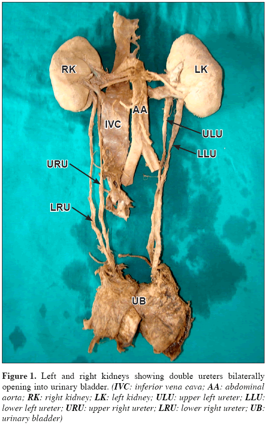
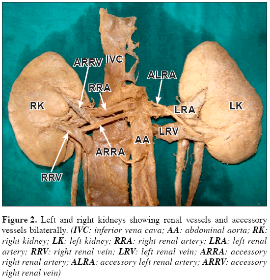
The present variation was observed during the abdomen dissection classes for the medical undergraduates, in the Department of Anatomy at International Medical and Technological University (IMTU). A male cadaver of approximately 40 years showed bilateral double ureters. In this case, the ureters originated from the upper and lower renal poles from the right and left kidneys (Figure 1). On both sides the ureters opened separately to the urinary bladder. In addition, we also detected in the same cadaver a bilateral lower polar accessory renal arteries and a right accessory renal vein (Figure 2). The lower polar accessory renal arteries took origin from the abdominal aorta to supply the respective poles of right and left kidneys. The right accessory renal vein opened directly into the inferior vena cava.
Figure 2: Left and right kidneys showing renal vessels and accessory vessels bilaterally. (IVC: inferior vena cava; AA: abdominal aorta; RK: right kidney; LK: left kidney; RRA: right renal artery; LRA: left renal artery; RRV: right renal vein; LRV: left renal vein; ARRA: accessory right renal artery; ALRA: accessory left renal artery; ARRV: accessory right renal vein)
Discussion
Double ureter is one of the variations of ureters that appear complete or incomplete [3,4]. Incomplete duplication of ureter is known as bifid ureter, this kind of variation may be formed due to some error or disturbance in development of the ureteric bud which arises from the mesonephric duct around the 5th week [2]. Double ureter may be found either bilaterally or unilaterally, more commonly the left. The ureter may join before reaching the bladder or remain separate and enter the bladder on the same side at two distinct points [5]. Incomplete ureteral duplication, in which one common ureter enters the bladder, is rarely clinically significant. Alternatively, complete ureteral duplication, in which 2 ureters ipsilaterally enter the urinary bladder, has a propensity for vesicoureteral reflux into the lower pole and obstruction of the upper pole, which can be problematic [6]. Duplex collecting systems can be associated with a variety of congenital genitourinary tract anomalies [7]. Most patients having double ureters are asymptomatic, with genitourinary tract anomalies being detected incidentally on imaging studies performed for other reasons. Symptomatic patients usually have complete ureteric duplication in which the ureters are prone to developing obstruction, reflux and infection. Ureteropelvic obstruction is more common when a duplex kidney exists and can be inherited in an autosomal dominant pattern [8].
Cases of double pelvis and double ureters may be found by chance on radiological investigation of the urinary tract. They are more liable to become infected or to be the seat of calculus formation than a normal ureter. The cause is a premature division of the ureteric bud near its termination [9,10].
The duplex ureters derive from two ureteric buds arising from the mesonephric duct. They are contained in a single fascia sheath and may fuse at any point along their course or may be separated until they insert through separate ureteric orifices into the urinary bladder. Care must be taken not to compromise the blood supply of the second ureter when excising or reimplanting a single ureter of a duplex [2].
The ureter from the upper pole of the kidney inserts more medially and caudally in the urinary bladder than the ureter from the lower pole. This reflects the embryological development of the ureter where by the ureteric bud which is initially more proximal on the mesonephric duct has a shorter time to be pulled cranially in the urinary bladder and so it inserts more distally in the mature urinary bladder. The ureter from the lower pole has a shorter intramural course than the longer ureter and is prone to reflux [2].
Lower division of the ureteric bud produce double ureters, which is due to premature division of the ureteric bud. The two ureters may branch from a single distal segment or they may open separately into the urinary bladder. In the later case, as dilatation of the distal ureteric segments incorporates them into the urinary bladder wall, rotation about each other results in the lower ureteric orifice draining the lower pole of the kidney [10]. 1 in 125 individuals, two ureters drain the renal pelvis on one side; this is termed a duplex system. Bilateral duplex ureters occur in approximately 1 in 800 cases [2].
Knowledge about the anatomical variations of the renal collecting system is of great importance for surgical approaches and radiologic and other evaluative methods, like cystoscopy and retrograde pyelography. Urologists, technicians and clinicians should keep in mind these anatomical variations as guidance for therapeutic and surgical interventions to avoid complications.
Acknowledgement
We, Rahabu Marwa-Morro and George J. Lufukuja are sincerely grateful to Prof. F. M. Fabian, our supervisor for her untiring support, constructive critics and encouragement during the preparation of this case report.
Special thanks should go to Mr. K. S. Varma our assistant supervisor for his great contribution in the image editorial.
Our heartfelt appreciation is also directed to all staff in the Anatomy and Histology department of International Medical and Technological University whose contribution and assistance have made this case report possible.
References
- Moore KL, Persaud TVN. The Developing Human. Clinically Oriented Embryology. 8th Ed., New Delhi, Elsevier. 2008; 244–245.
- Standring S, ed. Gray’s Anatomy. 40th Ed., Spain, Churchill Livingstone, Elsevier. 2008; 1238–1240.
- Khani S, Mahdavi K. A case report of a bifid ureter and renal pelvis. YAFT-E. 2004; 6: 59–62.
- Das S, Dhar P, Mehra RD. Unilateral isolated bifid ureter – a case report. J Anat Soc India. 2001; 50: 43–44.
- Summers JE. V. Double ureter. Report of a nephrectomy done upon a young child with this condition present. Ann Surg. 1901; 33: 39–41.
- Gatti JM. Ureteral Duplication, Ureteral Ectopia, and Ureterocele. http://emedicine.medscape.com/article/1017202-overview (accessed March 2011).
- Lee S, Kim W, Kang KP, Jeong YB, Kim YK, Jang YB, Park SK. Bilateral incomplete double ureters. Nephrol Dial Transplant. 2007; 22: 2720–2721.
- Khan AN, Chandramohan M, MacDonald S. Duplicated Collecting System. http://emedicine.medscape.com/article/378075-overview (accessed February 2011).
- Nishijima K, Iijima T, Hashimoto N. A case of right duplex kidney with double ureters and left dysplastic kidney with bifid ureter. Kaibogaku Zasshi. 1990; 65: 83–87. [Japanese]
- Last RJ. Anatomy. Regional and Applied. 4th Ed., London, Churchill LTD. 1966; 490.
Rahabu Marwa Morro*, George Joseph Lufukuja, Flora M. Fabian
Department of Anatomy and Histology, International Medical and Technological University (IMTU), Dar-Es-Salaam, Tanzania.
- *Corresponding Author:
- Rahabu Marwa Morro
Postgraduate Student in MS Anatomy, Department of Anatomy, International Medical and Technological University, P.O. Box: 77594, Dar-Es-Salaam, Tanzania.
Tel: +255 713 456775
E-mail: rahabudaniel@yahoo.com
Date of Received: April 18th, 2011
Date of Accepted: August 10th, 2011
Published Online: September 27th, 2011
© IJAV. 2011; 4: 164–166.
Abstract
We report a case of a male cadaver, of 40 years of age. We observed two ureters on the left kidney and two ureters on the right kidney. In this case, the ureters originated from the upper and lower poles, whereby, those from upper poles of the kidneys were longer than those from lower poles. On both sides the ureters opened separately into the urinary bladder. Bilateral double ureters are very rare anatomical variations. Knowledge of anatomical variations of the urinary system is of great importance for not only urological conditions but also in surgeries involving renal transplant and radiological examinations interpretation. If urologists and clinicians generally have a sound knowledge on anatomical variations it would ease management and surgical interventions, as this may reduce unnecessary complications.
-Keywords
human kidney, double ureters
Introduction
The human ureters are two muscular tubes that extend from each renal pelvis to the posterior surface of the urinary bladder. Embryologically, the ureters develop from the ureteric bud, a diverticulum from the mesonephric duct. The ureteric bud elongates to form the ureter and undergoes repeated branching to form the major and minor calyces [1,2].
The function of the ureter is to transport urine from the kidneys to the urinary bladder by peristaltic contractions of the muscular coat, assisted by the filtration pressure of the glomeruli. Each ureter measures 25 cm long, having three constrictions along its course; where the renal pelvis joins the ureter, where it is kinked as it crosses the pelvic brim, and where it pierces the wall of the urinary bladder. The renal pelvis is a funnel-shaped expanded upper end of the ureter. It lies within the hilum of the kidney and receives the major calyces and emerges from the hilum of the kidney and runs vertically downward behind the parietal peritoneum on the psoas muscle, which separates it from the tips of the transverse processes of the lumbar vertebrae. Normally it enters the pelvis by crossing the bifurcation of the common iliac artery anterior to the sacroiliac joint. The ureter then runs downwards the lateral wall of the pelvis to the region of the ischial spine and turns forward to enter the lateral angle of the urinary bladder. Near its termination in males it is crossed by the vas deferens and by the uterine artery in females. The ureter passes obliquely through the wall of the bladder for about 1.9 cm before opening into the urinary bladder [2]. Anatomical variations of ureters and their relationship to surrounding structures are therefore of particular importance from an academic point of view, a surgical perspective, radiological examinations and treatment to preserve renal functions.
Case Report
The present variation was observed during the abdomen dissection classes for the medical undergraduates, in the Department of Anatomy at International Medical and Technological University (IMTU). A male cadaver of approximately 40 years showed bilateral double ureters. In this case, the ureters originated from the upper and lower renal poles from the right and left kidneys (Figure 1). On both sides the ureters opened separately to the urinary bladder. In addition, we also detected in the same cadaver a bilateral lower polar accessory renal arteries and a right accessory renal vein (Figure 2). The lower polar accessory renal arteries took origin from the abdominal aorta to supply the respective poles of right and left kidneys. The right accessory renal vein opened directly into the inferior vena cava.
Figure 2: Left and right kidneys showing renal vessels and accessory vessels bilaterally. (IVC: inferior vena cava; AA: abdominal aorta; RK: right kidney; LK: left kidney; RRA: right renal artery; LRA: left renal artery; RRV: right renal vein; LRV: left renal vein; ARRA: accessory right renal artery; ALRA: accessory left renal artery; ARRV: accessory right renal vein)
Discussion
Double ureter is one of the variations of ureters that appear complete or incomplete [3,4]. Incomplete duplication of ureter is known as bifid ureter, this kind of variation may be formed due to some error or disturbance in development of the ureteric bud which arises from the mesonephric duct around the 5th week [2]. Double ureter may be found either bilaterally or unilaterally, more commonly the left. The ureter may join before reaching the bladder or remain separate and enter the bladder on the same side at two distinct points [5]. Incomplete ureteral duplication, in which one common ureter enters the bladder, is rarely clinically significant. Alternatively, complete ureteral duplication, in which 2 ureters ipsilaterally enter the urinary bladder, has a propensity for vesicoureteral reflux into the lower pole and obstruction of the upper pole, which can be problematic [6]. Duplex collecting systems can be associated with a variety of congenital genitourinary tract anomalies [7]. Most patients having double ureters are asymptomatic, with genitourinary tract anomalies being detected incidentally on imaging studies performed for other reasons. Symptomatic patients usually have complete ureteric duplication in which the ureters are prone to developing obstruction, reflux and infection. Ureteropelvic obstruction is more common when a duplex kidney exists and can be inherited in an autosomal dominant pattern [8].
Cases of double pelvis and double ureters may be found by chance on radiological investigation of the urinary tract. They are more liable to become infected or to be the seat of calculus formation than a normal ureter. The cause is a premature division of the ureteric bud near its termination [9,10].
The duplex ureters derive from two ureteric buds arising from the mesonephric duct. They are contained in a single fascia sheath and may fuse at any point along their course or may be separated until they insert through separate ureteric orifices into the urinary bladder. Care must be taken not to compromise the blood supply of the second ureter when excising or reimplanting a single ureter of a duplex [2].
The ureter from the upper pole of the kidney inserts more medially and caudally in the urinary bladder than the ureter from the lower pole. This reflects the embryological development of the ureter where by the ureteric bud which is initially more proximal on the mesonephric duct has a shorter time to be pulled cranially in the urinary bladder and so it inserts more distally in the mature urinary bladder. The ureter from the lower pole has a shorter intramural course than the longer ureter and is prone to reflux [2].
Lower division of the ureteric bud produce double ureters, which is due to premature division of the ureteric bud. The two ureters may branch from a single distal segment or they may open separately into the urinary bladder. In the later case, as dilatation of the distal ureteric segments incorporates them into the urinary bladder wall, rotation about each other results in the lower ureteric orifice draining the lower pole of the kidney [10]. 1 in 125 individuals, two ureters drain the renal pelvis on one side; this is termed a duplex system. Bilateral duplex ureters occur in approximately 1 in 800 cases [2].
Knowledge about the anatomical variations of the renal collecting system is of great importance for surgical approaches and radiologic and other evaluative methods, like cystoscopy and retrograde pyelography. Urologists, technicians and clinicians should keep in mind these anatomical variations as guidance for therapeutic and surgical interventions to avoid complications.
Acknowledgement
We, Rahabu Marwa-Morro and George J. Lufukuja are sincerely grateful to Prof. F. M. Fabian, our supervisor for her untiring support, constructive critics and encouragement during the preparation of this case report.
Special thanks should go to Mr. K. S. Varma our assistant supervisor for his great contribution in the image editorial.
Our heartfelt appreciation is also directed to all staff in the Anatomy and Histology department of International Medical and Technological University whose contribution and assistance have made this case report possible.
References
- Moore KL, Persaud TVN. The Developing Human. Clinically Oriented Embryology. 8th Ed., New Delhi, Elsevier. 2008; 244–245.
- Standring S, ed. Gray’s Anatomy. 40th Ed., Spain, Churchill Livingstone, Elsevier. 2008; 1238–1240.
- Khani S, Mahdavi K. A case report of a bifid ureter and renal pelvis. YAFT-E. 2004; 6: 59–62.
- Das S, Dhar P, Mehra RD. Unilateral isolated bifid ureter – a case report. J Anat Soc India. 2001; 50: 43–44.
- Summers JE. V. Double ureter. Report of a nephrectomy done upon a young child with this condition present. Ann Surg. 1901; 33: 39–41.
- Gatti JM. Ureteral Duplication, Ureteral Ectopia, and Ureterocele. http://emedicine.medscape.com/article/1017202-overview (accessed March 2011).
- Lee S, Kim W, Kang KP, Jeong YB, Kim YK, Jang YB, Park SK. Bilateral incomplete double ureters. Nephrol Dial Transplant. 2007; 22: 2720–2721.
- Khan AN, Chandramohan M, MacDonald S. Duplicated Collecting System. http://emedicine.medscape.com/article/378075-overview (accessed February 2011).
- Nishijima K, Iijima T, Hashimoto N. A case of right duplex kidney with double ureters and left dysplastic kidney with bifid ureter. Kaibogaku Zasshi. 1990; 65: 83–87. [Japanese]
- Last RJ. Anatomy. Regional and Applied. 4th Ed., London, Churchill LTD. 1966; 490.


