Bilateral duplicitous renal vasculature
Carleigh M. High*, Christopher M. Scott, Christopher M. Antonelli, Kellie M. Ruffer and Janet M. Cope
Department of Physical Therapy Education, Elon University, Elon, North Carolina, 27244, USA
- *Corresponding Author:
- Carleigh M. High
Dept. of Physical Therapy Education
762 E Haggard Ave Elon, NC 27244, USA
Tel: +1 (336) 278-6355
E-mail: chigh@elon.edu
Date of Received: October 19th, 2015
Date of Accepted: December 30th, 2016
Published Online: January 26th 2017
© Int J Anat Var (IJAV). 2016; 9: 67–69.
[ft_below_content] =>Keywords
bilateral, kidney, variation
Introduction
The kidneys are paired organs in the abdomen that are essential and responsible for filtering blood, removing waste, controlling body fluids, and regulating electrolyte and hormonal homeostasis. In order to properly perform these important functions the kidneys must have adequate blood supply. Typically, each kidney has a hilum that receives blood from a single renal artery from the aorta and one renal vein draining into the inferior vena cava. Buffoli and colleagues (2015) reported that renal variations are relatively common and make up about 30% of all congenital anomalies [1]. When additional renal vasculature is present, it may connect to the kidneys either through the hilum or at another point on the kidney’s surface. These anatomical variations are more prevalent in renal arteries compared to renal veins. According to Gupta and colleagues (2011) variations of renal veins tend to occur on the left side due to the complexity that arises from the presence of multiple veins, including the adrenal, gonadal, phrenic, and hemizygous vessels [2]. In contrast, Kaneko and colleagues (2008) report that multiple renal veins are relatively common on the right and rare on the left [3].
In this case report, the researchers discuss a previously unreported variant of bilateral renal veins and arteries in a single cadaver. In this individual, the renal vessels arose from each kidney at both the hilum and the inferior pole.
Case Report
During routine dissection of an 84-year-old female donor at Elon University, Elon, North Carolina, who had a recorded cause of death as dementia and Parkinson’s disease, the researchers observed an interesting variation in the cadaver’s renal arteries and veins.
The donor had worked as a waitress and during initial dissections it was noted that both of her shoulder joints had ossified and numerous bone spurs were present. The donor was also found to have hardware in her left ankle, and her left ovary was enlarged and calcified when compared to her right ovary.
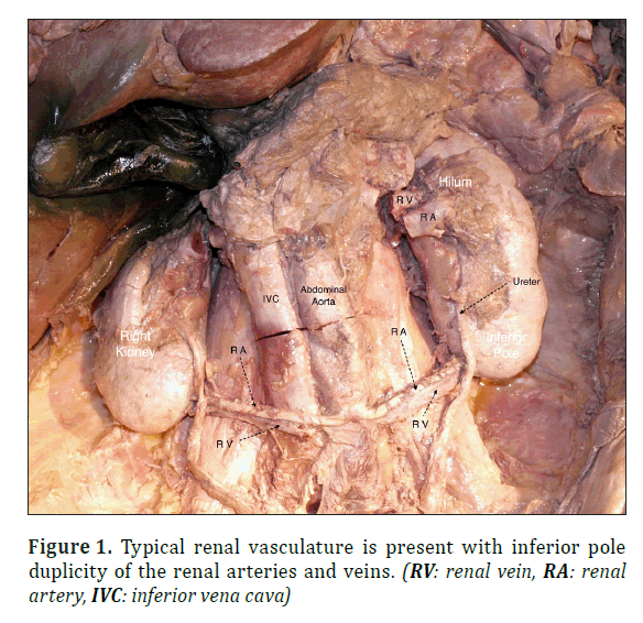
In this donor, the typical vasculature entering/exiting the kidney through the hilum was present in both kidneys. Further, it was observed that each kidney had an additional artery and vein that entered/exited the kidney via the inferior pole. These unusual renal vessels took unique routes to/from the inferior vena cava (IVC) and abdominal aorta. Both ureters were present and typical.
The supplemental vein of the right kidney originated at the inferior pole and attached directly to the anterior portion of the IVC. The paired artery also attached directly to the anterior portion of the abdominal aorta as seen in Figure 1.
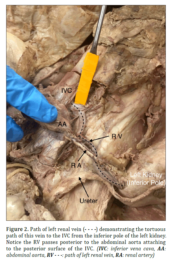
The left kidney had an ancillary vein that originated at the inferior pole and coursed medially, taking a sharp turn superiorly, and then passed behind the abdominal aorta before attaching to the IVC posteriorly. Similar to the right side, the left supplemental artery attached directly to the anterior portion of the abdominal aorta as seen in Figure 2.
Figure 2. Path of left renal vein (- - - -) demonstrating the tortuous path of this vein to the IVC from the inferior pole of the left kidney. Notice the RV passes posterior to the abdominal aorta attaching to the posterior surface of the IVC. (IVC: inferior vena cava, AA: abdominal aorta, RV - - -: path of left renal vein, RA: renal artery)
Discussion
The bilateral arterial and venous connections with the hilum and inferior pole of both kidneys to/from the aorta and inferior vena cava may not have caused any health issues for this individual. However, it is important to be aware of this and other renal vascular variations in patients with renal trauma or those requiring surgery in the retroperitoneal region of the abdomen. Gupta and colleagues (2011) report that multiple renal vasculature may present a threat during surgical intervention, and can impact the feasibility of surgeries (such as renal transplants) and restrict mobilization of the kidney [2]. While renal variations are common on the right, Anson and colleagues (1847) noted that duplication of the renal vein on the left side is exceedingly rare [4].
It is known that kidney vasculature can be quite variable. Kaneko and colleagues (2008) indicated that 47% of cadavers studied had multiple renal arteries, and 13% had multiple renal veins. However, it is important to note that bilateral multiple renal veins and arteries as seen in this individual are extremely rare. In the study by Kaneko and colleagues (2008), bilateral multiple veins were observed in 1 of the 170 cadavers examined (0.6%). Further, the presence of multiple bilateral veins did not coincide with the presence of multiple bilateral arteries [3].
In this donor, the left renal vein passed posterior to the aorta. This positioning may result in compromise of the vein due to compression between the aorta and vertebral bodies. Shah and colleagues (2013) describe the complications of Posterior Nutcracker Syndrome as leading to venous hypertension [5]. Imaging of patients who present with otherwise unexplained hematuria should be considered to rule out Posterior Nutcracker Syndrome as this condition may warrant surgical intervention.
Further documenting the uniqueness of this cadaver’s unusual renal vasculature, the central connections from the inferior poles to the aorta and inferior vena cava occur more distally than is typical. These unexpected, distal connections may cause complications with surgeries in the lower abdomen. It is important for clinicians and surgeons to be aware of these variations.
Acknowledgement
First we would like to thank our donor Joyce. Dr. Traci Little for her support and guidance throughout the dissection process. Dr. Kevin High for his editorial advisement. Elon University School of Health Sciences for the opportunity to participate in human dissection.
References
- Buffoli B, Franceschetti L, Belotti F, Ferrari M, Hirtler L, Tschabitscher M, Rodella LF. Multiple anatomical variations of the renal vessels associated with malrotated and unrotated kidneys: a case report. Surg Radiol Anat. 2015; 1-4.
- Gupta A, Singal R. Congenital variations of renal veins: embryological background and clinical implications. J Clin Diagn Res. 2011; 5(6):1140-1143.
- Kaneko N, Kobayashi Y, Okada Y. Anatomic variations of the renal vessels pertinent to transperitoneal vascular control in the management of trauma. Surgery. 2008; (143) 5: 616–622.
- Anson BJ, Cauldwell WE, Pick JW, Beaton LE. The blood supply of the kidney, suprarenal gland, and associated structures. Surg Gynecol Obstet. 1847; 84: 313-20.
- Shah D, Xiang Q, Shah A, Cao D. Posterior nutcracker syndrome with left renal vein duplication: An uncommon cause of hematuria. Inter J of Surg Case Reports. 2013; 4: 1142- 1144.
Carleigh M. High*, Christopher M. Scott, Christopher M. Antonelli, Kellie M. Ruffer and Janet M. Cope
Department of Physical Therapy Education, Elon University, Elon, North Carolina, 27244, USA
- *Corresponding Author:
- Carleigh M. High
Dept. of Physical Therapy Education
762 E Haggard Ave Elon, NC 27244, USA
Tel: +1 (336) 278-6355
E-mail: chigh@elon.edu
Date of Received: October 19th, 2015
Date of Accepted: December 30th, 2016
Published Online: January 26th 2017
© Int J Anat Var (IJAV). 2016; 9: 67–69.
Abstract
This case study documents a unique bilateral variation of renal vasculature. Renal arterial duplications are relatively common, whereas multiple renal veins occur much less commonly. In this case report the human donor had bilateral arterial and venous duplication. This variation has not been previously reported in the literature. It is important for clinicians to be aware of renal vessel variations for surgical procedures, particularly trauma and transplantation, and in other conditions that may lead to hypertension and/or urologic disorders.
-Keywords
bilateral, kidney, variation
Introduction
The kidneys are paired organs in the abdomen that are essential and responsible for filtering blood, removing waste, controlling body fluids, and regulating electrolyte and hormonal homeostasis. In order to properly perform these important functions the kidneys must have adequate blood supply. Typically, each kidney has a hilum that receives blood from a single renal artery from the aorta and one renal vein draining into the inferior vena cava. Buffoli and colleagues (2015) reported that renal variations are relatively common and make up about 30% of all congenital anomalies [1]. When additional renal vasculature is present, it may connect to the kidneys either through the hilum or at another point on the kidney’s surface. These anatomical variations are more prevalent in renal arteries compared to renal veins. According to Gupta and colleagues (2011) variations of renal veins tend to occur on the left side due to the complexity that arises from the presence of multiple veins, including the adrenal, gonadal, phrenic, and hemizygous vessels [2]. In contrast, Kaneko and colleagues (2008) report that multiple renal veins are relatively common on the right and rare on the left [3].
In this case report, the researchers discuss a previously unreported variant of bilateral renal veins and arteries in a single cadaver. In this individual, the renal vessels arose from each kidney at both the hilum and the inferior pole.
Case Report
During routine dissection of an 84-year-old female donor at Elon University, Elon, North Carolina, who had a recorded cause of death as dementia and Parkinson’s disease, the researchers observed an interesting variation in the cadaver’s renal arteries and veins.
The donor had worked as a waitress and during initial dissections it was noted that both of her shoulder joints had ossified and numerous bone spurs were present. The donor was also found to have hardware in her left ankle, and her left ovary was enlarged and calcified when compared to her right ovary.
In this donor, the typical vasculature entering/exiting the kidney through the hilum was present in both kidneys. Further, it was observed that each kidney had an additional artery and vein that entered/exited the kidney via the inferior pole. These unusual renal vessels took unique routes to/from the inferior vena cava (IVC) and abdominal aorta. Both ureters were present and typical.
The supplemental vein of the right kidney originated at the inferior pole and attached directly to the anterior portion of the IVC. The paired artery also attached directly to the anterior portion of the abdominal aorta as seen in Figure 1.
The left kidney had an ancillary vein that originated at the inferior pole and coursed medially, taking a sharp turn superiorly, and then passed behind the abdominal aorta before attaching to the IVC posteriorly. Similar to the right side, the left supplemental artery attached directly to the anterior portion of the abdominal aorta as seen in Figure 2.
Figure 2. Path of left renal vein (- - - -) demonstrating the tortuous path of this vein to the IVC from the inferior pole of the left kidney. Notice the RV passes posterior to the abdominal aorta attaching to the posterior surface of the IVC. (IVC: inferior vena cava, AA: abdominal aorta, RV - - -: path of left renal vein, RA: renal artery)
Discussion
The bilateral arterial and venous connections with the hilum and inferior pole of both kidneys to/from the aorta and inferior vena cava may not have caused any health issues for this individual. However, it is important to be aware of this and other renal vascular variations in patients with renal trauma or those requiring surgery in the retroperitoneal region of the abdomen. Gupta and colleagues (2011) report that multiple renal vasculature may present a threat during surgical intervention, and can impact the feasibility of surgeries (such as renal transplants) and restrict mobilization of the kidney [2]. While renal variations are common on the right, Anson and colleagues (1847) noted that duplication of the renal vein on the left side is exceedingly rare [4].
It is known that kidney vasculature can be quite variable. Kaneko and colleagues (2008) indicated that 47% of cadavers studied had multiple renal arteries, and 13% had multiple renal veins. However, it is important to note that bilateral multiple renal veins and arteries as seen in this individual are extremely rare. In the study by Kaneko and colleagues (2008), bilateral multiple veins were observed in 1 of the 170 cadavers examined (0.6%). Further, the presence of multiple bilateral veins did not coincide with the presence of multiple bilateral arteries [3].
In this donor, the left renal vein passed posterior to the aorta. This positioning may result in compromise of the vein due to compression between the aorta and vertebral bodies. Shah and colleagues (2013) describe the complications of Posterior Nutcracker Syndrome as leading to venous hypertension [5]. Imaging of patients who present with otherwise unexplained hematuria should be considered to rule out Posterior Nutcracker Syndrome as this condition may warrant surgical intervention.
Further documenting the uniqueness of this cadaver’s unusual renal vasculature, the central connections from the inferior poles to the aorta and inferior vena cava occur more distally than is typical. These unexpected, distal connections may cause complications with surgeries in the lower abdomen. It is important for clinicians and surgeons to be aware of these variations.
Acknowledgement
First we would like to thank our donor Joyce. Dr. Traci Little for her support and guidance throughout the dissection process. Dr. Kevin High for his editorial advisement. Elon University School of Health Sciences for the opportunity to participate in human dissection.
References
- Buffoli B, Franceschetti L, Belotti F, Ferrari M, Hirtler L, Tschabitscher M, Rodella LF. Multiple anatomical variations of the renal vessels associated with malrotated and unrotated kidneys: a case report. Surg Radiol Anat. 2015; 1-4.
- Gupta A, Singal R. Congenital variations of renal veins: embryological background and clinical implications. J Clin Diagn Res. 2011; 5(6):1140-1143.
- Kaneko N, Kobayashi Y, Okada Y. Anatomic variations of the renal vessels pertinent to transperitoneal vascular control in the management of trauma. Surgery. 2008; (143) 5: 616–622.
- Anson BJ, Cauldwell WE, Pick JW, Beaton LE. The blood supply of the kidney, suprarenal gland, and associated structures. Surg Gynecol Obstet. 1847; 84: 313-20.
- Shah D, Xiang Q, Shah A, Cao D. Posterior nutcracker syndrome with left renal vein duplication: An uncommon cause of hematuria. Inter J of Surg Case Reports. 2013; 4: 1142- 1144.


