Bilateral fibrous replacement of subclavius muscle in relation to nerve and artery compression of the upper limb
Georgi P. Georgiev* and Lazar Jelev
Department of Anatomy, Histology and Embryology, Medical University Sofia, Sofia, Bulgaria
- *Corresponding Author:
- Georgi P. Georgiev, MD
Department of Anatomy, Histology and Embryology, Medical University Sofia, Blvd. Sv. Georgi Sofiiski 1, BG-1431 Sofia, Bulgaria
Tel: +359 2 9172636
Fax: +359 2 8518783
E-mail: georgievgp@yahoo.com
Date of Received: January 12th, 2009
Date of Accepted: June 5th, 2009
Published Online: June 9th, 2009
© IJAV. 2009; 2: 57–59.
[ft_below_content] =>Keywords
variation, fibrous bands, costoclavicular space, thoracic outlet syndrome, human
Introduction
The upper part of brachial plexus and the subclavian/axillary vessels could be vulnerable in four main areas: the superior thoracic outlet, the scalene triangle, the costoclavicular interval (space) and the coracoid-pectoralis minor loop, and symptoms of compression syndromes may be developed [1]. According to Pratt [1], the most critical point appears to be the costoclavicular space and symptoms could be provoked by bone or soft tissue anomalies including different variant structures [2,3].
Case Report
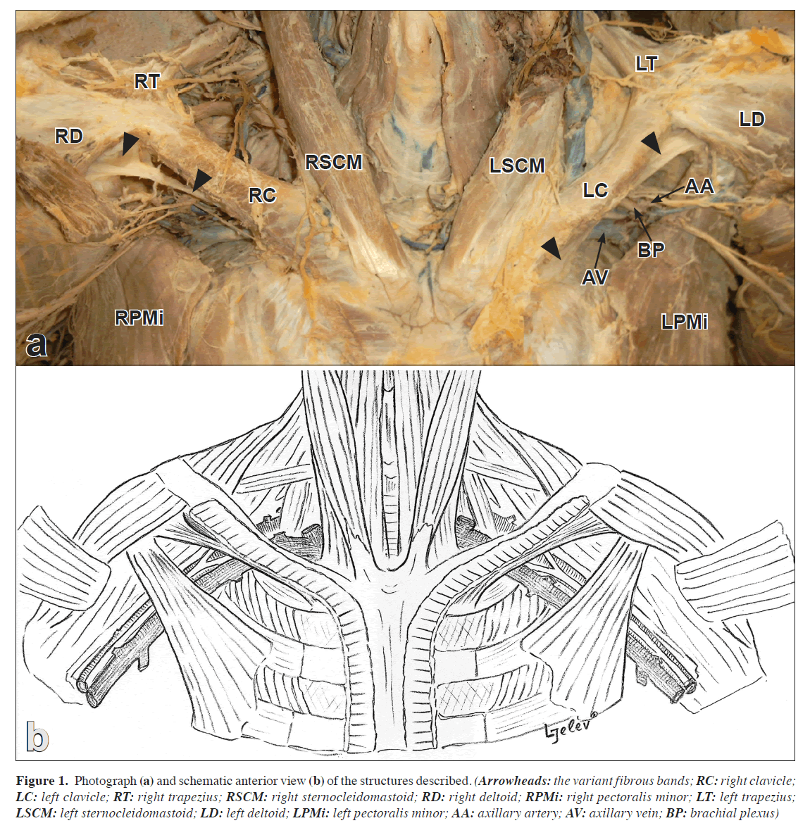
An interesting case of bilateral fibrous bands in the costoclavicular interval was discovered during routine anatomical dissection of a 58-year-old Caucasian male cadaver. It was the only case from a series of 56 dissected cadavers, examined over 8-year-period. In the dissected subject no signs of accidental or surgical trauma were observed; however, we were lack of information about previous diseases. On both sides, the fibrous bands replaced the usual subclavius muscle (Figure 1). On the right side, the fibrous band (7.2 cm in length) extended from the inferior surface of the acromial extremity of the clavicle and the coracoid process to the medial end of the first rib. Along its course through the costoclavicular space, the fibrous band passed obliquely downwards and crossed in front the subclavian vessels and brachial plexus. On the left side, a similar but stronger fibrous band (7.6 cm in length) was found. It extended between the inferior surface of the lateral two-thirds of the clavicle and the coracoid process to the medial end of the first rib. This variant fibrous band passed along the under surface of the clavicle, reducing vastly the costoclavicular interval.
Figure 1. Photograph (a) and schematic anterior view (b) of the structures described. (Arrowheads: the variant fibrous bands; RC: right clavicle; LC: left clavicle; RT: right trapezius; RSCM: right sternocleidomastoid; RD: right deltoid; RPMi: right pectoralis minor; LT: left trapezius; LSCM: left sternocleidomastoid; LD: left deltoid; LPMi: left pectoralis minor; AA: axillary artery; AV: axillary vein; BP: brachial plexus)
Discussion
Around the human clavicle, in relation to the subclavius muscle, a number of variant muscles have been described [2,4,5]. However, the subclavius itself shows a remarkable constancy of its structure, the trends of its variations being in the direction of duplicity, due essentially to anomalous differentiation of the primary pectoral muscle sheet [6]. Another subclavius muscle abnormality is its absence, defined as an excessively rare anatomic variant [6]. There may be a total subclavius absence [7] or the muscle body may be replaced by a fibrous band [4,5,8]. In the cases reported up to day, however, this fibrous replacement was detected only on one side. Here, we present a rare case of bilateral replacement of the subclavius by fibrous bands.
The present fibrous bands narrow the interval between the clavicle and first rib, which is essential for development of an entrapment of the brachial plexus and the subclavian/axillary vessels [1,9]. Symptoms characteristic of such entrapment is commonly diagnosed as thoracic outlet syndrome (TOS) [1,9]. Variant structures may provoke compression of the neuro-vascular structures in the axilla, especially in cases of depression and posterior displacement of the shoulder [1]; and symptom complex of TOS, including neurological symptoms, arterial stenosis and venous symptoms may be resulted [3]. There is no single criterion and no specific physical tests for TOS existence [1]. The diagnosis is based on a combination of clinical and electrophysiological criteria and the use of different imaging techniques [9,10]. Therefore, good knowledge of the variant structures, possibly compressing the neuro-vascular structures from the base of the neck to the axilla, including the here reported fibrous bands, may help clinicians identify the cause of TOS.
References
- Pratt NE. Neurovascular entrapment in the regions of the shoulder and posterior triangle of the neck. Phys Ther. 1986; 66: 1894–1900.
- Akita K, Ibukuro K, Yamaguchi K, Heima S, Sato T. The subclavius posticus muscle: a factor in arterial, venous or brachial plexus compression? Surg Radiol Anat. 2000; 22: 111–115.
- Degeorges R, Reynaud C, Becquemin JP. Thoracic outlet syndrome surgery: long-term functional results. Ann Vasc Surg. 2004; 18: 558–565.
- Bergman RA, Afifi AK, Miyauchi R. Part I: Muscular system. In: Illustrated encyclopedia of human anatomic variations. http://www.anatomyatlases.org/AnatomicVariants/AnatomyHP.shtml (accessed May 2009).
- Le Double A. Muscles des parois de la poitrine. In: Traité des Variations du Système Musculaire de l’Homme. Tome I, Paris, Schleicher Frères. 1897; 243–286.
- Cave AJE, Brown RW. On the tendon of the subclavius muscle. J Bone Joint Surg Br. 1952; 34: 466–469.
- Gruber W. Ueber congenitalen unvollständigen Radiusmangel. Virchows Arch. 1867; 40: 427–435.
- Crerar JW. Note on the absence of the subclavius muscle. J Anat Physiol. 1892; 26: 554.
- Le Forestier N, Moulonguet A, Maisonobe T, Leger JM, Bouche P. True neurogenic thoracic outlet syndrome: electrophysiological diagnosis in six cases. Muscle Nerve. 1998; 21: 1129–1134.
- Demondion X, Bacqueville E, Paul C, Duquesnoy B, Hachulla E, Cotten A. Thoracic outlet: assessment with MR imaging in asymptomatic and symptomatic populations. Radiology. 2003; 227: 461–468.
Georgi P. Georgiev* and Lazar Jelev
Department of Anatomy, Histology and Embryology, Medical University Sofia, Sofia, Bulgaria
- *Corresponding Author:
- Georgi P. Georgiev, MD
Department of Anatomy, Histology and Embryology, Medical University Sofia, Blvd. Sv. Georgi Sofiiski 1, BG-1431 Sofia, Bulgaria
Tel: +359 2 9172636
Fax: +359 2 8518783
E-mail: georgievgp@yahoo.com
Date of Received: January 12th, 2009
Date of Accepted: June 5th, 2009
Published Online: June 9th, 2009
© IJAV. 2009; 2: 57–59.
Abstract
Here, we report an interesting case of bilateral fibrous structures found in the costoclavicular space of a 58-year-old male cadaver during routine anatomical dissection. On the left side, a strong fibrous band extending between the inferior surface of the lateral two-thirds of the clavicle and the coracoid process to the medial end of the first rib was found. On the right side, a similar but thinner structure was discovered. On both sides the variant fibrous structures replacing the subclavius muscle, reduced the costoclavicular space thus led to a possible entrapment site for the brachial plexus and the subclavian/axillary vessels, and may be involved in the development of thoracic outlet syndrome.
-Keywords
variation, fibrous bands, costoclavicular space, thoracic outlet syndrome, human
Introduction
The upper part of brachial plexus and the subclavian/axillary vessels could be vulnerable in four main areas: the superior thoracic outlet, the scalene triangle, the costoclavicular interval (space) and the coracoid-pectoralis minor loop, and symptoms of compression syndromes may be developed [1]. According to Pratt [1], the most critical point appears to be the costoclavicular space and symptoms could be provoked by bone or soft tissue anomalies including different variant structures [2,3].
Case Report
An interesting case of bilateral fibrous bands in the costoclavicular interval was discovered during routine anatomical dissection of a 58-year-old Caucasian male cadaver. It was the only case from a series of 56 dissected cadavers, examined over 8-year-period. In the dissected subject no signs of accidental or surgical trauma were observed; however, we were lack of information about previous diseases. On both sides, the fibrous bands replaced the usual subclavius muscle (Figure 1). On the right side, the fibrous band (7.2 cm in length) extended from the inferior surface of the acromial extremity of the clavicle and the coracoid process to the medial end of the first rib. Along its course through the costoclavicular space, the fibrous band passed obliquely downwards and crossed in front the subclavian vessels and brachial plexus. On the left side, a similar but stronger fibrous band (7.6 cm in length) was found. It extended between the inferior surface of the lateral two-thirds of the clavicle and the coracoid process to the medial end of the first rib. This variant fibrous band passed along the under surface of the clavicle, reducing vastly the costoclavicular interval.
Figure 1. Photograph (a) and schematic anterior view (b) of the structures described. (Arrowheads: the variant fibrous bands; RC: right clavicle; LC: left clavicle; RT: right trapezius; RSCM: right sternocleidomastoid; RD: right deltoid; RPMi: right pectoralis minor; LT: left trapezius; LSCM: left sternocleidomastoid; LD: left deltoid; LPMi: left pectoralis minor; AA: axillary artery; AV: axillary vein; BP: brachial plexus)
Discussion
Around the human clavicle, in relation to the subclavius muscle, a number of variant muscles have been described [2,4,5]. However, the subclavius itself shows a remarkable constancy of its structure, the trends of its variations being in the direction of duplicity, due essentially to anomalous differentiation of the primary pectoral muscle sheet [6]. Another subclavius muscle abnormality is its absence, defined as an excessively rare anatomic variant [6]. There may be a total subclavius absence [7] or the muscle body may be replaced by a fibrous band [4,5,8]. In the cases reported up to day, however, this fibrous replacement was detected only on one side. Here, we present a rare case of bilateral replacement of the subclavius by fibrous bands.
The present fibrous bands narrow the interval between the clavicle and first rib, which is essential for development of an entrapment of the brachial plexus and the subclavian/axillary vessels [1,9]. Symptoms characteristic of such entrapment is commonly diagnosed as thoracic outlet syndrome (TOS) [1,9]. Variant structures may provoke compression of the neuro-vascular structures in the axilla, especially in cases of depression and posterior displacement of the shoulder [1]; and symptom complex of TOS, including neurological symptoms, arterial stenosis and venous symptoms may be resulted [3]. There is no single criterion and no specific physical tests for TOS existence [1]. The diagnosis is based on a combination of clinical and electrophysiological criteria and the use of different imaging techniques [9,10]. Therefore, good knowledge of the variant structures, possibly compressing the neuro-vascular structures from the base of the neck to the axilla, including the here reported fibrous bands, may help clinicians identify the cause of TOS.
References
- Pratt NE. Neurovascular entrapment in the regions of the shoulder and posterior triangle of the neck. Phys Ther. 1986; 66: 1894–1900.
- Akita K, Ibukuro K, Yamaguchi K, Heima S, Sato T. The subclavius posticus muscle: a factor in arterial, venous or brachial plexus compression? Surg Radiol Anat. 2000; 22: 111–115.
- Degeorges R, Reynaud C, Becquemin JP. Thoracic outlet syndrome surgery: long-term functional results. Ann Vasc Surg. 2004; 18: 558–565.
- Bergman RA, Afifi AK, Miyauchi R. Part I: Muscular system. In: Illustrated encyclopedia of human anatomic variations. http://www.anatomyatlases.org/AnatomicVariants/AnatomyHP.shtml (accessed May 2009).
- Le Double A. Muscles des parois de la poitrine. In: Traité des Variations du Système Musculaire de l’Homme. Tome I, Paris, Schleicher Frères. 1897; 243–286.
- Cave AJE, Brown RW. On the tendon of the subclavius muscle. J Bone Joint Surg Br. 1952; 34: 466–469.
- Gruber W. Ueber congenitalen unvollständigen Radiusmangel. Virchows Arch. 1867; 40: 427–435.
- Crerar JW. Note on the absence of the subclavius muscle. J Anat Physiol. 1892; 26: 554.
- Le Forestier N, Moulonguet A, Maisonobe T, Leger JM, Bouche P. True neurogenic thoracic outlet syndrome: electrophysiological diagnosis in six cases. Muscle Nerve. 1998; 21: 1129–1134.
- Demondion X, Bacqueville E, Paul C, Duquesnoy B, Hachulla E, Cotten A. Thoracic outlet: assessment with MR imaging in asymptomatic and symptomatic populations. Radiology. 2003; 227: 461–468.


