Bilateral incomplete superficial palmar arch
Lakshmiprabha R1*, Niranjan Murthy HL2, Komala B1, Sendil Kumaran D2
1Departments of Anatomy, Sree Siddhartha Medical College, Tumkur, Karnataka, India.
2Departments of Anatomy and Physiology, Sree Siddhartha Medical College, Tumkur, Karnataka, India.
- *Corresponding Author:
- Dr. Lakshmiprabha R
Professor and Head, Department of Anatomy, Sree Siddhartha Medical College. Tumkur, Karnataka, India.
Tel: +91 984 5708555
E-mail: lakshmiprabha100@yahoo.com
Date of Received: April 10th, 2009
Date of Accepted: August 5th, 2009
Published Online: August 17th, 2009
© IJAV. 2009; 2: 86–88.
[ft_below_content] =>Keywords
collateral, harvesting, palmar arch, radial artery, ulnar artery
Introduction
Superficial palmar arch (SPA) is the dominant vascular structure of the hand and is mainly formed by the anastomosis of ulnar artery (UA) with superficial branch of radial artery (RA), or arteria radialis indicis or arteria princeps pollicis from RA. A classical type of SPA is described as direct continuity between UA and superficial branch of RA confirms the presence of collateral supply in hand [1]. An incomplete arch has an absence of communication or anastomosis between the vessels constituting the arch [2]. Knowledge in variation of vascular pattern of hand gained more importance in microsurgical techniques, reconstructive hand surgeries, preoperative screening of RA harvesting for myocardial revascularization and also in arterial interventions that include RA cannulation and RA forearm flap.
In an incomplete SPA, UA do not anastomose with the RA or median artery (MA) and fails to reach the thumb and index finger. There were 4 main types of arch according to Coleman. Type A: Both superficial palmar branches of UA and RA take part in supplying the palm and fingers, but in doing so fail to anastomose and seen in 3.6% of cases. Type B: Only the UA forms the SPA, but the arch is incomplete in sense it does not supply thumb & index finger, and seen in 13.4% of cases. Type C: Superficial vessels receive contribution from both UA & MA, but without anastomosis, and seen in 3.8% of cases. Type D: RA, UA & MA all give origin to superficial vessels but do not anastomose and seen in 1.1% of cases. The present study belongs to type A, a rare and complex bilateral type [3].
Type A incomplete SPA leads to an increased vulnerability to digital ischemic changes following trauma or RA harvesting or RA interventions, and hence prior screening of presence of viable collateral circulation in hand is highly recommended [4]. This variation also assumes importance in obstruction of arteries at the level of wrist occurring in hypothenar hammer syndrome and in connective tissue diseases [3].
Case Report
A complex variation in the pattern of formation of SPA was bilaterally encountered in a 45-year-old female cadaver during routine dissection in the Department of Anatomy, SSMC, Tumkur.
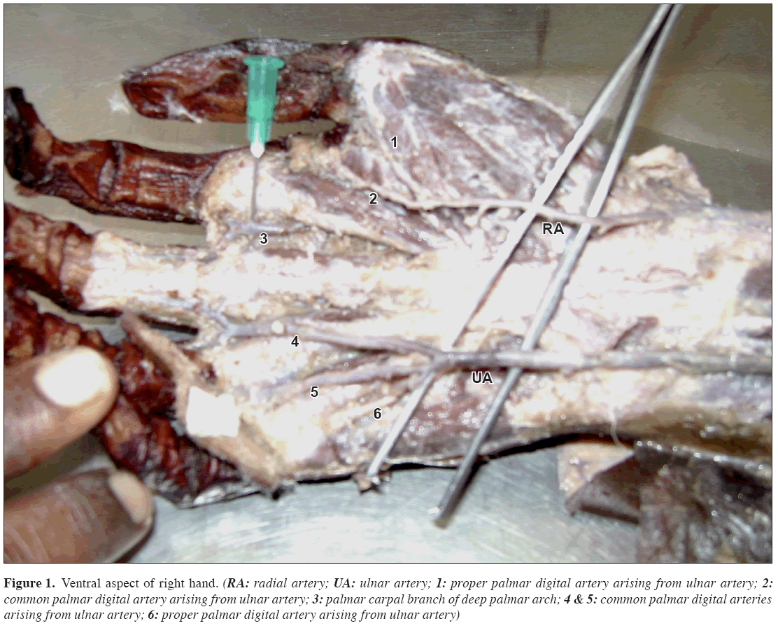
Right hand: The superficial branch of UA entered the palm of the hand superficial to flexor retinaculum and divided into 2 common palmar digital arteries (CPDAs) supplying contiguous sides of middle & ring fingers, and ring & little fingers (Figure 1). It also gave rise to one proper palmar digital artery (PPDA) to the ulnar side of little finger.
Figure 1: Ventral aspect of right hand. (RA: radial artery; UA: ulnar artery; 1: proper palmar digital artery arising from ulnar artery; 2: common palmar digital artery arising from ulnar artery; 3: palmar carpal branch of deep palmar arch; 4 & 5: common palmar digital arteries arising from ulnar artery; 6: proper palmar digital artery arising from ulnar artery)
The superficial palmar branch of RA entered the hand superficial to thenar muscles and gave rise to PPDA to the radial side of the thumb and one CPDA supplying the ulnar side of the thumb and radial side of the index finger. The interdigital space between the index and middle finger was supplied by the palmar carpal branch from deep palmar arch.
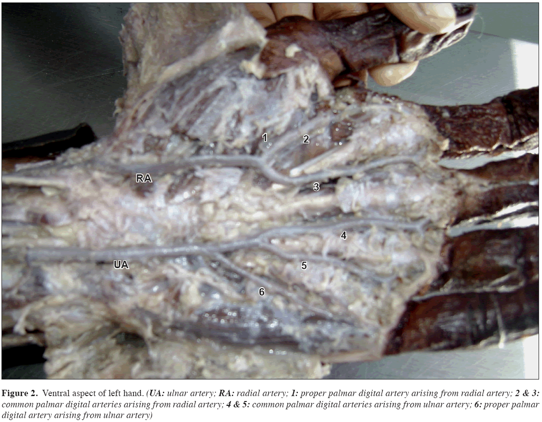
Left hand: The superficial branch of UA entered the palm superficial to flexor retinaculum and divided into 2 CPDAs supplying the ulnar side of middle & radial side of ring fingers, and ulnar side of ring & radial side of little fingers (Figure 2). One PPDA to ulnar side of little finger arose directly from the artery.
Figure 2: Ventral aspect of left hand. (UA: ulnar artery; RA: radial artery; 1: proper palmar digital artery arising from radial artery; 2 & 3: common palmar digital arteries arising from radial artery; 4 & 5: common palmar digital arteries arising from ulnar artery; 6: proper palmar digital artery arising from ulnar artery)
Superficial palmar branch of RA entered the hand deep to thenar muscles and divided into 2 CPDAs supplying the adjacent sides of thumb & index finger and index & middle fingers. The branch to radial side of the thumb arose from first CPDA.
There was no anastomosis between the RA and UA and as a whole on both the sides the fingers were supplied by their branches. The UA and RA arose from brachial artery normally at the level of neck of the radius and their course in forearm on both sides was normal.
Discussion
The superficial arteries of the hand formed several diversified patterns that permitted into well-defined categories. In 52% of the subjects the arterial pattern in right and left hands were different with respect to one or more arteries and while 48% were identical in both the hands [5]. About a third of the SPA is formed by the UA alone; a further third are completed by the superficial palmar branch of the RA and a third either by the arteria radialis indicis or by princeps pollicis or by median artery [6]. A classic type of SPA in which the superficial branch of RA joins the superficial branch of UA is found in 34.5% of cases [6,7]. The complete arch was found in 78.5% of cases and incomplete arch in 21.5% of cases and is a major underlying factor in the etiology of digital ischemia [3]. Ikeda et al. conducted stereoscopic arteriography of 220 cadaver hands and reported complete SPA in 96.4% of cases and 3.6% as incomplete arch [8]. Gellman et al. reported 84.4% of cases as complete SPA and 15.5% of cases as incomplete SPA [2]. Al Turk and Metcalf showed complete SPA in 84% of cases and incomplete in 16% using Doppler flowmeter [9]. Harvesting RA for use as arterial by-pass conduits needs to look specifically for variation in collateral circulation like presence of incomplete SPA [4].
The patients with coronary artery disease should be screened before RA harvesting to confirm the presence of viable collateral circulation. Currently the methods of assessing hand circulation include the modified Allen test, Doppler ultrasonography, photoplethysmography. Doppler study is a useful tool in preoperative screening for RA harvesting for myocardial revascularization [10].
Although ipsilateral incomplete SPA is mentioned in literature frequently, bilateral arch is a rare event and this variant vascular anatomy of hand can be identified with a careful preoperative examination and failure to appropriately manage these variations may result in a compromised surgical outcome.
References
- Agur AM, Lee MJ. Grant’s Atlas of Anatomy. 9th Ed., Philadelphia, Lippincott, Williams and Wilkins. 1999; 419.
- Gellman H, Botte MJ, Shankwiler J, Gelberman RH. Arterial patterns of the deep and superficial palmar arches. Clin Orthop Relat Res. 2001; 383: 41–46.
- Coleman SS, Anson BJ. Arterial patterns in the hand based upon a study of 650 specimens. Surg Gynecol Obstet. 1961; 113: 409–424.
- Ruengsakulrach P, Brooks M, Hare DL, Gordon I, Buxton BF. Preoperative assessment of hand circulation by means of Doppler ultrasonography and the modified Allen test. J Thorac Cardiovasc Surg. 2001; 121: 526–531.
- Mozersky DJ, Buckley CJ, Hagood CO Jr, Capps WF Jr, Dannemiller FJ Jr. Ultrasonic evaluation of the palmar circulation. A useful adjunct to radial artery cannulation. Am J Surg. 1973; 126: 810–812.
- Willams PL, Bannister LG, Berry MM, eds. Gray’s Anatomy. 38th Ed., New York, Churchill Livingstone. 2000; 1544.
- Moore KL, Dalley AF. Clinically Oriented Anatomy. 10th Ed., Philadelphia, Lippincott, Williams & Wilkins. 1999; 751.
- Ikeda A, Ugawa A, Kazihara Y, Hamada N. Arterial patterns in the hand based on a three-dimensional analysis of 220 cadaver hands. J Hand Surg Am. 1988; 13: 501–509.
- Al-Turk M, Metcalf WK. A study of the superficial palmar arteries using the Doppler Ultrasonic Flowmeter. J Anat. 1984; 138: 27–32.
- Pola P, Serricchio M, Flore R, Manasse E, Favuzzi A, Possati GF. Safe removal of the radial artery for myocardial revascularization: a Doppler study to prevent ischemic complications to the hand. J Thorac Cardiovasc Surg. 1996; 112: 737–744.
Lakshmiprabha R1*, Niranjan Murthy HL2, Komala B1, Sendil Kumaran D2
1Departments of Anatomy, Sree Siddhartha Medical College, Tumkur, Karnataka, India.
2Departments of Anatomy and Physiology, Sree Siddhartha Medical College, Tumkur, Karnataka, India.
- *Corresponding Author:
- Dr. Lakshmiprabha R
Professor and Head, Department of Anatomy, Sree Siddhartha Medical College. Tumkur, Karnataka, India.
Tel: +91 984 5708555
E-mail: lakshmiprabha100@yahoo.com
Date of Received: April 10th, 2009
Date of Accepted: August 5th, 2009
Published Online: August 17th, 2009
© IJAV. 2009; 2: 86–88.
Abstract
A rare variation of bilateral incomplete palmar arch was observed during routine dissection of a cadaver. Ulnar arteries gave rise to two common palmar digital arteries supplying the ring and little fingers and one proper palmar digital artery, which supplied ulnar side of little finger. Radial artery gave rise to two common palmar digital arteries that supplied the remaining fingers. Right side also had a palmar carpal branch from deep palmar arch.
-Keywords
collateral, harvesting, palmar arch, radial artery, ulnar artery
Introduction
Superficial palmar arch (SPA) is the dominant vascular structure of the hand and is mainly formed by the anastomosis of ulnar artery (UA) with superficial branch of radial artery (RA), or arteria radialis indicis or arteria princeps pollicis from RA. A classical type of SPA is described as direct continuity between UA and superficial branch of RA confirms the presence of collateral supply in hand [1]. An incomplete arch has an absence of communication or anastomosis between the vessels constituting the arch [2]. Knowledge in variation of vascular pattern of hand gained more importance in microsurgical techniques, reconstructive hand surgeries, preoperative screening of RA harvesting for myocardial revascularization and also in arterial interventions that include RA cannulation and RA forearm flap.
In an incomplete SPA, UA do not anastomose with the RA or median artery (MA) and fails to reach the thumb and index finger. There were 4 main types of arch according to Coleman. Type A: Both superficial palmar branches of UA and RA take part in supplying the palm and fingers, but in doing so fail to anastomose and seen in 3.6% of cases. Type B: Only the UA forms the SPA, but the arch is incomplete in sense it does not supply thumb & index finger, and seen in 13.4% of cases. Type C: Superficial vessels receive contribution from both UA & MA, but without anastomosis, and seen in 3.8% of cases. Type D: RA, UA & MA all give origin to superficial vessels but do not anastomose and seen in 1.1% of cases. The present study belongs to type A, a rare and complex bilateral type [3].
Type A incomplete SPA leads to an increased vulnerability to digital ischemic changes following trauma or RA harvesting or RA interventions, and hence prior screening of presence of viable collateral circulation in hand is highly recommended [4]. This variation also assumes importance in obstruction of arteries at the level of wrist occurring in hypothenar hammer syndrome and in connective tissue diseases [3].
Case Report
A complex variation in the pattern of formation of SPA was bilaterally encountered in a 45-year-old female cadaver during routine dissection in the Department of Anatomy, SSMC, Tumkur.
Right hand: The superficial branch of UA entered the palm of the hand superficial to flexor retinaculum and divided into 2 common palmar digital arteries (CPDAs) supplying contiguous sides of middle & ring fingers, and ring & little fingers (Figure 1). It also gave rise to one proper palmar digital artery (PPDA) to the ulnar side of little finger.
Figure 1: Ventral aspect of right hand. (RA: radial artery; UA: ulnar artery; 1: proper palmar digital artery arising from ulnar artery; 2: common palmar digital artery arising from ulnar artery; 3: palmar carpal branch of deep palmar arch; 4 & 5: common palmar digital arteries arising from ulnar artery; 6: proper palmar digital artery arising from ulnar artery)
The superficial palmar branch of RA entered the hand superficial to thenar muscles and gave rise to PPDA to the radial side of the thumb and one CPDA supplying the ulnar side of the thumb and radial side of the index finger. The interdigital space between the index and middle finger was supplied by the palmar carpal branch from deep palmar arch.
Left hand: The superficial branch of UA entered the palm superficial to flexor retinaculum and divided into 2 CPDAs supplying the ulnar side of middle & radial side of ring fingers, and ulnar side of ring & radial side of little fingers (Figure 2). One PPDA to ulnar side of little finger arose directly from the artery.
Figure 2: Ventral aspect of left hand. (UA: ulnar artery; RA: radial artery; 1: proper palmar digital artery arising from radial artery; 2 & 3: common palmar digital arteries arising from radial artery; 4 & 5: common palmar digital arteries arising from ulnar artery; 6: proper palmar digital artery arising from ulnar artery)
Superficial palmar branch of RA entered the hand deep to thenar muscles and divided into 2 CPDAs supplying the adjacent sides of thumb & index finger and index & middle fingers. The branch to radial side of the thumb arose from first CPDA.
There was no anastomosis between the RA and UA and as a whole on both the sides the fingers were supplied by their branches. The UA and RA arose from brachial artery normally at the level of neck of the radius and their course in forearm on both sides was normal.
Discussion
The superficial arteries of the hand formed several diversified patterns that permitted into well-defined categories. In 52% of the subjects the arterial pattern in right and left hands were different with respect to one or more arteries and while 48% were identical in both the hands [5]. About a third of the SPA is formed by the UA alone; a further third are completed by the superficial palmar branch of the RA and a third either by the arteria radialis indicis or by princeps pollicis or by median artery [6]. A classic type of SPA in which the superficial branch of RA joins the superficial branch of UA is found in 34.5% of cases [6,7]. The complete arch was found in 78.5% of cases and incomplete arch in 21.5% of cases and is a major underlying factor in the etiology of digital ischemia [3]. Ikeda et al. conducted stereoscopic arteriography of 220 cadaver hands and reported complete SPA in 96.4% of cases and 3.6% as incomplete arch [8]. Gellman et al. reported 84.4% of cases as complete SPA and 15.5% of cases as incomplete SPA [2]. Al Turk and Metcalf showed complete SPA in 84% of cases and incomplete in 16% using Doppler flowmeter [9]. Harvesting RA for use as arterial by-pass conduits needs to look specifically for variation in collateral circulation like presence of incomplete SPA [4].
The patients with coronary artery disease should be screened before RA harvesting to confirm the presence of viable collateral circulation. Currently the methods of assessing hand circulation include the modified Allen test, Doppler ultrasonography, photoplethysmography. Doppler study is a useful tool in preoperative screening for RA harvesting for myocardial revascularization [10].
Although ipsilateral incomplete SPA is mentioned in literature frequently, bilateral arch is a rare event and this variant vascular anatomy of hand can be identified with a careful preoperative examination and failure to appropriately manage these variations may result in a compromised surgical outcome.
References
- Agur AM, Lee MJ. Grant’s Atlas of Anatomy. 9th Ed., Philadelphia, Lippincott, Williams and Wilkins. 1999; 419.
- Gellman H, Botte MJ, Shankwiler J, Gelberman RH. Arterial patterns of the deep and superficial palmar arches. Clin Orthop Relat Res. 2001; 383: 41–46.
- Coleman SS, Anson BJ. Arterial patterns in the hand based upon a study of 650 specimens. Surg Gynecol Obstet. 1961; 113: 409–424.
- Ruengsakulrach P, Brooks M, Hare DL, Gordon I, Buxton BF. Preoperative assessment of hand circulation by means of Doppler ultrasonography and the modified Allen test. J Thorac Cardiovasc Surg. 2001; 121: 526–531.
- Mozersky DJ, Buckley CJ, Hagood CO Jr, Capps WF Jr, Dannemiller FJ Jr. Ultrasonic evaluation of the palmar circulation. A useful adjunct to radial artery cannulation. Am J Surg. 1973; 126: 810–812.
- Willams PL, Bannister LG, Berry MM, eds. Gray’s Anatomy. 38th Ed., New York, Churchill Livingstone. 2000; 1544.
- Moore KL, Dalley AF. Clinically Oriented Anatomy. 10th Ed., Philadelphia, Lippincott, Williams & Wilkins. 1999; 751.
- Ikeda A, Ugawa A, Kazihara Y, Hamada N. Arterial patterns in the hand based on a three-dimensional analysis of 220 cadaver hands. J Hand Surg Am. 1988; 13: 501–509.
- Al-Turk M, Metcalf WK. A study of the superficial palmar arteries using the Doppler Ultrasonic Flowmeter. J Anat. 1984; 138: 27–32.
- Pola P, Serricchio M, Flore R, Manasse E, Favuzzi A, Possati GF. Safe removal of the radial artery for myocardial revascularization: a Doppler study to prevent ischemic complications to the hand. J Thorac Cardiovasc Surg. 1996; 112: 737–744.


