Bilateral malrotated kidney with major venous variant: a cadaver case report
Joe Iwanaga*, Koichi Watanabe, Tsuyoshi Saga and Koh-ichi Yamaki
Department of Anatomy, Kurume University School of Medicine, 67 Asahi-machi, Kurume, Fukuoka 830-0011, Japan.
- *Corresponding Author:
- Joe Iwanaga
Department of Anatomy, Kurume University School of Medicine, 67 Asahi-machi, Kurume Fukuoka 830-0011, Japan
Tel: +81-942-31-7540
E-mail: iwanaga_jyou@med.kurume-u.ac.jp
Date of Received: May 20th, 2016
Date of Accepted: December 24th, 2016
Published Online: January 1st, 2017
© Int J Anat Var (IJAV). 2016; 9: 43–45.
[ft_below_content] =>Keywords
kidney, anatomy, cadaver, iliac vein, inferior vena cava
Introduction
Congenital anomalies of the kidney exhibit great variety. The major anomalies are fused kidney, ectopic kidney, and malrotated kidney [1]. Because many affected patients are asymptomatic, some anomalies are incidentally found by computed tomography and others are found after death. Recent popularization of kidney transplantation requests has led to an increase in research of the vascular anatomy in patients with many kinds of kidney anomalies. In particular, the right common iliac vein is a very important vessel for angiostomy. Moreover, rotational anomalies of the kidney definitely affect the vascular structure. We herein report a case of bilateral malrotated kidneys with a variation of the common iliac vein found during a gross anatomical dissection course for students in 2015.
Case Report
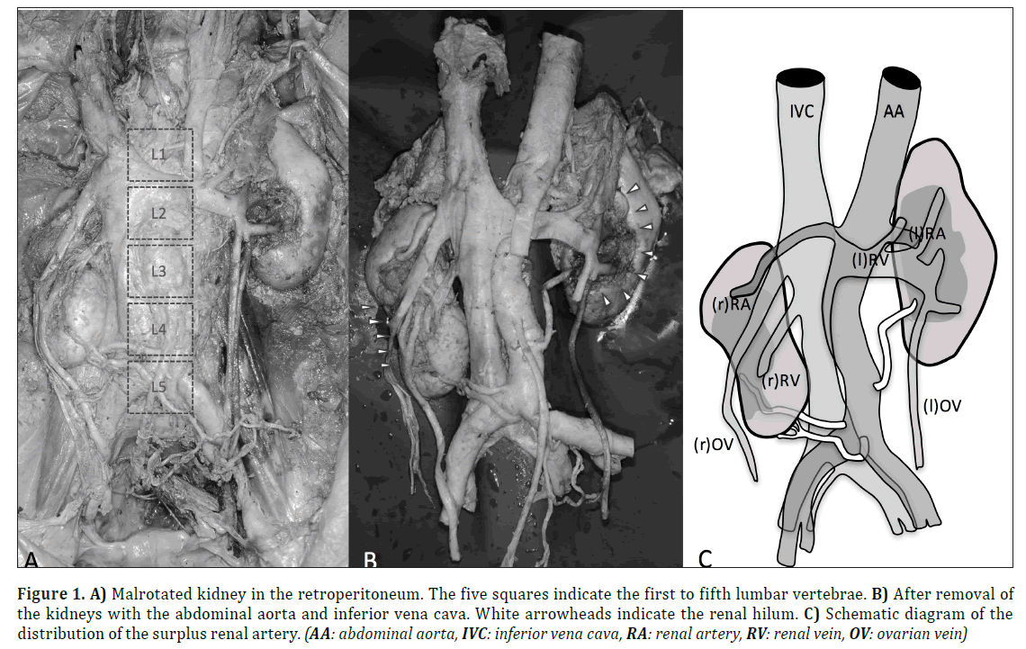
Bilateral malrotated kidneys were found in an 85-year-old woman who died of cardiac arrest (Figs. 1A and 1B). The hilum of the left kidney and tissue surrounding the kidney were filled with coagulated blood.
The superior border of the right kidney was at the level of the lower part of the second lumbar vertebra, and the inferior border was at located between the fourth and fifth lumbar vertebrae; the kidney was thus positioned lower than the normal kidney. The long and short axis of the right kidney measured 9.9 and 5.5 cm, respectively. The right hilum opened laterally. Three surplus renal arteries (SRAs) from the lower part of the abdominal aorta (AA) entered renal parenchyma ventral and dorsal to the kidney, respectively (Fig. 1C). The first SRA arose from the right side of the abdominal aorta and drained into the dorsal side of the right kidney. The second SRA arose from the dorsal side of the abdominal aorta, which was almost the bifurcation point of common iliac arteries, and drained into the lower border of the right kidney. The right ureter descended ventral to the renal parenchyma with a shallow groove. The superior border of the left kidney was at the level of the lower part of the 12th thoracic vertebra, and the inferior border was at the level of the lower part of the 3rd lumbar vertebra. The long and short axis of the left kidney measured 10.9 and 5.6 cm, respectively. The left hilum opened ventrally. The third SRA arose from the left side of the abdominal aorta and drained into the renal hilum of the left kidney (Fig. 1C).
Figure 1: A) Malrotated kidney in the retroperitoneum. The five squares indicate the first to fifth lumbar vertebrae. B) After removal of the kidneys with the abdominal aorta and inferior vena cava. White arrowheads indicate the renal hilum. C) Schematic diagram of the distribution of the surplus renal artery. (AA: abdominal aorta, IVC: inferior vena cava, RA: renal artery, RV: renal vein, OV: ovarian vein)
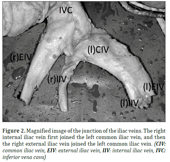
The celiac trunk, superior mesenteric artery, and inferior mesenteric artery branched off from the ventral side of the AA as usual. In the venous system, the right internal iliac vein (IIV) and external iliac vein (EIV) joined the left common iliac vein (CIV) separately, forming the inferior vena cava (IVC) (Fig. 2). Renal veins (RVs) were unremarkable and two left ovarian veins (OVs) were observed.
Figure 2: Magnified image of the junction of the iliac veins. The right internal iliac vein first joined the left common iliac vein, and then the right external iliac vein joined the left common iliac vein. (CIV: common iliac vein, EIV: external iliac vein, IIV: internal iliac vein, IVC: inferior vena cava)
This study was performed in accordance with the requirements of the Declaration of Helsinki (64th WMA General Assembly, Fortaleza, Brazil, October 2013).
Discussion
Kidney malrotation is classified into four groups: the non-rotation, incomplete rotation, reverse rotation, and hyper-rotation types, which are characterized by ventral, ventromedial, lateral, and dorsal or lateral opening of the hilum, respectively.
The incidence of kidney malrotation is 1 in 939 cases [1]. Lateral malrotation is the least common type, and its true incidence is unknown. According to the classification proposed by Shapiro et al. [1], the right kidney in the present case was of the reverse rotation type because its hilum opened laterally and the normal renal artery passed ventral to the parenchyma. The left kidney in the present case was of the incomplete rotation type because its hilum opened ventrally and the normal renal artery passed ventral to the kidney. However, the classic embryological theory for kidney rotation is only speculative without valid evidence. Additionally, there are some unexplained inquiries regarding the classic theory, such as whether one artery feeds the kidney during rotation and why one of the surplus renal arteries of the right kidney passes dorsal to the parenchyma. Some cases of the sagittally malrotated kidney has been also reported [2,3], which cannot be explained by classic theory. Theodore [4] reported a malrotated kidney as a complication of partical nephrectomy. They hypothesized that mobilization of the kidney during surgery resulted in malrotation. Thus, surgical procedure may affect the orientation of the kidney in some cases. There was no evidence of surgery in the present case.
Major venous variations are also challenging problems. In our previous report, an L-shaped kidney which has lower right kidney showed a venous variant similar to that in the present case (Iwanaga J, Watanabe K, Saga S, Tahara N, Tabira Y, Sakuragi A, Kaji K, Takahashi K, Yamaki K, unpublished data, 2016). The lower position of the kidney may have caused the major venous variation, or the major venous anomaly might have affected the rotation and ascension of the kidney.
Variations of the renal arteries with malrotated kidneys have been reported [5]. However, approximately 28% of normal kidneys also have surplus renal arteries [6]. Therefore, the presence of surplus renal arteries in a malrotated kidney is not surprising. Notably, few reports of bilateral malrotated kidneys have been reported. Gopinath et al. [7] described bilateral outward-facing hila. Patil et al. [8] described a right lateral renal hilum and left anterior renal hilum. Singh et al. [9] described a case involving bilateral anterior renal hila. However, a concurrent anomaly of the common iliac vein has never been reported.
This is the first reported case of bilateral malrotated kidneys with an anomaly of the common iliac vein.
Acknowledgements
The authors wish to thank those individuals who have consented to donate their bodies and tissues for the advancement of education and research.
Disclosures
There is no conflict of interest to declare.
References
- Shapiro E, Bauer S, Chow J. Anomalies of the upper urinary tract. In: Wein A, Kavoussi L, Novick A, Partin A, Peters C, eds. Campbell-Walsh Urology. 10th Ed., Philadelphia, Elsevier Saunders. 2011; 3149–3150.
- Tsai HY, Lee MH, Chen HC, Chen HC, Guh JY. Sagittally malrotated kidney: a case series of two patients. Surg Radiol Anat. 2015; 37: 551-553.
- Lim TJ, Choi SK, You HW, Kim MJ, Ahn JS, Kim TG, et al. Renal cell carcinoma in a right malrotated kidney. Korean J Urol. 2011; 52: 792-794.
- Theodore JE, Paterdis J. Malrotated kidney as a complication of partial nephrectomy. ANZ J Surg. 2015; 85: 287-289.
- Nathan H, Glezer I. Right and left accessory renal arteries arising from a common trunk associated with unrotated kidneys. J Urol. 1984; 132: 7-9.
- Satyapal KS, Haffejee AA, Singh B, Ramsaroop L, Robbs JV, Kalideen JM. Additional renal arteries: incidence and morphometry. Surg Radiol Anat. 2001; 23: 33-38.
- Gopinath V, Mookambika R, Nair V, Nair RV, Rao M. Bilateral lateral rotation of the kidneys with their hilum facing laterally (outwards)-Rare anatomical variations. Asian Journal of Medical Sciences (E-ISSN 2091-0576; P-ISSN 2467-9100). 2015; 6: 82-84.
- Patil ST, Meshram MM, Kasote AP. Bilateral malrotation and lobulation of kidney with altered hilar anatomy: a rare congenital variation. Surg Radiol Anat. 2011; 33: 941-944.
- Singh J, Singh N, Kapoor K, Sharma M. Bilateral Malrotation and a Congenital Pelvic Kidney with Varied Vasculature and Altered Hilar Anatomy. Case Rep Med. 2015; 2015:848949.
Joe Iwanaga*, Koichi Watanabe, Tsuyoshi Saga and Koh-ichi Yamaki
Department of Anatomy, Kurume University School of Medicine, 67 Asahi-machi, Kurume, Fukuoka 830-0011, Japan.
- *Corresponding Author:
- Joe Iwanaga
Department of Anatomy, Kurume University School of Medicine, 67 Asahi-machi, Kurume Fukuoka 830-0011, Japan
Tel: +81-942-31-7540
E-mail: iwanaga_jyou@med.kurume-u.ac.jp
Date of Received: May 20th, 2016
Date of Accepted: December 24th, 2016
Published Online: January 1st, 2017
© Int J Anat Var (IJAV). 2016; 9: 43–45.
Abstract
A laterally malrotated kidney, which is a variation of kidney malrotation, is a rare congenital kidney anomaly. Kidney malrotation is often accompanied by vascular variations such as the renal arteries and veins and common iliac arteries and veins, for surgical procedures. A cadaver with bilateral malrotated kidneys with variations of the common iliac veins was found during a dissection course for students. The right kidney was laterally malrotated and positioned lower than the normal kidney, and the left kidney was ventrally malrotated. The right internal and external iliac veins joined the left common iliac vein separately, forming the inferior vena cava. Such cases are very rare and can provide important information for considering the genesis of kidney.
-Keywords
kidney, anatomy, cadaver, iliac vein, inferior vena cava
Introduction
Congenital anomalies of the kidney exhibit great variety. The major anomalies are fused kidney, ectopic kidney, and malrotated kidney [1]. Because many affected patients are asymptomatic, some anomalies are incidentally found by computed tomography and others are found after death. Recent popularization of kidney transplantation requests has led to an increase in research of the vascular anatomy in patients with many kinds of kidney anomalies. In particular, the right common iliac vein is a very important vessel for angiostomy. Moreover, rotational anomalies of the kidney definitely affect the vascular structure. We herein report a case of bilateral malrotated kidneys with a variation of the common iliac vein found during a gross anatomical dissection course for students in 2015.
Case Report
Bilateral malrotated kidneys were found in an 85-year-old woman who died of cardiac arrest (Figs. 1A and 1B). The hilum of the left kidney and tissue surrounding the kidney were filled with coagulated blood.
The superior border of the right kidney was at the level of the lower part of the second lumbar vertebra, and the inferior border was at located between the fourth and fifth lumbar vertebrae; the kidney was thus positioned lower than the normal kidney. The long and short axis of the right kidney measured 9.9 and 5.5 cm, respectively. The right hilum opened laterally. Three surplus renal arteries (SRAs) from the lower part of the abdominal aorta (AA) entered renal parenchyma ventral and dorsal to the kidney, respectively (Fig. 1C). The first SRA arose from the right side of the abdominal aorta and drained into the dorsal side of the right kidney. The second SRA arose from the dorsal side of the abdominal aorta, which was almost the bifurcation point of common iliac arteries, and drained into the lower border of the right kidney. The right ureter descended ventral to the renal parenchyma with a shallow groove. The superior border of the left kidney was at the level of the lower part of the 12th thoracic vertebra, and the inferior border was at the level of the lower part of the 3rd lumbar vertebra. The long and short axis of the left kidney measured 10.9 and 5.6 cm, respectively. The left hilum opened ventrally. The third SRA arose from the left side of the abdominal aorta and drained into the renal hilum of the left kidney (Fig. 1C).
Figure 1: A) Malrotated kidney in the retroperitoneum. The five squares indicate the first to fifth lumbar vertebrae. B) After removal of the kidneys with the abdominal aorta and inferior vena cava. White arrowheads indicate the renal hilum. C) Schematic diagram of the distribution of the surplus renal artery. (AA: abdominal aorta, IVC: inferior vena cava, RA: renal artery, RV: renal vein, OV: ovarian vein)
The celiac trunk, superior mesenteric artery, and inferior mesenteric artery branched off from the ventral side of the AA as usual. In the venous system, the right internal iliac vein (IIV) and external iliac vein (EIV) joined the left common iliac vein (CIV) separately, forming the inferior vena cava (IVC) (Fig. 2). Renal veins (RVs) were unremarkable and two left ovarian veins (OVs) were observed.
Figure 2: Magnified image of the junction of the iliac veins. The right internal iliac vein first joined the left common iliac vein, and then the right external iliac vein joined the left common iliac vein. (CIV: common iliac vein, EIV: external iliac vein, IIV: internal iliac vein, IVC: inferior vena cava)
This study was performed in accordance with the requirements of the Declaration of Helsinki (64th WMA General Assembly, Fortaleza, Brazil, October 2013).
Discussion
Kidney malrotation is classified into four groups: the non-rotation, incomplete rotation, reverse rotation, and hyper-rotation types, which are characterized by ventral, ventromedial, lateral, and dorsal or lateral opening of the hilum, respectively.
The incidence of kidney malrotation is 1 in 939 cases [1]. Lateral malrotation is the least common type, and its true incidence is unknown. According to the classification proposed by Shapiro et al. [1], the right kidney in the present case was of the reverse rotation type because its hilum opened laterally and the normal renal artery passed ventral to the parenchyma. The left kidney in the present case was of the incomplete rotation type because its hilum opened ventrally and the normal renal artery passed ventral to the kidney. However, the classic embryological theory for kidney rotation is only speculative without valid evidence. Additionally, there are some unexplained inquiries regarding the classic theory, such as whether one artery feeds the kidney during rotation and why one of the surplus renal arteries of the right kidney passes dorsal to the parenchyma. Some cases of the sagittally malrotated kidney has been also reported [2,3], which cannot be explained by classic theory. Theodore [4] reported a malrotated kidney as a complication of partical nephrectomy. They hypothesized that mobilization of the kidney during surgery resulted in malrotation. Thus, surgical procedure may affect the orientation of the kidney in some cases. There was no evidence of surgery in the present case.
Major venous variations are also challenging problems. In our previous report, an L-shaped kidney which has lower right kidney showed a venous variant similar to that in the present case (Iwanaga J, Watanabe K, Saga S, Tahara N, Tabira Y, Sakuragi A, Kaji K, Takahashi K, Yamaki K, unpublished data, 2016). The lower position of the kidney may have caused the major venous variation, or the major venous anomaly might have affected the rotation and ascension of the kidney.
Variations of the renal arteries with malrotated kidneys have been reported [5]. However, approximately 28% of normal kidneys also have surplus renal arteries [6]. Therefore, the presence of surplus renal arteries in a malrotated kidney is not surprising. Notably, few reports of bilateral malrotated kidneys have been reported. Gopinath et al. [7] described bilateral outward-facing hila. Patil et al. [8] described a right lateral renal hilum and left anterior renal hilum. Singh et al. [9] described a case involving bilateral anterior renal hila. However, a concurrent anomaly of the common iliac vein has never been reported.
This is the first reported case of bilateral malrotated kidneys with an anomaly of the common iliac vein.
Acknowledgements
The authors wish to thank those individuals who have consented to donate their bodies and tissues for the advancement of education and research.
Disclosures
There is no conflict of interest to declare.
References
- Shapiro E, Bauer S, Chow J. Anomalies of the upper urinary tract. In: Wein A, Kavoussi L, Novick A, Partin A, Peters C, eds. Campbell-Walsh Urology. 10th Ed., Philadelphia, Elsevier Saunders. 2011; 3149–3150.
- Tsai HY, Lee MH, Chen HC, Chen HC, Guh JY. Sagittally malrotated kidney: a case series of two patients. Surg Radiol Anat. 2015; 37: 551-553.
- Lim TJ, Choi SK, You HW, Kim MJ, Ahn JS, Kim TG, et al. Renal cell carcinoma in a right malrotated kidney. Korean J Urol. 2011; 52: 792-794.
- Theodore JE, Paterdis J. Malrotated kidney as a complication of partial nephrectomy. ANZ J Surg. 2015; 85: 287-289.
- Nathan H, Glezer I. Right and left accessory renal arteries arising from a common trunk associated with unrotated kidneys. J Urol. 1984; 132: 7-9.
- Satyapal KS, Haffejee AA, Singh B, Ramsaroop L, Robbs JV, Kalideen JM. Additional renal arteries: incidence and morphometry. Surg Radiol Anat. 2001; 23: 33-38.
- Gopinath V, Mookambika R, Nair V, Nair RV, Rao M. Bilateral lateral rotation of the kidneys with their hilum facing laterally (outwards)-Rare anatomical variations. Asian Journal of Medical Sciences (E-ISSN 2091-0576; P-ISSN 2467-9100). 2015; 6: 82-84.
- Patil ST, Meshram MM, Kasote AP. Bilateral malrotation and lobulation of kidney with altered hilar anatomy: a rare congenital variation. Surg Radiol Anat. 2011; 33: 941-944.
- Singh J, Singh N, Kapoor K, Sharma M. Bilateral Malrotation and a Congenital Pelvic Kidney with Varied Vasculature and Altered Hilar Anatomy. Case Rep Med. 2015; 2015:848949.


