Bilateral middle concha bullosa mucopyocele connecting to headache disablement: a case study
2 Department of Radiology, Sifa Hospital, 07070 Antalya, Turkey
3 Department of Ear Nose and Throat, Sifa Hospital, 07070 Antalya, Turkey, Email: kandemirs@gmail.com
4 Department of Anatomy, Akdeniz University, 07070 Antalya, Turkey, Email: sindelmuzaffer@gmail.com
Citation: Kandemir YB, Ergin I, Kandemir S, et al. Bilateral middle concha bullosa mucopyocele connecting to headache disablement: a case study. Int J Anat Var. 2017;10(S1):75-76.
This open-access article is distributed under the terms of the Creative Commons Attribution Non-Commercial License (CC BY-NC) (http://creativecommons.org/licenses/by-nc/4.0/), which permits reuse, distribution and reproduction of the article, provided that the original work is properly cited and the reuse is restricted to noncommercial purposes. For commercial reuse, contact reprints@pulsus.com
Abstract
Concha Bullosa (CB) is the most common anatomic variations of sinonasal anatomy in which the middle nasal turbinate contains pneumatized cells. The most usual variation is concha bullosa in nasal cavity. Sometimes the cause of headache or nasal obstruction is CB. This study describes the case of Computed Tomography (CT) findings of bilateral middle and inferior concha bullosa in a 38-year-old female with nasal obstruction and headache. We obtained paranasal sinus CT examinations from the Department of Radiology, Ear Nose and Throat of Sifa Hospital in Antalya. The patient was operated victoriously with endoscopicsurgery of conchas by Ear Nose and Throat surgeon.
Keywords
Concha bullosa; Sinonasal anatomy; Mucocele
Introduction
Anatomic variations are crucial in preendoscopic computed tomography assessment of the paranasal sinuses. Concha bullosa is a well-known anatomic variant of sinonasal anatomy. CB is defined as an air-filled cavity within the nasal turbinate. Which is the most prevalently present in the inferior turbinate followed by the superior turbinate [1-3]. Nasal obstructions or headache can ensue from pressure on the nasal mucosa due to CB which the contact with the lateral nasal wall [4]. Obstruction of a CB can ensue in a mucocele. Secondary infections that develop over a mucocele are assigned to as mucopyoceles [5]. We indicated in this case a concha bullosa associated mucopyocele with a large hyperemic nasal mass that induced nasal obstruction and headache. It was successfully treated endoscopically. We also review the utilizable literature.
Case Report
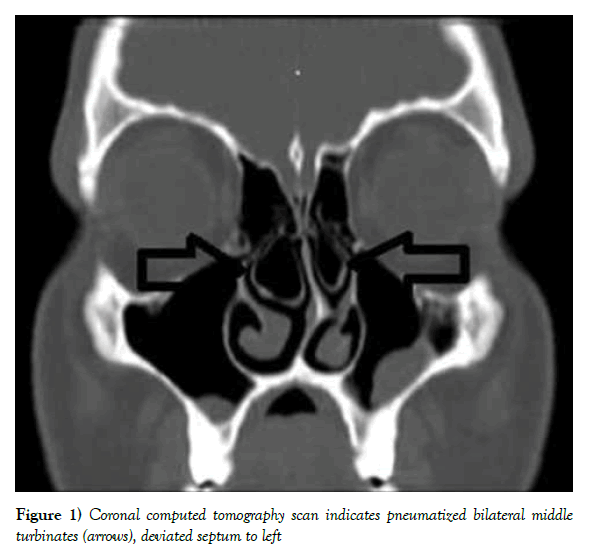
A 38-year-old female with a history of progressive headache and nasal obstruction which had persisted for several years. She was referred to Ear Nose and Throat department of Sifa Hospital in Antalya. The patient that has not got any history of nasal allergy, trauma, or sinus surgery. On nasal endoscopic examination observed enlargement of left and right middle turbinate. Two concha bullosa were placed at the postero inferior portions of the middle turbinate. Computed Tomography (CT) showed bilateral pneumatization of the middle turbinate and hypertrophy of the left and right middle turbinate. Two concha bullosa were located at the postero-superior and postero inferior portions of the left inferior turbinate. Paranasal sinus tomography (CT) observed a lesion in the left and right nasal cavity similar a mucopyocele in the middle turbinate. A large quantity of purulent was involved in the middle turbinate. The purulent was afterwards aspirated. Staphylococcus Aureus was detected on the culture.
The patient clavulanic acid/amoxicillin treatment was applied for 10 days. Cultures were obtained during the operation. As a result of pathological examination, excision material was described as mucocele. The patient imposed endoscopic middle turbinectomy surgery under general anesthesia. During the following 7 months post-surgery, the patient’s symptoms had significantly developed and she was no recurrent pathology on her nasal endoscopic examination (Figure 1).
Figure 1) Coronal computed tomography scan indicates pneumatized bilateral middle turbinates (arrows), deviated septum to left
Discussion and Conclusion
Rhinogenic Headache (RH) is among the most common of headaches [6-8]. Concha bullosa is the well-known induced of RH and there is frequently an accompaniment septal deviation [9]. The term concha bullosa is principally used to characterize pneumatization of the middle turbinate occasionally, of the superioror inferior turbinates [10]. Concha bullosa is come acrossed both unilaterally and bilaterally and the incidence of it ranges from 14 to 53% [11].
The development of paranasal sinus CT has provided us particular information about this unapproachable area of the middle nasal cavity. Some studies have given an idea that nasal endoscopy disallows ready approach to this area [12]. Though the endoscopic examination of the nasal cavity was not indicant of pneumatization of the middle turbinates in our patient, coronal plain paranasal sinus CT imaging permitted us to diagnose the pathology.
A symptomatic pneumatization of the middle turbinate is observed usually uncommon. If the pneumatization is common, because of mucosal contact and mechanic obstruction, it may induce important symptoms such as nasal obstruction and headache [13]. The principal symptoms were nasal obstruction and headache in our patient. The first person to indicate that middle turbinate was a headache source that needed counseling was Clerio [12]. A massive pneumatized middle concha related with headache is very rare. Massive pneumatization of middle turbinates with accompaniment to mucosal contact can be the induce of headache even in the lack of sinonasal inflammation [14]. The aerobic bacteria Staphylococcus Aureus was recognized and it was treated by clavulanic acid/amoxicillin. Additionally, endoscopic concha bullosa resection and drainage were the best alternatives for mucopyoceles treatment at the same time and consequently, the lateral border of the concha bullosa was excised and aspirated the mucopurulent secretion in our case.
Finally bilateral middle concha bullosa is not a common anatomic variation. Middle turbinate may not be noticed by endoscopic examination but may be easily recognized on paranasal sinus CT. Paranasal sinus CT scans perform a crucial role in diagnosis and treatment planning because a coronal plain CT of the paranasal sinuses can decided definitely the anatomic variations in this area and their connection to mucosal pathologies. We propose that concha bullosa mucopyoceles should be noted in patients with a large hyperemic nasal mass.
REFERENCES
- Yang BT, Chong VF, Wang ZC, et al. CT appearance of pneumatized inferior turbinate. Clin Radiol. 2008;63(8):901-5.
- Ozturk A, Alatas N, Ozturk E, et al. Pneumatization of the inferior turbinates: incidence and radiologic appearance. J Comput Assist Tomogr. 2005;29(3):311-4.
- Ariyurek OM, Balkanci F, Aydingoz U, et al. Pneumatised superior turbinate: a common anatomic variation? Surg Radiol Anat. 1996;18(2):137-9.
- Peric A, Baletic N, Sotirovic J. A case of an uncommon anatomic variation of the middle turbinate associated with headache. Acta Otorhinolaryngol Ital. 2010;30(3):156-9.
- Shihada R, Luntz M. A concha bullosa mucopyocele manifesting as migraine headaches: a case report and literature review. Ear Nose Throat J. 2012;91(5):E16-8.
- Peric A, Rasic D, Grgurevic U. Surgical Treatment of Rhinogenic Contact Point Headache: An Experience from a Tertiary Care Hospital. Int Arch Otorhinolaryngol. 2016;20(2):166-71.
- Cantone E, Castagna G, Ferranti I, et al. Concha bullosa related headache disability. Eur Rev Med Pharmacol Sci. 2015;19(13):2327-30.
- Yarmohammadi ME, Ghasemi H, Pourfarzam S, et al. Effect of turbinoplasty in concha bullosa induced rhinogenic headache, a randomized clinical trial. J Res Med Sci. 2012;17(3):229-34.
- Roozbahany NA, Nasri S. Nasal and paranasal sinus anatomical variations in patients with rhinogenic contact point headache. Auris Nasus Larynx. 2013;40(2):177-83.
- Bolger WE. Anatomy of the paranasal sinuses. In: Kennedy DW, Bolger WE, Zinreich SJ, editors. Diseases of the sinuses. London: BC, Decker. 2001;1-12.
- Stallman JS, Lobo JN, Som PM. The incidence of concha bullosa and its relationship to nasal septal deviation and paranasal sinus disease. AJNR Am J Neuroradiol. 2004;25(9):1613-8.
- Clerico DM. Pneumatized superior turbinate as a cause of referred migraine headache. Laryngoscope. 1996;106(7):874-9.
- Unlu HH, Altuntas A, Aslan A, et al. Inferior concha bullosa. J Otolaryngol. 2002;31(1):62-4.
- Kantarci M, Karasen RM, Alper F, et al. Remarkable anatomic variations in paranasal sinus region and their clinical importance. Eur J Radiol. 2004;50(3):296-302.


