Bilateral pectoralis major and pectoralis quartus variants: A conjoined tendon passing through the inter tubercular groove
Received: 11-Sep-2017 Accepted Date: Sep 21, 2017; Published: 28-Sep-2017
Citation: Hunt JD. Bilateral pectoralis major and pectoralis quartus variants: A conjoined tendon passing through the intertubercular groove. Int J Anat Var. 2017;10(S1): 88-90.
This open-access article is distributed under the terms of the Creative Commons Attribution Non-Commercial License (CC BY-NC) (http://creativecommons.org/licenses/by-nc/4.0/), which permits reuse, distribution and reproduction of the article, provided that the original work is properly cited and the reuse is restricted to noncommercial purposes. For commercial reuse, contact reprints@pulsus.com
Abstract
The pectoralis major has been described to insert on to the lateral lip of the intertubercular groove of the humerus. The pectoralis quartus is a variant that runs parallel to the lateral border of the pectoralis major. It has been described with insertions in to the pectoralis major at the lateral lip of intertubercular groove, short head of the biceps, and coracobrachialis. This case report describes additional bilateral variants found in an elderly male cadaver.
The first variant included a conjoined tendon of the pectoralis major and quartus that passed superiorly through the intubercular groove to insert to the blended fascia of the superolateral glenohumeral joint capsule. The second variant included different insertions between the left and right pectoralis quartus in to pectoralis major fascia. These findings have not been previously reported in the literature. The bilateral presence of such variants also makes this case unique. Clinical and surgical considerations are included in the discussion of this case report.
Keywords
Pectoralis quartus; Pectoralis major; Accessory pectoralis; Axillary arch; Pectoralis variant
Introduction
The pectoralis major has been described to insert on to the lateral lip of the intertubercular groove of the humerus. The pectoralis quartus is considered to be a variant of the pectoralis major muscle. It is reported to present in as much as 11-16% in the literature [1]. There have been other authors who have described the presence and attachments of the pectoralis quartus [1-4]. Although the origin has remained consistent between cases the insertion has been variable. This case report describes a variant of the insertion of the pectoralis major/quartus that could have surgical implications for procedures involving the thoracic wall and the long head of the biceps, as well as clinical implications for the examination/rehabilitation of injuries to the area. Considering the frequency of injuries and surgical interventions to this area it’s important to have knowledge of anatomical variations. To this author’s knowledge, there are no other variants such as described in this case report available in the literature.
Case Report
During a routine cadaver dissection of an elderly Caucasian male pectoralis major muscle variants were found in bilateral upper extremities. Age and medical history were unknown due to this state maintaining confidentiality of such information. There were no surgical identifying characteristics during dissection of the involved areas. The variants appeared consistent with other literature citing the presence of a pectoralis quartus. Each pectoralis quartus was observed following typical reflection of the pectoralis major from the clavicle, sternal and costal attachments. The pectoralis quartus were positioned in between the pectoralis major and minor with fascial attachments to the undersurface of the pectoralis major. The origin of bilateral pectoralis quartus was the fascia of the rectus abdominus, and 5th and 6th costal cartilage.
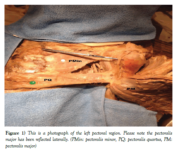
The insertion of the left pectoralis quartus remained as an untwisted tendon attaching immediately superior to the most superior edge of the pectoralis major along extended fascia from pectoralis major and the proximal lateral lip of the intertubercular groove (Figure 1). The dimensions from the origin to humeral insertion of the pectoralis quartus were 17.3 cm in length, and 4-5.3 cm in width.
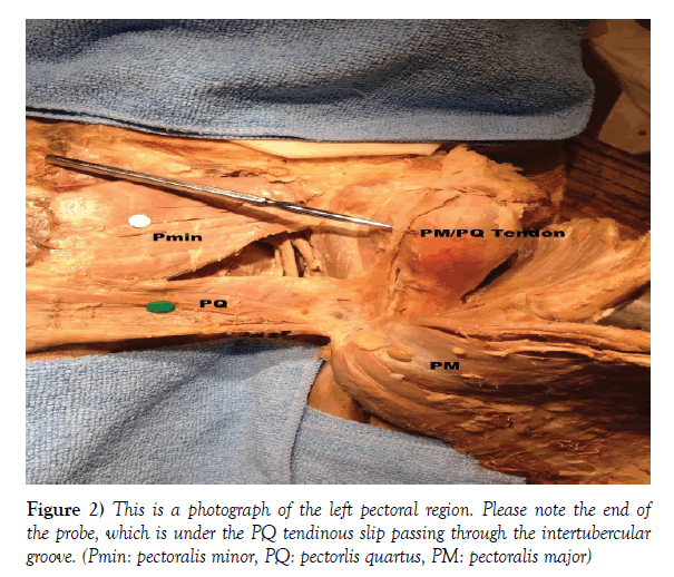
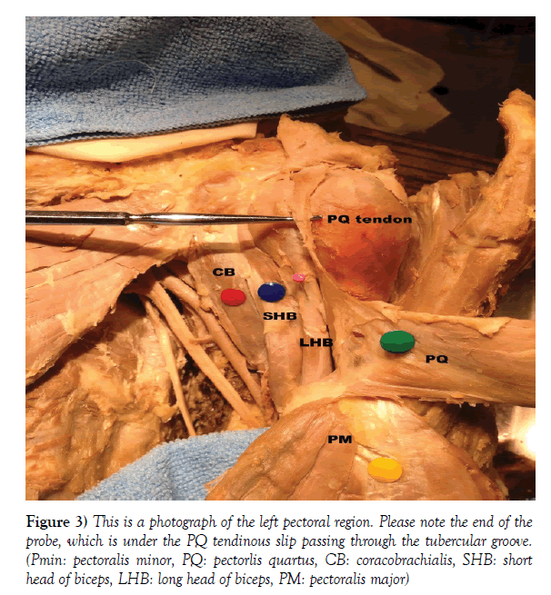
The second variant on the left side was a conjoined tendinous slip of the pectoralis major and quartus. This tendon was an extension of the fascial connection between the pectoralis major and quartus (Figure 2). It continued superiorly through the intertubercular groove, however remaining superficial to the long head of the biceps, before blending in with the fibrous layer of the superolateral glenohumeral joint and blending fascia (Figure 3).
Figure 3) This is a photograph of the left pectoral region. Please note the end of the probe, which is under the PQ tendinous slip passing through the tubercular groove. (Pmin: pectoralis minor, PQ: pectorlis quartus, CB: coracobrachialis, SHB: short head of biceps, LHB: long head of biceps, PM: pectoralis major)
An additional variant noted during this dissection was the distal insertion point of the pectoralis major on to the lateral lip of the intertubercular groove of the humerus. This distance measured 7.6 cm from the most superomedial point of the greater tuberosity of the humerus.
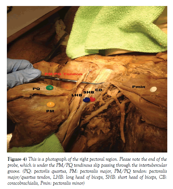
The insertion of the right pectoralis major was as typically described attaching in to the lateral lip of the intertubercular groove. The variant of the pectoralis major was the tendinous slip that arose from the undersurface of the pectoralis major and passed superior. This tendon conjoined with the pectoralis quartus and continued through the interbucular groove, however remaining as a separate tendon superficial to the long head of the biceps, before blending with the fibrous layer of the superolateral glenohumeral joint and blending fascia (Figure 4).
Figure 4) This is a photograph of the right pectoral region. Please note the end of the probe, which is under the PM/PQ tendinous slip passing through the intertubercular groove. (PQ: pectorlis quartus, PM: pectoralis major, PM/PQ tendon: pectoralis major/quartus tendon, LHB: long head of biceps, SHB: short head of biceps, CB: coracobrachialis, Pmin: pectoralis minor)
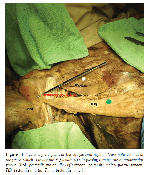
The insertion of the right pectoralis quartus also remained as an untwisted tendon. Rather than attaching to the lateral lip of the intertubercular groove it attached directly in to the tendinous extension of the pectoralis major (Figure 5). The dimensions from the origin to humeral insertion were 20 cm in length, and 3-4 cm in width.
Discussion
Muscular variants of the pectoralis major have been reported before, however it remains important to report rare variations when they occur. The presence of the pectoralis quartus has been reported since the 1800’s. Birmingham reported that it attached in to the lower ribs and lateral thoracic fascia, running on the inferior border of the pectoralis major to insert on to the humerus with, or below the major, or only to fascia of medial arm [2]. The pectoralis quartus has been reported to originate from the lateral border of the pectoralis major, costal cartilage of 5th and 6th ribs, and rectus sheath. It’s been described to insert to the fascia of the coracobrachialis deep to the pectoralis major [1,3,4]. Verma et al. reported it inserting to the fascia of the short head of the biceps and coracobrachialis just lateral to the coracoid process [5]. Bonastre et al. reported an interesting variant of the pectoralis quartus inserting in to the fibrous band of the axillary arch with final insertion on to the coracoid process [6]. The pectoralis quartus is also known to insert to the axillary arch and sternalis when both of those muscles are present [6,7]. Barcia, et al reported another variant of the pectoralis quartus insertion with an attachment in to the fascia of the medial arm without attachment to the humerus on one side of the body while on the other side the insertion was to the medial epicondyle [8]. In this case the right pectoralis quartus attached directly to the variant tendinous slip of the pectoralis major to become a conjoined tendon. Although the left pectoralis quartus did insert on to the lateral lip of the intertubercular groove it also became a conjoined tendon with the pectoralis major.
The insertion of the pectoralis major has been described to be the lateral lip of the intertubercular groove of the humerus [9]. It’s been shown that the most superior extent of this insertion is roughly 4.2cm inferior to the superomedial corner of the greater tuberosity [9-11]. In this case the insertion point on the left was more inferior to accommodate for the presence of the pectorlis quartus insertion. The right maintained its typical distance. Bilaterally, a tendinous slip then conjoined with the pectoralis quartus and continued superior through the intertubercular groove, however superficial to the long head of the biceps, attaching to the fascia of the superololateral aspect of glenohumeral joint.
According to embryologic studies the pectoral muscles are derived from the panniculus carnosus, which is a large sheet of muscle in lower animals [5,12]. The panniculus carnosus is considered to be vestigial in humans due to the amount of upper limb mobility humans possess and the vestigial remnants present as pectoral muscle variants [12].
The long head of the biceps has been reported to originate on to the supraglenoid tubercle and posterior glenoid labrum in approx. 75% of cases and to only the supraglenoid tubercle in 25% of cases [10]. It then traverses through the intertubercular groove of the humerus extending down the arm to insert as a flat tendon to the radial tuberosity, and bicipital aponeurosis, which blends with the antebrachial fascia. It is known to be the only tendon to be located within the intertubercular groove of the humerus.
In this case the long head of the biceps was located in the intertubercular groove of the humerus as previously stated in the literature. However, the conjoined tendinous slip of the pectoralis major and quartus passed superficial to the long head of the biceps, within the intertubercular groove, to insert in to the fascia of the superolateral aspect of the glenohumeral joint. This is the first case to describe such a variant.
It’s important for rehabilitation professionals, and orthopedic physicians to be aware of this type of variant as it may have clinical and surgical implications.
Clinically, the superficial location of the tendinous slip of the pectoralis major and quartus within the intertubercular groove may obscure the long head of the biceps during imaging studies leading to misdiagnosis of pathology to the long head of the biceps.
A clinician may also consider pain in the intertubercular groove to be pathology of long head of the biceps when it’s actually the pectoralis major/ quartus. This may lead to ineffective conservative treatment.
This variant may complicate surgical intervention in this area and possibly lead to mistakenly identifying the wrong tendon. The presence of a pectoralis quartus has been associated with complications during axillary lymphadenectomy due to it reducing the area of surgical field [5,11]. Knowledge of this variant will assist surgeons who perform surgery to this area.
The presence of a muscular variant may present challenges to clinicians who are unfamiliar with such variants. However, presence of multiple variants causes more complications as they may affect multiple regions of the body. Therefore, it’s important to perform anatomic study and report such variants to educate clinicians in order to improve patient outcomes.
REFERENCES
- Terfera DR, Kelliher KR. Unilateral presentation of three muscle variants in the pectoral region. Eur J Anat. 2014;18:335-9.
- Birmingham A. Homology and Innervation of the Ashselbogen and Pectoralis Quartus, and the Natoure of the Lateral Cutaneous Nerve of the Thorax. J Anat. 1889;23:206-223.
- Fabrizio PA, Hardy MA. An accessory muscle of the thoracic wall. Int J Anat Var. 2009;2:93-5.
- Natsis K, Vlasis K, Totlis T, et al. Abnormal muscles that may affect axillary lymphade- nectomy: surgical anatomy. Breast Cancer Res Treat. 2010;120:77-82.
- Verma S, Sakthivel S. Concomitant pectoralis minor, pectoralis quartus and axillary arch. Eur J Phar and Med Res. 2017;4:570-2.
- Bonastre V, Rodriguez-Niedenfuhr M, Choi D, Sanudo JR. Coexistence of a pectoralis quartus muscle and unusual axillary arch: Case report and review. Clin Anat. 2002;15:366-70.
- Bergman RA. Doubled pectoralis quartus, axillary arch, chondroepitrochlearis, and the twist of the tendon of pectoralis major. Anat Anz. 1991;173: 23-6.
- Barcia J, Genoves J. Chondrofascialis Versus Pectoralis Quartus. Clin Anat. 2009;22:871-2.
- Carey P, Owens BD. Insertional footprint anatomy of the pectoralis major tendon. Orthopedics.
- Jain P, Tuli A, Raheja S, et al. Morphological description of variations of the origin of long head of biceps brachii: An evolutionary and embryological interpretation. Int Med J. 2014;21: 123-5.
- Bergman RA, Afifi AK, Miyauchi R. Illustrated Encyclopedia of Human Anatomic Variation.
- Bergman RA, Afifi AK, Miyauchi R. Illustrated Encyclopedia of Human Anatomic Variation.


