Bilateral superficial brachial artery and high origin of common interosseous artery – a rare variation
Shilpa Sonare* and Praful Nikam
Department of Anatomy, Jawaharlal Nehru Medical College, Sawangi (Meghe), Wardha, India
- *Corresponding Author:
- Dr. Shilpa Sonare
Assistant Professor, Department of Anatomy, Jawaharlal Nehru Medical College Sawangi (Meghe), Wardha, Maharashtra, India
Tel: +91 (992) 2953708
E-mail: drshilpasonare@gmail.com
Date of Received: July 5th, 2011
Date of Accepted: July 31st, 2012
Published Online: December 15th, 2012
© Int J Anat Var (IJAV). 2012; 5: 102–103.
[ft_below_content] =>Keywords
superficial brachial artery, common interosseous artery, median nerve, pronator teres muscle
Introduction
Brachial artery, the major artery of upper limb is continuation of the axillary artery and extends from lower border of the teres major to the neck of the radius in the cubital fossa where it terminates by dividing into the ulnar and the radial arteries. In the arm, it gives off the profunda brachii artery, unnamed muscular branches, the nutrient artery, the superior and the inferior ulnar collateral arteries.
The ulnar artery which is the terminal branch begins in the cubital fossa and leaves the cubital fossa by passing deep to both heads of the pronator teres muscle and then in the forearm, the ulnar artery gives off the common interosseous, the anterior ulnar recurrent and the posterior ulnar recurrent branches. The common interosseous artery divides into anterior and posterior interosseus arteries.
The radial artery, another terminal branch of the brachial artery, is smaller and superficial as compared to the ulnar artery. It runs downwards to the wrist joint with lateral convexity and leaves the forearm by entering into the anatomical snuffbox.
Case Report
During routine dissection for undergraduate students in a 65-year-old male cadaver without any congenital anomaly, we found bilateral variations in the branching pattern of the brachial artery. Same variation was observed on both sides.
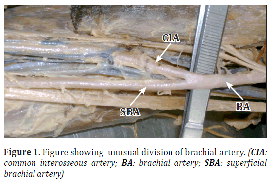
The brachial artery, about 5 cm below the lower border of the teres major muscle, instead of giving the profunda brachii, the superior and the inferior ulnar collateral arteries ran towards the cubital fossa, and divided into two branches. One branch was superficial and termed as superficial brachial artery [1], while the other branch was deep to the biceps brachii muscle. The superficial brachial artery ran along the medial side of the arm, superficial to the median nerve and divided into ulnar and radial arteries in the cubital fossa.
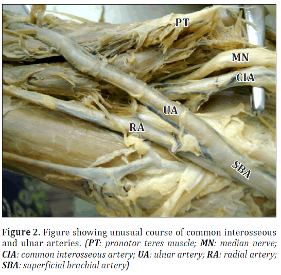
The ulnar artery, instead of passing deep to both heads of the pronator teres, passed superficial to both heads of the pronator teres, and also superficial to the flexor carpi radialis, the palmaris longus and the flexor digitorum superficialis muscles. The course, relations and distribution of the ulnar artery were as usual in the distal part of the forearm.
The radial artery followed the usual course in the forearm.
The deep branch of the brachial artery passed along with the median nerve. The median nerve passed medial to the artery throughout its course in the arm. In the cubital fossa, the median nerve was medial to the artery. The artery and the median nerve passed deep to both the heads of the pronator teres and the artery branched into anterior and posterior interosseus arteries, which followed the usual course thereafter. This deep branch was the common interosseus artery that was originated high in the arm and was larger and deep to the superficial brachial artery.
Discussion
In the present study, we got a rare branching pattern of the brachial artery into superficial brachial artery and deep common interosseous artery, just below the lower border of the teres major muscle. Similar findings were observed by Cherian et al. in 2009 [2]. De Garis and Swartley reported the origin of superficial brachial in the axilla in 17 out of 512 limbs while Muller found it in 1 out of 960 limbs [1].
Similarly, in our study, the ulnar artery was the terminal branch of the superficial brachial artery and passed superficial to the pronator teres muscle. Similar finding was observed by Sennayake et al. (incidence is 0.7 to 7%)[3], and Dartnel et al. (incidence 4.2%) [4].
Previous studies showed that the common interosseous artery arises from the axillary artery or the brachial artery high in the arm. In our study, the common interosseous artery was the terminal branch of the brachial artery, and it was arisen in the arm.
Another important finding of our study was that the median nerve passed deep to both heads of the pronator teres muscle along with the common interosseous artery, instead of passing between its two heads.
These variations of brachial artery branching pattern, the unusual relation of its branches with the median nerve as well as the pronator teres muscle are very useful for the surgeons while performing various surgeries on the upper limb. The surgeons should consider this variation and avoid any trouble to the patient.
References
- Hollinshead HW. Anatomy for Surgeons. Vol. 3. 3rd Ed., Philadelphia, Harper and Row. 1982; 362.
- Cherian SB, Nayak SB, Somayaji N. Superficial course of brachial and ulnar arteries and high origin of common interosseous artery. Int J Anat Var (IJAV). 2009; 2: 4–6.
- Senanayake KJ, Salgado S, Rathnayake MJ, Fernando R, Somarathne K. A rare variant of the superficial ulnar artery and its implications: a case report. J Med Case Rep. 2007; 1:128.
- Dartnell J, Sekaran P, Ellis H. The superficial ulnar artery: incidence and calibre in 95 cadaveric specimens. Clin Anat. 2007; 20: 929–932.
Shilpa Sonare* and Praful Nikam
Department of Anatomy, Jawaharlal Nehru Medical College, Sawangi (Meghe), Wardha, India
- *Corresponding Author:
- Dr. Shilpa Sonare
Assistant Professor, Department of Anatomy, Jawaharlal Nehru Medical College Sawangi (Meghe), Wardha, Maharashtra, India
Tel: +91 (992) 2953708
E-mail: drshilpasonare@gmail.com
Date of Received: July 5th, 2011
Date of Accepted: July 31st, 2012
Published Online: December 15th, 2012
© Int J Anat Var (IJAV). 2012; 5: 102–103.
Abstract
Brachial artery is the continuation of axillary artery just below the lower border of teres major muscle and then by giving various branches in the arm, it get divided into two terminal branches in the cubital fossa –the radial and the ulnar arteries. In the present study, we got a rare variation of branching pattern of brachial artery. The brachial artery just below the lower border of teres major muscle get divided into superficial brachial artery and deep branch that is common interosseous artery. The ulnar artery, instead of passing deep to both heads of the pronator teres, passed superficial to both heads of the pronator teres, and also superficial to the flexor carpi radialis, the palmaris longus and the flexor digitorum superficialis muscles. The deep branch of the brachial artery passed along with the median nerve. The artery and the median nerve passed deep to both the heads of the pronator teres muscle and the artery branched into anterior and posterior interosseus arteries that followed the usual course thereafter. This deep branch was the common interosseus artery which originated high in the arm and was larger and deep to the superficial brachial artery.
-Keywords
superficial brachial artery, common interosseous artery, median nerve, pronator teres muscle
Introduction
Brachial artery, the major artery of upper limb is continuation of the axillary artery and extends from lower border of the teres major to the neck of the radius in the cubital fossa where it terminates by dividing into the ulnar and the radial arteries. In the arm, it gives off the profunda brachii artery, unnamed muscular branches, the nutrient artery, the superior and the inferior ulnar collateral arteries.
The ulnar artery which is the terminal branch begins in the cubital fossa and leaves the cubital fossa by passing deep to both heads of the pronator teres muscle and then in the forearm, the ulnar artery gives off the common interosseous, the anterior ulnar recurrent and the posterior ulnar recurrent branches. The common interosseous artery divides into anterior and posterior interosseus arteries.
The radial artery, another terminal branch of the brachial artery, is smaller and superficial as compared to the ulnar artery. It runs downwards to the wrist joint with lateral convexity and leaves the forearm by entering into the anatomical snuffbox.
Case Report
During routine dissection for undergraduate students in a 65-year-old male cadaver without any congenital anomaly, we found bilateral variations in the branching pattern of the brachial artery. Same variation was observed on both sides.
The brachial artery, about 5 cm below the lower border of the teres major muscle, instead of giving the profunda brachii, the superior and the inferior ulnar collateral arteries ran towards the cubital fossa, and divided into two branches. One branch was superficial and termed as superficial brachial artery [1], while the other branch was deep to the biceps brachii muscle. The superficial brachial artery ran along the medial side of the arm, superficial to the median nerve and divided into ulnar and radial arteries in the cubital fossa.
The ulnar artery, instead of passing deep to both heads of the pronator teres, passed superficial to both heads of the pronator teres, and also superficial to the flexor carpi radialis, the palmaris longus and the flexor digitorum superficialis muscles. The course, relations and distribution of the ulnar artery were as usual in the distal part of the forearm.
The radial artery followed the usual course in the forearm.
The deep branch of the brachial artery passed along with the median nerve. The median nerve passed medial to the artery throughout its course in the arm. In the cubital fossa, the median nerve was medial to the artery. The artery and the median nerve passed deep to both the heads of the pronator teres and the artery branched into anterior and posterior interosseus arteries, which followed the usual course thereafter. This deep branch was the common interosseus artery that was originated high in the arm and was larger and deep to the superficial brachial artery.
Discussion
In the present study, we got a rare branching pattern of the brachial artery into superficial brachial artery and deep common interosseous artery, just below the lower border of the teres major muscle. Similar findings were observed by Cherian et al. in 2009 [2]. De Garis and Swartley reported the origin of superficial brachial in the axilla in 17 out of 512 limbs while Muller found it in 1 out of 960 limbs [1].
Similarly, in our study, the ulnar artery was the terminal branch of the superficial brachial artery and passed superficial to the pronator teres muscle. Similar finding was observed by Sennayake et al. (incidence is 0.7 to 7%)[3], and Dartnel et al. (incidence 4.2%) [4].
Previous studies showed that the common interosseous artery arises from the axillary artery or the brachial artery high in the arm. In our study, the common interosseous artery was the terminal branch of the brachial artery, and it was arisen in the arm.
Another important finding of our study was that the median nerve passed deep to both heads of the pronator teres muscle along with the common interosseous artery, instead of passing between its two heads.
These variations of brachial artery branching pattern, the unusual relation of its branches with the median nerve as well as the pronator teres muscle are very useful for the surgeons while performing various surgeries on the upper limb. The surgeons should consider this variation and avoid any trouble to the patient.
References
- Hollinshead HW. Anatomy for Surgeons. Vol. 3. 3rd Ed., Philadelphia, Harper and Row. 1982; 362.
- Cherian SB, Nayak SB, Somayaji N. Superficial course of brachial and ulnar arteries and high origin of common interosseous artery. Int J Anat Var (IJAV). 2009; 2: 4–6.
- Senanayake KJ, Salgado S, Rathnayake MJ, Fernando R, Somarathne K. A rare variant of the superficial ulnar artery and its implications: a case report. J Med Case Rep. 2007; 1:128.
- Dartnell J, Sekaran P, Ellis H. The superficial ulnar artery: incidence and calibre in 95 cadaveric specimens. Clin Anat. 2007; 20: 929–932.


