Bilateral variant thyroid arteries Introduction This
Arjun Burlakoti* and Nicola Massy-Westropp
Division of Health Sciences, University of South Australia (UniSA), School of Health Sciences, Adelaide,Australia.
- *Corresponding Author:
- Dr. Arjun Burlakoti
Lecturer in Human Anatomy, Division of Health Sciences University of South Australia School of Health Sciencesm City East Campus, Room: BJ2-40, North Terrace, Adelaide SA 5000 Australia.
Tel: +61 (08) 883021206
E-mail: Arjun.Burlakoti@unisa.edu.a
Date of Received: March 11th, 2014
Date of Accepted: September 19th, 2015
Published Online: December 31st, 2015
© Int J Anat Var (IJAV). 2015; 8: 43–46.
[ft_below_content] =>Keywords
variant,superior thyroid artery,middle thyroid artery
Introduction
This paper aims to present a rare case of bilateral thyroid arterial variations, which include variant right superior thyroid artery (STA), right middle thyroid artery (MTA), left thyrolingual trunk (TLT) and left STA.
There are two common carotid arteries (CCAs) one on each side of the neck. Normally each common carotid artery does not give any branches in this region and terminates by bifurcating into external (ECA) and internal carotid artery (ICA) at variable labels around the thyroid cartilage and hyoid bone [1,2]. More commonly the left and right common carotid arteries bifurcate at the level of hyoid bone (27 out of 67; 40%), upper border of the thyroid cartilage (26 out of 67; 39%) and the body of the thyroid cartilage (4 out of 67; 6%)[1].
Normally there are two thyroid arteries on each side, the superior and inferior thyroid arteries. In most of the cases STA branches off as the first branch of the external carotid artery (ECA)[3], and the ITA branches off the corresponding thyrocervical trunk [4]. However studies revealed that the STA commonly arises from CCA [1,5]. In a small number of individuals (10%), an unpaired arteria thyroidea ima may arise from the brachiocephalic trunk or aorta and supply the isthmic part of the thyroid gland [6].
Case Report
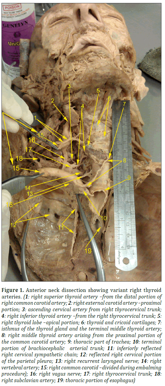
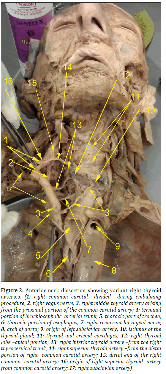
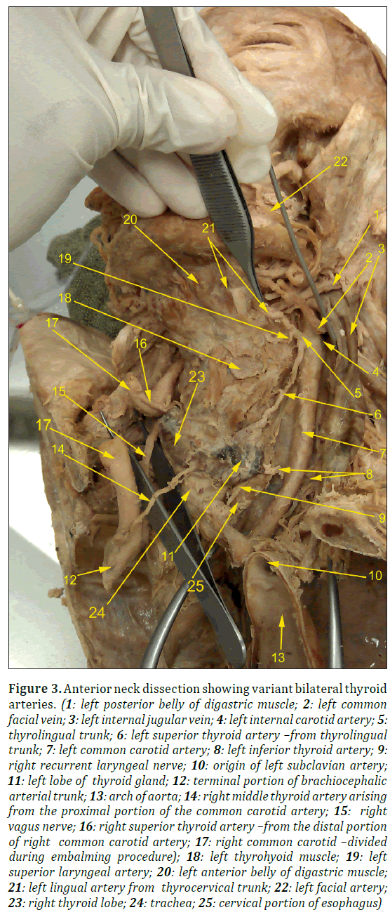
While dissecting the neck of a donated cadaver of an elderly female who died of congestive cardiac failure, we noticed a variant left thyrolingual artery branching off the left ECA, and right STA and MTA coming off the right common carotid artery (Figures 1, 2, 3).
Figure 1: Anterior neck dissection showing variant right thyroid arteries. (1: right superior thyroid artery –from the distal portion of right common carotid artery; 2: right external carotid artery –proximal portion; 3: ascending cervical artery from right thyrocervical trunk; 4: right inferior thyroid artery –from the right thyrocervical trunk; 5: right thyroid lobe –apical portion; 6: thyroid and cricoid cartilages; 7: isthmus of the thyroid gland and the terminal middle thyroid artery; 8: right middle thyroid artery arising from the proximal portion of the common carotid artery; 9: thoracic part of trachea; 10: terminal portion of brachiocephalic arterial trunk; 11: inferiorly reflected right cervical sympathetic chain; 12: reflected right cervical portion of the parietal pleura; 13: right recurrent laryngeal nerve; 14: right vertebral artery; 15: right common carotid –divided during embalming procedure); 16: right vagus nerve; 17: right thyrocervical trunk; 18: right subclavian artery; 19: thoracic portion of esophagus)
Figure 2: Anterior neck dissection showing variant right thyroid arteries. (1: right common carotid –divided during embalming procedure; 2: right vagus nerve; 3: right middle thyroid artery arising from the proximal portion of the common carotid artery; 4: terminal portion of brachiocephalic arterial trunk; 5: thoracic part of trachea; 6: thoracic portion of esophagus; 7: right recurrent laryngeal nerve; 8: arch of aorta; 9: origin of left subclavian artery; 10: isthmus of the thyroid gland; 11: thyroid and cricoid cartilages; 12: right thyroid lobe –apical portion; 13: right inferior thyroid artery –from the right thyrocervical trunk; 14: right superior thyroid artery –from the distal portion of right common carotid artery; 15: distal end of the right common carotid artery; 16: origin of right superior thyroid artery from common carotid artery; 17: right subclavian artery)
Figure 3: Anterior neck dissection showing variant bilateral thyroid arteries. (1: left posterior belly of digastric muscle; 2: left common facial vein; 3: left internal jugular vein; 4: left internal carotid artery; 5: thyrolingual trunk; 6: left superior thyroid artery –from thyrolingual trunk; 7: left common carotid artery; 8: left inferior thyroid artery; 9: right recurrent laryngeal nerve; 10: origin of left subclavian artery; 11: left lobe of thyroid gland; 12: terminal portion of brachiocephalic arterial trunk; 13: arch of aorta; 14: right middle thyroid artery arising from the proximal portion of the common carotid artery; 15: right vagus nerve; 16: right superior thyroid artery –from the distal portion of right common carotid artery; 17: right common carotid –divided during embalming procedure); 18: left thyrohyoid muscle; 19: left superior laryngeal artery; 20: left anterior belly of digastric muscle; 21: left lingual artery from thyrocervical trunk; 22: left facial artery; 23: right thyroid lobe; 24: trachea; 25: cervical portion of esophagus)
The right superior thyroid artery measured 30 millimeters (mm) in length and 2 mm in external diameter at its origin. It was supplying the upper pole of the right thyroid pyramid and arose 28 mm proximal to the point of final bifurcation of the right common carotid.
Similarly, the right middle thyroid artery (MTA) measured 56 mm in length and 2 mm in external diameter at its point of origin. It was supplying the isthmus of the thyroid gland and arose 8 mm distal to the origin of the right common carotid artery (Figures 1, 2, 3)
On the left side, the superior and inferior thyroid arteries were the major branches supplying the left lobe of the thyroid gland. The left STA, measured 50 mm in length and 2 mm in external diameter, and originated as a branch of the left thyrolingual artery, an unusual branch of the external carotid artery. We noticed three distinct branches of the left thyrolingual artery; the left STA, the lingual artery and the smallest superior laryngeal branch (13mm x 1 mm), (Figures 1, 2, 3).
The left inferior thyroid artery (40 mm in length x 3 mm in external diameter) and the right inferior thyroid artery (60 mm in length and 2.5 mm in external diameter) showed textbook origin from the corresponding thyrocervical trunks.
In this case, right CCA measured 76 mm in length and 8 mm internal diameter (at the divided stumps) likewise Left CCA measured 91 mm in length and 9 mm external diameter.
Discussion
Variations in the origin of thyroid arteries have been found in the literature [1,7,8]. A case paper published in early 2011 had described the left MTA arising from the left CCA and STA branching off the corresponding CCAs [7]. The variation on right STA was reported in a cadaveric neck dissection as a branch arising from the right ICA in a 68 year old women [8]. Another rare variation reported in journals published in 2012 [2,9] was the thyrolinguofacial trunk branching off as the common arterial trunk for STA, facial and lingual arteries. A meta-analysis of data collected from 330 embalmed human cadavers showed 55 out of 207 cases had STA coming off the CCA. It also mentioned about the rare occurrence of thyrolingual trunk (0.6% incidence, 2 out of 330 cases)[5]. Japanese scientists [10] reported a case of a variant left CCA which had four branches including STA, lingual, facial and posterior auricular artery. Superior thyroid artery branching off the internal carotid artery on the left side has been mentioned in the journal [11] published in 2006 and a wide range of variations about arteries supplying the thyroid have been observed however this case not only had atypical origin of right STA and MTA from right CCA but also additional unique site of origin of left STA via thyrolingual arterial trunk from the proximal left ECA. Rare variants of thyrolingual artery have been reported in papers published in 2005 [2], 2000, and 2011[12,13].
This paper focused on the variant right superior and middle thyroid arteries arising from the right common carotid artery and left thyrolingual trunk arising from the left ECA in the same individual. There is a current gap in knowledge about these variations in the origin of thyroid arteries bilaterally. There has been no clear mention of this type of multiple bilateral thyroid arterial variations in a single individual. Our case is very peculiar because of the presence of rare bilateral thyroid arterial variations as described above.
Clinical importance
There are reported cases of life threatening post thyroidectomy hemorrhage and hematoma [11-16] and post tracheostomy bleeding [17,18] which could be associated with the thyroid arterial variations.
Knowing the variations in the origin and branching pattern of thyroid arteries has important implications during thyroid and tracheostomy surgeries. It can prevent accidental damage of these arteries and their complications such as excessive bleeding and hematoma formation. Preoperative ultrasound examination may be helpful find these types of arterial variations.
All in all, the presence of additional arterial branches supplying the thyroid gland would have predisposed this person to surgical complications if he had had undergone thyroid surgery; those variant arteries would have been liable to be damaged during thyroid surgery.
Acknowledgements
We would like to thank and express our utmost respect to the generous body donors to the University of Adelaide, South Australia Body Donor Program. This work would not have been possible without their great generosity. We would also like to give thanks to our Anatomy Team, laboratory manager Mr. Brad Jeffrey for helping with the photographs.
References
- Lo A, Oehley M, Bartlett A, Adams D, Blyth P, Al-Ali S. Anatomical variations of the common carotid artery bifurcation. ANZ J Surg. 2006; 76: 970–972.
- Zumre O, Salbacak A, Cicekcibasi AE, Tuncer I, Seker M. Investigation of the bifurcation level of the common carotid artery and variations of the branches of the external carotid artery in human fetuses. Ann Anat. 2005; 187: 361–369.
- Bliss RD, Gauger PG, Delbridge LW. Surgeon’s approach to the thyroid gland: surgical anatomy and the importance of technique. World J Surg. 2000; 24: 891–897.
- Standring S, ed. Gray’s Anatomy. The Anatomical Basis of Clinical Practice. 40th Ed., Spain, Churchill Livingstone. 2008; 462.
- Vázquez T, Cobiella R, Maranillo E, Valderrama FJ, McHanwell S, Parkin I, Sañudo JR.Anatomical variations of the superior thyroid and superior laryngeal arteries. Head Neck.2009; 31: 1078-1085.
- Yilmaz E, Celik HH, Durgun B, Atasever A, Ilgi S. Arteria thyroidea ima arising from the brachiocephalic trunk with bilateral absence of inferior thyroid arteries: a case report.Surg Radiol Anat. 1993; 15: 197–199.
- Won HS, Han SH, Oh CS, Chung IH. Superior and middle thyroid arteries arising from the common carotid artery. Surg Radiol Anat. 2011; 33: 645–647.
- Lemaire V, Jacquemin G, Medot M, Fissette J. Thyrolingual trunk arising from the common carotid artery: A case report. Surg Radiol Anat. 2001; 23: 135–137.
- Iwai T, Izumi T, Inoue T, Maegawa J, Fuwa N, Mitsudo K, Tohnai I. Thyrolingual trunk arising from the common carotid artery identified by three-dimensional computed tomography angiography. Surg Radiol Anat. 2012; 34: 85–88.
- Iwai T, Izumi T, Inoue T, Fuwa N, Shibasaki M, Oguri S, Iwai T, Izumi T, Inoue T, Fuwa N, Shibasaki M, Oguri S. Thyrolinguofacial trunk arising from the carotid bifurcation determined by three-dimensional computed tomography angiography. Surg Radiol Anat.2013; 35: 75–78.
- Aggarwal NR, Krishnamoorthy T, Devasia B, Menon G, Chandrasekhar K. Variant origin of superior thyroid artery, occipital artery and ascending pharyngeal artery from a common trunk from the cervical segment of internal carotid artery. Surg Radiol Anat. 2006; 28:650–653.
- Calò PG, Erdas E, Medas F, Pisano G, Barbarossa M, Pomata M, Nicolosi A. Late bleeding after total thyroidectomy: Report of two cases occurring 13 days after operation. Clin Med Insights Case Rep. 2013; 6: 165–170.
- Calò PG, Pisano G, Piga G, Meadas F, Tatti A, Donati M, Nicolosi A. Postoperative hematomas after thyroid surgery: Incidence and risk factors in our experience. Ann Ital Chir. 2010; 81: 343–347.
- Bergenfelz A, Jansson S, Kristoffersson A, Mårtensson H, Reihnér E, Wallin G, Lausen I. Complications to thyroid surgery: Results as reported in a database from a multicenter audit comprising 3,660 patients. Langenbeck’s Arch Surg. 2008; 393: 667–673.
- Morton RP, Mak V, Moss D, Ahmad Z, Sevao J. Risk of bleeding after thyroid surgery: Matched pairs analysis. J Laryngol Otol. 2012; 126: 285–288.
- Torfs A, Laureyns G, Lemkens P. Outpatient hemithyroidectomy: safety and feasibility. B-ENT. 2012; 8: 279–283.
- Praveen CV, Martin A. A rare case of fatal haemorrhage after tracheostomy. Ann R Coll Surg Engl. 2007; 89: W6–8.
- Shahabi I, Zada B, Imad, Ali M. Complications of conventional tracheostomy. J Postgrad Med Ins. 2005; 19: 187–191.
Arjun Burlakoti* and Nicola Massy-Westropp
Division of Health Sciences, University of South Australia (UniSA), School of Health Sciences, Adelaide,Australia.
- *Corresponding Author:
- Dr. Arjun Burlakoti
Lecturer in Human Anatomy, Division of Health Sciences University of South Australia School of Health Sciencesm City East Campus, Room: BJ2-40, North Terrace, Adelaide SA 5000 Australia.
Tel: +61 (08) 883021206
E-mail: Arjun.Burlakoti@unisa.edu.a
Date of Received: March 11th, 2014
Date of Accepted: September 19th, 2015
Published Online: December 31st, 2015
© Int J Anat Var (IJAV). 2015; 8: 43–46.
Abstract
Three variant thyroid arteries were found in a cadaver during routine dissection at the University of South Australia Human Anatomy cadaveric laboratory.
Three thyroid arteries were detected in the right side, two of them (superior and middle thyroid arteries) came off the right common carotid artery (CCA) and the third one (inferior thyroid artery) arose from the right thyrocervical trunk.
In the same specimen, the left superior thyroid artery (STA) had an unusual origin from the left thyrolingual trunk, which in turn originated as a branch of the left external carotid artery (ECA). The left and right inferior thyroid artery (ITA) showed a normal origin from the corresponding thyrocervical trunks.
-Keywords
variant,superior thyroid artery,middle thyroid artery
Introduction
This paper aims to present a rare case of bilateral thyroid arterial variations, which include variant right superior thyroid artery (STA), right middle thyroid artery (MTA), left thyrolingual trunk (TLT) and left STA.
There are two common carotid arteries (CCAs) one on each side of the neck. Normally each common carotid artery does not give any branches in this region and terminates by bifurcating into external (ECA) and internal carotid artery (ICA) at variable labels around the thyroid cartilage and hyoid bone [1,2]. More commonly the left and right common carotid arteries bifurcate at the level of hyoid bone (27 out of 67; 40%), upper border of the thyroid cartilage (26 out of 67; 39%) and the body of the thyroid cartilage (4 out of 67; 6%)[1].
Normally there are two thyroid arteries on each side, the superior and inferior thyroid arteries. In most of the cases STA branches off as the first branch of the external carotid artery (ECA)[3], and the ITA branches off the corresponding thyrocervical trunk [4]. However studies revealed that the STA commonly arises from CCA [1,5]. In a small number of individuals (10%), an unpaired arteria thyroidea ima may arise from the brachiocephalic trunk or aorta and supply the isthmic part of the thyroid gland [6].
Case Report
While dissecting the neck of a donated cadaver of an elderly female who died of congestive cardiac failure, we noticed a variant left thyrolingual artery branching off the left ECA, and right STA and MTA coming off the right common carotid artery (Figures 1, 2, 3).
Figure 1: Anterior neck dissection showing variant right thyroid arteries. (1: right superior thyroid artery –from the distal portion of right common carotid artery; 2: right external carotid artery –proximal portion; 3: ascending cervical artery from right thyrocervical trunk; 4: right inferior thyroid artery –from the right thyrocervical trunk; 5: right thyroid lobe –apical portion; 6: thyroid and cricoid cartilages; 7: isthmus of the thyroid gland and the terminal middle thyroid artery; 8: right middle thyroid artery arising from the proximal portion of the common carotid artery; 9: thoracic part of trachea; 10: terminal portion of brachiocephalic arterial trunk; 11: inferiorly reflected right cervical sympathetic chain; 12: reflected right cervical portion of the parietal pleura; 13: right recurrent laryngeal nerve; 14: right vertebral artery; 15: right common carotid –divided during embalming procedure); 16: right vagus nerve; 17: right thyrocervical trunk; 18: right subclavian artery; 19: thoracic portion of esophagus)
Figure 2: Anterior neck dissection showing variant right thyroid arteries. (1: right common carotid –divided during embalming procedure; 2: right vagus nerve; 3: right middle thyroid artery arising from the proximal portion of the common carotid artery; 4: terminal portion of brachiocephalic arterial trunk; 5: thoracic part of trachea; 6: thoracic portion of esophagus; 7: right recurrent laryngeal nerve; 8: arch of aorta; 9: origin of left subclavian artery; 10: isthmus of the thyroid gland; 11: thyroid and cricoid cartilages; 12: right thyroid lobe –apical portion; 13: right inferior thyroid artery –from the right thyrocervical trunk; 14: right superior thyroid artery –from the distal portion of right common carotid artery; 15: distal end of the right common carotid artery; 16: origin of right superior thyroid artery from common carotid artery; 17: right subclavian artery)
Figure 3: Anterior neck dissection showing variant bilateral thyroid arteries. (1: left posterior belly of digastric muscle; 2: left common facial vein; 3: left internal jugular vein; 4: left internal carotid artery; 5: thyrolingual trunk; 6: left superior thyroid artery –from thyrolingual trunk; 7: left common carotid artery; 8: left inferior thyroid artery; 9: right recurrent laryngeal nerve; 10: origin of left subclavian artery; 11: left lobe of thyroid gland; 12: terminal portion of brachiocephalic arterial trunk; 13: arch of aorta; 14: right middle thyroid artery arising from the proximal portion of the common carotid artery; 15: right vagus nerve; 16: right superior thyroid artery –from the distal portion of right common carotid artery; 17: right common carotid –divided during embalming procedure); 18: left thyrohyoid muscle; 19: left superior laryngeal artery; 20: left anterior belly of digastric muscle; 21: left lingual artery from thyrocervical trunk; 22: left facial artery; 23: right thyroid lobe; 24: trachea; 25: cervical portion of esophagus)
The right superior thyroid artery measured 30 millimeters (mm) in length and 2 mm in external diameter at its origin. It was supplying the upper pole of the right thyroid pyramid and arose 28 mm proximal to the point of final bifurcation of the right common carotid.
Similarly, the right middle thyroid artery (MTA) measured 56 mm in length and 2 mm in external diameter at its point of origin. It was supplying the isthmus of the thyroid gland and arose 8 mm distal to the origin of the right common carotid artery (Figures 1, 2, 3)
On the left side, the superior and inferior thyroid arteries were the major branches supplying the left lobe of the thyroid gland. The left STA, measured 50 mm in length and 2 mm in external diameter, and originated as a branch of the left thyrolingual artery, an unusual branch of the external carotid artery. We noticed three distinct branches of the left thyrolingual artery; the left STA, the lingual artery and the smallest superior laryngeal branch (13mm x 1 mm), (Figures 1, 2, 3).
The left inferior thyroid artery (40 mm in length x 3 mm in external diameter) and the right inferior thyroid artery (60 mm in length and 2.5 mm in external diameter) showed textbook origin from the corresponding thyrocervical trunks.
In this case, right CCA measured 76 mm in length and 8 mm internal diameter (at the divided stumps) likewise Left CCA measured 91 mm in length and 9 mm external diameter.
Discussion
Variations in the origin of thyroid arteries have been found in the literature [1,7,8]. A case paper published in early 2011 had described the left MTA arising from the left CCA and STA branching off the corresponding CCAs [7]. The variation on right STA was reported in a cadaveric neck dissection as a branch arising from the right ICA in a 68 year old women [8]. Another rare variation reported in journals published in 2012 [2,9] was the thyrolinguofacial trunk branching off as the common arterial trunk for STA, facial and lingual arteries. A meta-analysis of data collected from 330 embalmed human cadavers showed 55 out of 207 cases had STA coming off the CCA. It also mentioned about the rare occurrence of thyrolingual trunk (0.6% incidence, 2 out of 330 cases)[5]. Japanese scientists [10] reported a case of a variant left CCA which had four branches including STA, lingual, facial and posterior auricular artery. Superior thyroid artery branching off the internal carotid artery on the left side has been mentioned in the journal [11] published in 2006 and a wide range of variations about arteries supplying the thyroid have been observed however this case not only had atypical origin of right STA and MTA from right CCA but also additional unique site of origin of left STA via thyrolingual arterial trunk from the proximal left ECA. Rare variants of thyrolingual artery have been reported in papers published in 2005 [2], 2000, and 2011[12,13].
This paper focused on the variant right superior and middle thyroid arteries arising from the right common carotid artery and left thyrolingual trunk arising from the left ECA in the same individual. There is a current gap in knowledge about these variations in the origin of thyroid arteries bilaterally. There has been no clear mention of this type of multiple bilateral thyroid arterial variations in a single individual. Our case is very peculiar because of the presence of rare bilateral thyroid arterial variations as described above.
Clinical importance
There are reported cases of life threatening post thyroidectomy hemorrhage and hematoma [11-16] and post tracheostomy bleeding [17,18] which could be associated with the thyroid arterial variations.
Knowing the variations in the origin and branching pattern of thyroid arteries has important implications during thyroid and tracheostomy surgeries. It can prevent accidental damage of these arteries and their complications such as excessive bleeding and hematoma formation. Preoperative ultrasound examination may be helpful find these types of arterial variations.
All in all, the presence of additional arterial branches supplying the thyroid gland would have predisposed this person to surgical complications if he had had undergone thyroid surgery; those variant arteries would have been liable to be damaged during thyroid surgery.
Acknowledgements
We would like to thank and express our utmost respect to the generous body donors to the University of Adelaide, South Australia Body Donor Program. This work would not have been possible without their great generosity. We would also like to give thanks to our Anatomy Team, laboratory manager Mr. Brad Jeffrey for helping with the photographs.
References
- Lo A, Oehley M, Bartlett A, Adams D, Blyth P, Al-Ali S. Anatomical variations of the common carotid artery bifurcation. ANZ J Surg. 2006; 76: 970–972.
- Zumre O, Salbacak A, Cicekcibasi AE, Tuncer I, Seker M. Investigation of the bifurcation level of the common carotid artery and variations of the branches of the external carotid artery in human fetuses. Ann Anat. 2005; 187: 361–369.
- Bliss RD, Gauger PG, Delbridge LW. Surgeon’s approach to the thyroid gland: surgical anatomy and the importance of technique. World J Surg. 2000; 24: 891–897.
- Standring S, ed. Gray’s Anatomy. The Anatomical Basis of Clinical Practice. 40th Ed., Spain, Churchill Livingstone. 2008; 462.
- Vázquez T, Cobiella R, Maranillo E, Valderrama FJ, McHanwell S, Parkin I, Sañudo JR.Anatomical variations of the superior thyroid and superior laryngeal arteries. Head Neck.2009; 31: 1078-1085.
- Yilmaz E, Celik HH, Durgun B, Atasever A, Ilgi S. Arteria thyroidea ima arising from the brachiocephalic trunk with bilateral absence of inferior thyroid arteries: a case report.Surg Radiol Anat. 1993; 15: 197–199.
- Won HS, Han SH, Oh CS, Chung IH. Superior and middle thyroid arteries arising from the common carotid artery. Surg Radiol Anat. 2011; 33: 645–647.
- Lemaire V, Jacquemin G, Medot M, Fissette J. Thyrolingual trunk arising from the common carotid artery: A case report. Surg Radiol Anat. 2001; 23: 135–137.
- Iwai T, Izumi T, Inoue T, Maegawa J, Fuwa N, Mitsudo K, Tohnai I. Thyrolingual trunk arising from the common carotid artery identified by three-dimensional computed tomography angiography. Surg Radiol Anat. 2012; 34: 85–88.
- Iwai T, Izumi T, Inoue T, Fuwa N, Shibasaki M, Oguri S, Iwai T, Izumi T, Inoue T, Fuwa N, Shibasaki M, Oguri S. Thyrolinguofacial trunk arising from the carotid bifurcation determined by three-dimensional computed tomography angiography. Surg Radiol Anat.2013; 35: 75–78.
- Aggarwal NR, Krishnamoorthy T, Devasia B, Menon G, Chandrasekhar K. Variant origin of superior thyroid artery, occipital artery and ascending pharyngeal artery from a common trunk from the cervical segment of internal carotid artery. Surg Radiol Anat. 2006; 28:650–653.
- Calò PG, Erdas E, Medas F, Pisano G, Barbarossa M, Pomata M, Nicolosi A. Late bleeding after total thyroidectomy: Report of two cases occurring 13 days after operation. Clin Med Insights Case Rep. 2013; 6: 165–170.
- Calò PG, Pisano G, Piga G, Meadas F, Tatti A, Donati M, Nicolosi A. Postoperative hematomas after thyroid surgery: Incidence and risk factors in our experience. Ann Ital Chir. 2010; 81: 343–347.
- Bergenfelz A, Jansson S, Kristoffersson A, Mårtensson H, Reihnér E, Wallin G, Lausen I. Complications to thyroid surgery: Results as reported in a database from a multicenter audit comprising 3,660 patients. Langenbeck’s Arch Surg. 2008; 393: 667–673.
- Morton RP, Mak V, Moss D, Ahmad Z, Sevao J. Risk of bleeding after thyroid surgery: Matched pairs analysis. J Laryngol Otol. 2012; 126: 285–288.
- Torfs A, Laureyns G, Lemkens P. Outpatient hemithyroidectomy: safety and feasibility. B-ENT. 2012; 8: 279–283.
- Praveen CV, Martin A. A rare case of fatal haemorrhage after tracheostomy. Ann R Coll Surg Engl. 2007; 89: W6–8.
- Shahabi I, Zada B, Imad, Ali M. Complications of conventional tracheostomy. J Postgrad Med Ins. 2005; 19: 187–191.


