Bilateral variations in renal vasculature
Vanita Gupta1*, Sheetal Kotgirwar1, Soumitra Trivedi1, Rashmi Deopujari1 and Vikrant Singh2
1Department of Anatomy, Peoples College of Medical Sciences and Research Center, Bhopal, India
2Bhopal Memorial Hospital and Research Center, Bhopal, India
- *Corresponding Author:
- Dr. Vanita Gupta
Assistant Professor, Department of Anatomy, Peoples College of Medical Sciences and Research Center, Bhopal, 462038, India
Tel: +91 755 2742348
E-mail: doctorvanita@yahoo.co.in
Date of Received: August 13th, 2009
Date of Accepted: February 26th, 2010
Published Online: April 23rd, 2010
© IJAV. 2010; 3: 53–55.
[ft_below_content] =>Keywords
kidney, anatomical variant, vasculature, renal artery, renal vein
Introduction
The renal arteries arise from abdominal aorta below the origin of superior mesenteric artery, on each side. Near the hilum of the kidney, each renal artery divides into anterior and posterior branch, which in turn divides into number of segmental arteries supplying the different renal segments. The presence of unusual branching patterns of the renal arteries is not uncommon. In 70% of cases there is a single renal artery supplying each kidney [1]. Anatomical variations and congenital anomalies of the renal veins have been well described. Numerous reports have appeared in the literature describing variations in renal vascular anatomy. Knowledge of the variations of renal vascular anatomy has importance in exploration and treatment of renal trauma, renal transplantation, renovascular hypertension, renal artery embolization, angioplasty or vascular reconstruction for congenital and acquired lesions, surgery for abdominal aortic aneurysm and conservative or radical renal surgery [2].
The objective of the case report and review of literature is to bring awareness to clinicians about the variations in the blood supply of the kidney especially those who are performing invasive procedures and vascular surgeries on kidney.
Case Report
In routine dissection of 67-year-old male cadaver, certain variations in the renal vasculature were observed.
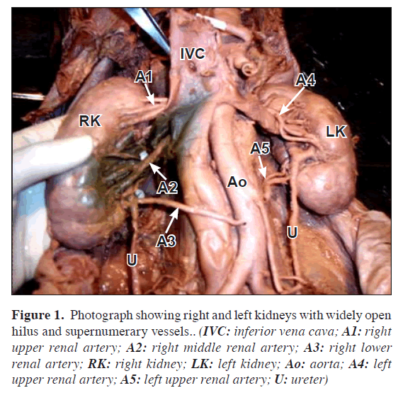
The right kidney was well lobulated and measured 11 x 5 x 3 cm. It was supplied by three renal arteries and was drained by two renal veins. The upper renal artery arose at the level just below the superior mesenteric artery (SMA), before going to the kidney it divided into three branches in a looped pattern. The middle renal artery arose just 1 cm below the first. The lower renal artery was seen arising from the aorta just below the origin of inferior mesenteric artery (IMA). Out of the two veins draining the parenchyma, upper vein arose as two tributaries and then taking a straight course between the upper and the middle renal artery, drained into the inferior vena cava (IVC). The lower vein also drained into the IVC. Two pairs of minor calyces formed one major calyx to open into the ureter (Figure 1).
Figure 1. Photograph showing right and left kidneys with widely open hilus and supernumerary vessels.. (IVC: inferior vena cava; A1: right upper renal artery; A2: right middle renal artery; A3: right lower renal artery; RK: right kidney; LK: left kidney; Ao: aorta; A4: left upper renal artery; A5: left upper renal artery; U: ureter)
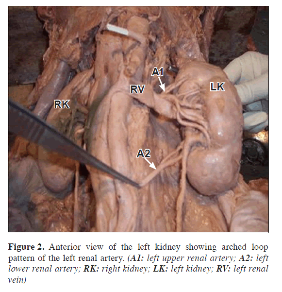
The left kidney measured 10 x 6.5 x 4 cm. The hilum was 4 cm wide. Well-defined lobules were seen. There were two renal arteries. The upper renal artery arose from the aorta and taking a curved path it divided into five branches, out of which three branches again divided into two before entering the kidney. The lower renal artery took origin from aorta just below the origin of IMA and divided into two branches at the hilum. There was a single vein draining the kidney, coursing in between the two renal arteries. Two pairs of minor calyces formed one major calyx to open into the ureter (Figure 2).
Discussion
The variations in the renal arteries are considered critical issues that surgeons should have a thorough envision and appreciation of the condition. Accessory renal arteries constitute the most common, clinically important vascular variant and are seen in up to one-third of patients. Multiple renal arteries are unilateral in approximately 30% of patients and bilateral in approximately 10%. Accessory renal arteries usually arise from the aorta or iliac arteries anywhere from the level of T11 to the level of L4 vertebra. In rare cases, they can arise from the lower thoracic aorta or from lumbar or mesenteric arteries. Usually, the accessory artery courses into the renal hilum to perfuse the upper or lower renal poles. Accessory vessels to the polar regions are usually smaller than accessory hilar renal arteries, which are typically equal in size to a single renal artery [3].
The kidneys begin their development in the pelvic cavity. During further development, they ascend to their final position in the lumbar region. When the kidneys are located in the pelvis, they are supplied by the branches of internal iliac or common iliac arteries. While the kidneys ascend to lumbar region, their arterial supply also shifts from common iliac artery to the abdominal aorta. Accessory renal arteries originate from the abdominal aorta either above or below the main renal artery and reach the hilum. In recent years, interest in the surgical and medical aspects of accessory renal arteries has been high. One has to keep in mind that transplanting a kidney with accessory renal arteries has several theoretical disadvantages – acute tubular necrosis and rejection episodes, decreased graft function, and prolonged hospitalization [4]. The renal veins show less variation than the renal arteries; the right renal vein may be doubled, even though the left renal vein is usually single. Bergman et al. observed double renal and testicular arteries in a well-developed 69-year-old Caucasian male. The right kidney had two renal arteries, one at its usual midorgan (hilar) position and one inferior polar. One testicular artery arose from the mid-point of the usual renal artery. The second testicular artery arose from the inferior polar renal artery near its origin from the abdominal aorta. The two testicular arteries remained doubled throughout their course and both entered the right testis at separate sites on the organ [5]. Rusu reported bilateral doubled renal arteries on the right side as superior hilar and inferior hilar renal arteries, and on the left side as superior hilar and inferior polar renal arteries. All these renal arteries emerged from the abdominal aorta, as in our case [6]. Bayramoglu et al. reported a variant which consisted of bilateral additional renal arteries originating from the abdominal aorta and an additional right renal vein accompanying the additional right renal artery. These anomalies were associated with unrotated kidneys with extrarenal calices and pelvis. All the additional vessels were located posterior to the ureter with a close relationship to the ureteropelvic junction on the right side [7]. Bulic et al. reported that the right kidney received two renal arteries from the aorta that were similar in diameter, both entering through the hilum. The left kidney had three arteries originating from the aorta, one at its usual hilar position and two entering the renal cortex at its upper and lower poles. The upper pole of the left kidney also gave rise to an additional tributary of the renal vein [8]. Kaneko et al. from their study of 170 cases, found that 36 of 170 subjects (21.2%) had multiple arterial origins on the left or right side, and 8 subjects (4.7%) had bilateral multiple arterial origins. Multiple renal veins were present in 22 cadavers (12.9%), and bilateral multiple veins were observed in 1 cadaver (0.6%) [9]. Singh et al. reported the presence of accessory renal arteries during routine dissection in an elderly female cadaver. The uniqueness in the variations noted included (1) a dual relationship of the ureters to the accessory renal arteries and (2) both the right and left ovarian arteries originating from their respective accessory arteries [10].
Tanyeli et al. described a common trunk from the right side of the aorta ramifying into suprarenal and two renal hilar arteries in a 40-year-old male cadaver. The suprarenal branch divided into several smaller branches to supply the gland. The superior renal hilar artery gave origin to the right testicular artery and an additional suprarenal artery. The inferior renal hilar artery gave rise to one more additional suprarenal artery. The superior renal hilar artery crossed the inferior renal hilar artery. Right renal veins were also double [11].
The variations described in the current observation present a unique pattern of congenital renal vascular variants having surgical and radiological importance.
References
- Standring S. Gray’s Anatomy. The Anatomical Basis of Clinical Practice. 39th Ed. London, Elseiver Churchill Livingstone Publishers. 2005; 1274–1275.
- Fernandes RMP, Conte FHP, Favorito LA, Abidu-Figueiredo M, Babinski MA. Triple right renal vein: an uncommon variation. Int J Morphol. 2005; 23: 231–233.
- Kadir S. Angiography of the kidneys. In: Kadir S, ed. Diagnostic angiography. Philadelphia, Saunders. 1986; 445–495.
- Harrison LH Jr, Flye MW, Seigler HF. Incidence of anatomical variants in renal vasculature in the presence of normal renal function. Ann Surg. 1978; 188: 83–89.
- Bergman RA, Cassell MD, Sahinoglu K, Heidger PM Jr. Human doubled renal and testicular arteries. Ann Anat. 1992; 174: 313–315.
- Rusu MC. Human bilateral doubled renal and testicular arteries with a left testicular arterial arch around the left renal vein. Rom J Morphol Embryol. 2006; 47: 197–200.
- Bayramoglu A, Demiryurek D, Erbil KM. Bilateral additional renal arteries and an additional right renal vein associated with unrotated kidneys. Saudi Med J. 2003; 24: 535–537.
- Bulic K, Ivkic G, Pavic T. A case of duplicated right renal artery and triplicated left renal artery. Ann Anat. 1996; 178: 281–283.
- Kaneko N, Kobayashi Y, Okada Y. Anatomic variations of the renal vessels pertinent to transperitoneal vascular control in the management of trauma. Surgery. 2008; 143: 616–622.
- Singh G, Ng YK, Bay BH. Bilateral accessory renal arteries associated with some anomalies of the ovarian arteries: a case study. Clin Anat. 1998; 11: 417–420.
- Tanyeli E, Uzel M, Soyluoglu AI. Complex renal vascular variation: a case report. Ann Anat. 2006; 188: 455–458.
Vanita Gupta1*, Sheetal Kotgirwar1, Soumitra Trivedi1, Rashmi Deopujari1 and Vikrant Singh2
1Department of Anatomy, Peoples College of Medical Sciences and Research Center, Bhopal, India
2Bhopal Memorial Hospital and Research Center, Bhopal, India
- *Corresponding Author:
- Dr. Vanita Gupta
Assistant Professor, Department of Anatomy, Peoples College of Medical Sciences and Research Center, Bhopal, 462038, India
Tel: +91 755 2742348
E-mail: doctorvanita@yahoo.co.in
Date of Received: August 13th, 2009
Date of Accepted: February 26th, 2010
Published Online: April 23rd, 2010
© IJAV. 2010; 3: 53–55.
Abstract
Routine dissection of a 67-year-old male cadaver, revealed a complex anatomical variation of the renal vasculature. Right kidney was multilobulated measuring 11 x 5 x 3 cm, with the hilum containing three renal arteries and two renal veins. The upper renal artery arose from aorta just below origin of superior mesenteric artery, middle renal artery arose from 1 cm below the upper artery and the lower renal artery arose just below the origin of inferior mesenteric artery, respectively. Two veins drained the right kidney into inferior vena cava. Left kidney measured 10 x 6.5 x 4 cm. The hilum contained two renal arteries. The upper renal artery arose from the aorta just below the origin of superior mesenteric artery, the lower renal artery arose from aorta just below the origin of inferior mesenteric artery. There was a single vein draining the left kidney. Knowledge of the variations of renal vascular anatomy has importance in exploration and treatment of renal trauma, renal transplantation, renal artery embolization, surgery for abdominal aortic aneurysm and conservative or radical renal surgery.
-Keywords
kidney, anatomical variant, vasculature, renal artery, renal vein
Introduction
The renal arteries arise from abdominal aorta below the origin of superior mesenteric artery, on each side. Near the hilum of the kidney, each renal artery divides into anterior and posterior branch, which in turn divides into number of segmental arteries supplying the different renal segments. The presence of unusual branching patterns of the renal arteries is not uncommon. In 70% of cases there is a single renal artery supplying each kidney [1]. Anatomical variations and congenital anomalies of the renal veins have been well described. Numerous reports have appeared in the literature describing variations in renal vascular anatomy. Knowledge of the variations of renal vascular anatomy has importance in exploration and treatment of renal trauma, renal transplantation, renovascular hypertension, renal artery embolization, angioplasty or vascular reconstruction for congenital and acquired lesions, surgery for abdominal aortic aneurysm and conservative or radical renal surgery [2].
The objective of the case report and review of literature is to bring awareness to clinicians about the variations in the blood supply of the kidney especially those who are performing invasive procedures and vascular surgeries on kidney.
Case Report
In routine dissection of 67-year-old male cadaver, certain variations in the renal vasculature were observed.
The right kidney was well lobulated and measured 11 x 5 x 3 cm. It was supplied by three renal arteries and was drained by two renal veins. The upper renal artery arose at the level just below the superior mesenteric artery (SMA), before going to the kidney it divided into three branches in a looped pattern. The middle renal artery arose just 1 cm below the first. The lower renal artery was seen arising from the aorta just below the origin of inferior mesenteric artery (IMA). Out of the two veins draining the parenchyma, upper vein arose as two tributaries and then taking a straight course between the upper and the middle renal artery, drained into the inferior vena cava (IVC). The lower vein also drained into the IVC. Two pairs of minor calyces formed one major calyx to open into the ureter (Figure 1).
Figure 1. Photograph showing right and left kidneys with widely open hilus and supernumerary vessels.. (IVC: inferior vena cava; A1: right upper renal artery; A2: right middle renal artery; A3: right lower renal artery; RK: right kidney; LK: left kidney; Ao: aorta; A4: left upper renal artery; A5: left upper renal artery; U: ureter)
The left kidney measured 10 x 6.5 x 4 cm. The hilum was 4 cm wide. Well-defined lobules were seen. There were two renal arteries. The upper renal artery arose from the aorta and taking a curved path it divided into five branches, out of which three branches again divided into two before entering the kidney. The lower renal artery took origin from aorta just below the origin of IMA and divided into two branches at the hilum. There was a single vein draining the kidney, coursing in between the two renal arteries. Two pairs of minor calyces formed one major calyx to open into the ureter (Figure 2).
Discussion
The variations in the renal arteries are considered critical issues that surgeons should have a thorough envision and appreciation of the condition. Accessory renal arteries constitute the most common, clinically important vascular variant and are seen in up to one-third of patients. Multiple renal arteries are unilateral in approximately 30% of patients and bilateral in approximately 10%. Accessory renal arteries usually arise from the aorta or iliac arteries anywhere from the level of T11 to the level of L4 vertebra. In rare cases, they can arise from the lower thoracic aorta or from lumbar or mesenteric arteries. Usually, the accessory artery courses into the renal hilum to perfuse the upper or lower renal poles. Accessory vessels to the polar regions are usually smaller than accessory hilar renal arteries, which are typically equal in size to a single renal artery [3].
The kidneys begin their development in the pelvic cavity. During further development, they ascend to their final position in the lumbar region. When the kidneys are located in the pelvis, they are supplied by the branches of internal iliac or common iliac arteries. While the kidneys ascend to lumbar region, their arterial supply also shifts from common iliac artery to the abdominal aorta. Accessory renal arteries originate from the abdominal aorta either above or below the main renal artery and reach the hilum. In recent years, interest in the surgical and medical aspects of accessory renal arteries has been high. One has to keep in mind that transplanting a kidney with accessory renal arteries has several theoretical disadvantages – acute tubular necrosis and rejection episodes, decreased graft function, and prolonged hospitalization [4]. The renal veins show less variation than the renal arteries; the right renal vein may be doubled, even though the left renal vein is usually single. Bergman et al. observed double renal and testicular arteries in a well-developed 69-year-old Caucasian male. The right kidney had two renal arteries, one at its usual midorgan (hilar) position and one inferior polar. One testicular artery arose from the mid-point of the usual renal artery. The second testicular artery arose from the inferior polar renal artery near its origin from the abdominal aorta. The two testicular arteries remained doubled throughout their course and both entered the right testis at separate sites on the organ [5]. Rusu reported bilateral doubled renal arteries on the right side as superior hilar and inferior hilar renal arteries, and on the left side as superior hilar and inferior polar renal arteries. All these renal arteries emerged from the abdominal aorta, as in our case [6]. Bayramoglu et al. reported a variant which consisted of bilateral additional renal arteries originating from the abdominal aorta and an additional right renal vein accompanying the additional right renal artery. These anomalies were associated with unrotated kidneys with extrarenal calices and pelvis. All the additional vessels were located posterior to the ureter with a close relationship to the ureteropelvic junction on the right side [7]. Bulic et al. reported that the right kidney received two renal arteries from the aorta that were similar in diameter, both entering through the hilum. The left kidney had three arteries originating from the aorta, one at its usual hilar position and two entering the renal cortex at its upper and lower poles. The upper pole of the left kidney also gave rise to an additional tributary of the renal vein [8]. Kaneko et al. from their study of 170 cases, found that 36 of 170 subjects (21.2%) had multiple arterial origins on the left or right side, and 8 subjects (4.7%) had bilateral multiple arterial origins. Multiple renal veins were present in 22 cadavers (12.9%), and bilateral multiple veins were observed in 1 cadaver (0.6%) [9]. Singh et al. reported the presence of accessory renal arteries during routine dissection in an elderly female cadaver. The uniqueness in the variations noted included (1) a dual relationship of the ureters to the accessory renal arteries and (2) both the right and left ovarian arteries originating from their respective accessory arteries [10].
Tanyeli et al. described a common trunk from the right side of the aorta ramifying into suprarenal and two renal hilar arteries in a 40-year-old male cadaver. The suprarenal branch divided into several smaller branches to supply the gland. The superior renal hilar artery gave origin to the right testicular artery and an additional suprarenal artery. The inferior renal hilar artery gave rise to one more additional suprarenal artery. The superior renal hilar artery crossed the inferior renal hilar artery. Right renal veins were also double [11].
The variations described in the current observation present a unique pattern of congenital renal vascular variants having surgical and radiological importance.
References
- Standring S. Gray’s Anatomy. The Anatomical Basis of Clinical Practice. 39th Ed. London, Elseiver Churchill Livingstone Publishers. 2005; 1274–1275.
- Fernandes RMP, Conte FHP, Favorito LA, Abidu-Figueiredo M, Babinski MA. Triple right renal vein: an uncommon variation. Int J Morphol. 2005; 23: 231–233.
- Kadir S. Angiography of the kidneys. In: Kadir S, ed. Diagnostic angiography. Philadelphia, Saunders. 1986; 445–495.
- Harrison LH Jr, Flye MW, Seigler HF. Incidence of anatomical variants in renal vasculature in the presence of normal renal function. Ann Surg. 1978; 188: 83–89.
- Bergman RA, Cassell MD, Sahinoglu K, Heidger PM Jr. Human doubled renal and testicular arteries. Ann Anat. 1992; 174: 313–315.
- Rusu MC. Human bilateral doubled renal and testicular arteries with a left testicular arterial arch around the left renal vein. Rom J Morphol Embryol. 2006; 47: 197–200.
- Bayramoglu A, Demiryurek D, Erbil KM. Bilateral additional renal arteries and an additional right renal vein associated with unrotated kidneys. Saudi Med J. 2003; 24: 535–537.
- Bulic K, Ivkic G, Pavic T. A case of duplicated right renal artery and triplicated left renal artery. Ann Anat. 1996; 178: 281–283.
- Kaneko N, Kobayashi Y, Okada Y. Anatomic variations of the renal vessels pertinent to transperitoneal vascular control in the management of trauma. Surgery. 2008; 143: 616–622.
- Singh G, Ng YK, Bay BH. Bilateral accessory renal arteries associated with some anomalies of the ovarian arteries: a case study. Clin Anat. 1998; 11: 417–420.
- Tanyeli E, Uzel M, Soyluoglu AI. Complex renal vascular variation: a case report. Ann Anat. 2006; 188: 455–458.


