Bilateral variations in the formation of sural nervetibial
Rengin Kosif*,Yasin Arifoglu and Murat Diramali
Abant Izzet Baysal University, Faculty of Medicine, Department of Anatomy, Bolu, TURKEY.
- *Corresponding Author:
- Dr. Rengin Kosif
Assistant Professor, Abant Izzet Baysal University, Faculty of Medicine, Department of Anatomy, Bolu, 14280,Turkey.
Tel: +90 374 253 46 56 / 3043
E-mail: rengink@yahoo.com
Date of Received: March 8th 2010
Date of Accepted: March 18th, 2010
Published Online: August 19th, 2010
© IJAV. 2010; 3: 118–121.
[ft_below_content] =>Keywords
sural nerve,variation,human cadaver,leg
Introduction
The sural nerve (SN) is a sensory nerve supplying the skin of the lateral and posterior part of the inferior third of the leg and lateral side of the foot and easily located in the leg [1,2]. The sural nerve is generally formed by the union of the medial sural cutaneous nerve derived from the tibial nerve, and the communicating fibular branch of the lateral sural cutaneous nerve, a branch derived from the common fibular nerve [3,4]. The union that results in the formation of the sural nerve occurs in the middle or lower third of the leg, at the popliteal fossa, at or just below the ankle [5]. Although the sural nerve has a constant topographical localization, anatomical variation is frequent [6].
Case Report
During routine cadaver dissection performed in the laboratory of the Department of Anatomy of the Faculty of Medicine at Abant Izzet Baysal University, two different sural nerve (SN) formations were observed separately in the right and left leg of a 64-year-old male cadaver. SN is usually formed when medial sural cutaneous nerve (MSCN) arising from tibial nerve (TN) unites with lateral sural cutaneous nerve (LSCN) arising from common fibular nerve (CFN) around the middle of posterior portion of leg [2,5]. Our case exhibited the following SN formations in cadaver’s right and left leg:
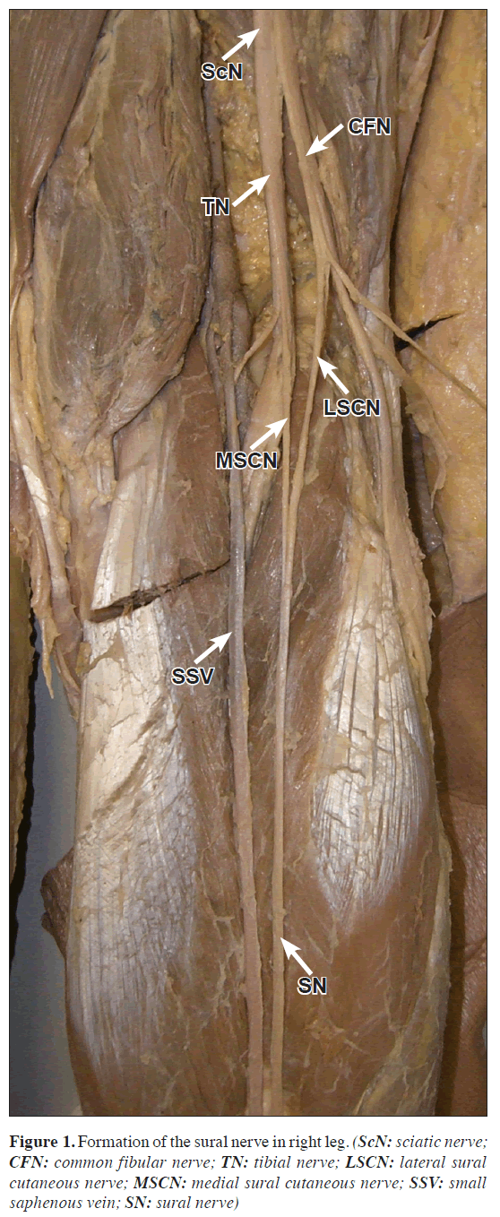
Formation of sural nerve in the right leg: Sciatic nerve (SC) was divided into its two terminal branches as nervetibial and common fibular nerves in the upper corner of popliteal fossa. In popliteal fossa, the level where small saphenous vein (SSV) ended was at the same point as the origin of gastrocnemius muscle (GM), and also at this level, MSCN was separated from TN. LSCN was separated from CFN 39.71 mm above this level. These two branches united in the 1/6 upper portion of the leg and 74.44 mm down to the origin of the GM, forming the SN. A branch originated from LSCN supplied sensation for the lateral portion of the leg (Figure 1).
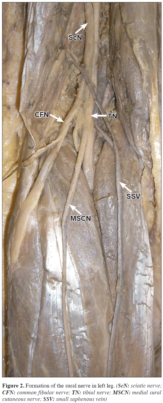
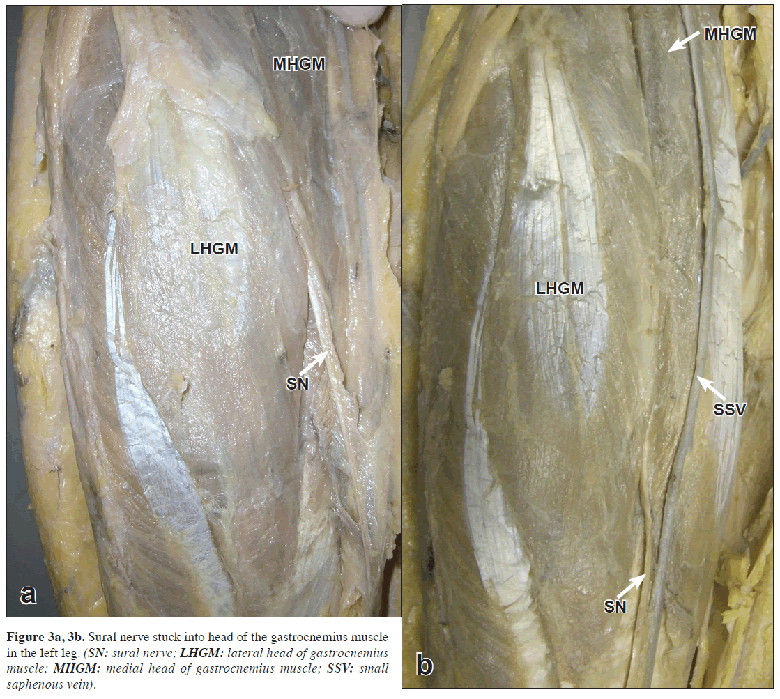
Formation of sural nerve in the left leg: SC was divided into its two terminal branches at the level where SSV came to the end. SSV ended 27.37 mm below to the origin of the GM, at the point where CFN was formed. SN was separated from the TN in the form of MSCN 14.23 mm below this level, and no branch of the CFN was involved (Figure 2). SN was stuck in the fascia between the heads of GM, however, it did not pierce the muscle (Figures 3a and 3b).
When compared to the right side, the left GM was atrophic. LSCN was separated from CFN 25.37 mm from its lateral side without giving any connecting branch to the SN. CFN was thicker when compared to the other side. There was no difference between the diameters of both sural nerves.
According to measurements performed with digital caliper which has the accuracy of 0.01 mm/0.0005’’, the medial head of the right GM was 63.52 mm and lateral head was 57.37 mm; while medial head of the left GM was 58.49 mm and lateral head was 44.24 mm, when the measurement was taken at the widest section of the muscle. The length of the right CFN was 4.35 mm, the left CFN was 8.32 mm, the right SN was 2.06 mm, and the left SN was 2.50 mm, respectively. The diameters of the TN were similar.
Discussion
The SN is a sensory nerve supplying the skin of the lateral and posterior part of the leg which is located at the lower 1/3 part and lateral side of the foot. Generally, it is formed by the union of the MSCN, a branch of the TN, and the LSCN, a branch of the CFN [1,7].
There may be numerous variations for the division of the SC into terminal branches. The division of the SC into the tibial and the common fibular nerves demonstrated a high variability in the branch level [8]. The nerve usually divides into the CFN and the TN in the popliteal fossa [9]. Three levels of SC division into terminal branches were reported: high (pelvic) level, intermediate level (SC divides at lower 2/3 of femur) and low (popliteal) level, in the literature [10]. It is reported that distances from the division of the SC to the popliteal fossa crease did not differ between the left and right legs of the cadavers [11].
In our case, division levels of SC in the popliteal fossa differed between the right and left legs. On right side SC is branched in the middle of the popliteal fossa, whereas, in the left side at the upper corner of the popliteal fossa.
According to a study of the SN, authors reported that in 80% of the cases it was formed by the union of the MSCN and LSCN, and in 20% of the cases it was a direct continuation of the MSCN [12]. Furthermore, a study showed that the SN was formed by the union of the MSCN and the LSCN in 67.1% of cases and the it was a continuation of the MSCN alone in 32.2% of cases [1]. The medial sural cutaneous nerve was reported to originate as a branch of the tibial nerve in the popliteal fossa at the left side [4,13].
In our case, the SN formation in the right leg was similar to the cases reported as 80%, whereas the left leg was similar to the 20% groups. In our cadaver, the MSNC formed the sural nerve by itself only in the left leg as indicated in the Pimental et al. and Bryan et al.’s studies.
The site where the MSCN and LSCN were united to form the sural nerve is highly variable. Previous studies reported that it may happen in the popliteal fossa, at the lower 1/3 of the leg or at the ankle [1,13-15]. In another study, the SN was formed by the union of the MSCN and LSCN in the popliteal fossa in 12% of cases, and the union occurred in the the lower 1/3 of the leg in 84% of cases [16]. In our case, the SN in the right leg was formed in the 1/6 upper part of the leg, whereas, in the left leg it was formed in the popliteal fossa and only by MSCN.
According to George and Nayak, the SN was reduced in size and pierced the GM instead of passing superficial to it, in the left leg of a male cadaver [17]. In our study, however, the SN was passing subfascial, although it was stuck into heads of GM it was thicker than that of the right leg. The diameter of the SN in the right leg was 2.06 mm, whereas, it was measured as 2.50 mm in the left leg. The diameter of CFN in the left side was interestingly higher (the right CFN was 4.35 mm and the left CFN was 8.32 mm).
Studies showed that the SN may course either intramuscularly or subfascially. During the regular dissection, a precise assessment of the frequency of this muscular course is important because of the possibility of this nerve being confused with included fascia instead of the muscular course [1]. As shown by the foregoing literature reports, there are many cases in which the SN or its branches are surrounded by fascia or scar tissue the sural nerve followed a transmuscular course, which corresponded to a frequency of 6.7% of all legs and 10% of the cadavers [16]. The SN pierced the GM along with the SSV instead of passing superficial to it. This variation was found in the left leg of a male cadaver and was unilateral [17,18].
Although only the left SN coursed subfascially and was stuck in between the lateral and medial heads of GM in our case, the left SN was thicker than the right. Moreover, the left GM was atrophic. This variant course of the SN might produce pain during the contraction of the GM or altered sensation over the area of its distribution. Due to the generated pain, GM might be less frequently used than it supposed to be, and this may cause atrophy. Though the SN is considered to be a sensory nerve, motor fibers have been found in 4.5% of cases [19]. However, motor fiber from SN to GM was not observed in our cadaver.
Conclusion
The variation of SN is an important surgical consideration when it is used as an autograft for peripheral nerve reconstruction. The knowledge of kind of entrapment of SN is very important in plastic surgery, sports medicine, physical therapy, clinical and surgical procedures. Clinically, the SN is widely used for both diagnostic (biopsy and nerve conduction velocity studies) and therapeutic purposes (nerve grafting). Thus, a detailed knowledge of the anatomy of the SN and its contributing nerves are important in carrying out these and other procedures.
References
- Moore KL, Dalley AF. Clinically oriented anatomy. 4th Ed. Philadelphia, Lippincott Williams & Wilkins. 1999; 572, 601.
- Baqai HZ, Din AM, Irshad M, Tariq M, Khawaja I. Sural nerve conduction; Age related variation studies in our normal population. Professional Med J. 2001; 8: 439–444.
- Mestdagh H, Drizenko A, Maynou C, Demondion X, Monier R. Origin and make up of the human sural nerve. Surg Radiol Anat. 2001; 23: 307–312.
- Bryan BM, Lutz GE, O’Brien SJ. Sural nerve entrapment after injury to the gastrocnemius: a case report. Arch Phys Med Rehabil. 1999; 80: 604–606.
- Mahakkanukrauh P, Chomsung R. Anatomical variations of the sural nerve. Clin Anat. 2002; 15: 263–266.
- Solomon LB , Ferris L, Tedman R, Henneberg M. Surgical anatomy of the sural and superficial fibular nerves with an emphasis on the approach to the lateral malleolus. J Anat. 2001; 199: 717–723.
- Standring S, ed. Gray’s Anatomy. The Anatomical Basis of Clinical Practice. 40th Ed., Edinburg, Churchill-Livingstone. 2008; 1427.
- Driban JB, Swanik CB, Barbe MF. Anatomical evaluation of the tibial nerve within the popliteal fossa. Clin Anat. 2007; 20: 694–698.
- Babinski MA, Machado FA, Costa WS. A rare variation in the high division of the sciatic nerve surrounding the superior gemellus muscle. Eur J Morphol. 2003; 41: 41–42.
- Okraszewska E, Migdalski L, Jedrzejewski KS, Bolanowski W. Sciatic nerve variations in some studies on the Polish population and its statistical significance. Folia Morphol (Warsz). 2002; 61: 277–282.
- Vloka JD, Hadzic A, April E, Thys DM. The division of the sciatic nerve in the popliteal fossa: anatomical implications for popliteal nerve blockade. Anesth Analg. 2001; 92: 215–217
- Ortiguela ME, Wood MB, Cahill DR. Anatomy of the sural nerve complex. J Hand Surg Am. 1987; 12: 1119–1123.
- Pimentel ML, Fernandes RMP, Babinski MA. Anomalous course of the medial sural cutaneous nerve and its clinical implications. Braz J Morphol Sci. 2005; 22: 179–182.
- McMinn RMH. Last’s anatomy regional and applied. 9th Ed. Edinburgh, Churchill Livingstone. 1994; 174.
- Woodburne RT, Burkel WE. Essentials of human anatomy. 9th Ed. New York, Oxford University Press.1994; 582–583.
- Coert JH, Dellon AL. Clinical implications of the surgical anatomy of the sural nerve. Plast Reconstr Surg. 1994; 94: 850–855.
- George B, Nayak S. Sural nerve entrapment in gastrocnemius muscle – a case report. Neuroanatomy. 2007; 6: 41–42.
- Nayak SB. Sural nerve and short saphenous vein entrapment–a case report. Indian J Plast Surg. 2005; 38: 171–172.
- Amoiridis G, Schols L, Ameridis N, Przuntek H. Motor fibers in the sural nerve of humans. Neurology. 1997; 49: 1725–1728.
Rengin Kosif*,Yasin Arifoglu and Murat Diramali
Abant Izzet Baysal University, Faculty of Medicine, Department of Anatomy, Bolu, TURKEY.
- *Corresponding Author:
- Dr. Rengin Kosif
Assistant Professor, Abant Izzet Baysal University, Faculty of Medicine, Department of Anatomy, Bolu, 14280,Turkey.
Tel: +90 374 253 46 56 / 3043
E-mail: rengink@yahoo.com
Date of Received: March 8th 2010
Date of Accepted: March 18th, 2010
Published Online: August 19th, 2010
© IJAV. 2010; 3: 118–121.
Abstract
During routine cadaver dissection two different sural nerve formations were observed in the 64-year-old male cadaver. Clinically, sural nerve is largely used in biopsy and as a graft in nerve transplantations. Therefore, knowing about the course, formation pattern and variations of sural nerve are important for the abovementioned procedures, as well as explaining the different clinical findings.
-Keywords
sural nerve,variation,human cadaver,leg
Introduction
The sural nerve (SN) is a sensory nerve supplying the skin of the lateral and posterior part of the inferior third of the leg and lateral side of the foot and easily located in the leg [1,2]. The sural nerve is generally formed by the union of the medial sural cutaneous nerve derived from the tibial nerve, and the communicating fibular branch of the lateral sural cutaneous nerve, a branch derived from the common fibular nerve [3,4]. The union that results in the formation of the sural nerve occurs in the middle or lower third of the leg, at the popliteal fossa, at or just below the ankle [5]. Although the sural nerve has a constant topographical localization, anatomical variation is frequent [6].
Case Report
During routine cadaver dissection performed in the laboratory of the Department of Anatomy of the Faculty of Medicine at Abant Izzet Baysal University, two different sural nerve (SN) formations were observed separately in the right and left leg of a 64-year-old male cadaver. SN is usually formed when medial sural cutaneous nerve (MSCN) arising from tibial nerve (TN) unites with lateral sural cutaneous nerve (LSCN) arising from common fibular nerve (CFN) around the middle of posterior portion of leg [2,5]. Our case exhibited the following SN formations in cadaver’s right and left leg:
Formation of sural nerve in the right leg: Sciatic nerve (SC) was divided into its two terminal branches as nervetibial and common fibular nerves in the upper corner of popliteal fossa. In popliteal fossa, the level where small saphenous vein (SSV) ended was at the same point as the origin of gastrocnemius muscle (GM), and also at this level, MSCN was separated from TN. LSCN was separated from CFN 39.71 mm above this level. These two branches united in the 1/6 upper portion of the leg and 74.44 mm down to the origin of the GM, forming the SN. A branch originated from LSCN supplied sensation for the lateral portion of the leg (Figure 1).
Formation of sural nerve in the left leg: SC was divided into its two terminal branches at the level where SSV came to the end. SSV ended 27.37 mm below to the origin of the GM, at the point where CFN was formed. SN was separated from the TN in the form of MSCN 14.23 mm below this level, and no branch of the CFN was involved (Figure 2). SN was stuck in the fascia between the heads of GM, however, it did not pierce the muscle (Figures 3a and 3b).
When compared to the right side, the left GM was atrophic. LSCN was separated from CFN 25.37 mm from its lateral side without giving any connecting branch to the SN. CFN was thicker when compared to the other side. There was no difference between the diameters of both sural nerves.
According to measurements performed with digital caliper which has the accuracy of 0.01 mm/0.0005’’, the medial head of the right GM was 63.52 mm and lateral head was 57.37 mm; while medial head of the left GM was 58.49 mm and lateral head was 44.24 mm, when the measurement was taken at the widest section of the muscle. The length of the right CFN was 4.35 mm, the left CFN was 8.32 mm, the right SN was 2.06 mm, and the left SN was 2.50 mm, respectively. The diameters of the TN were similar.
Discussion
The SN is a sensory nerve supplying the skin of the lateral and posterior part of the leg which is located at the lower 1/3 part and lateral side of the foot. Generally, it is formed by the union of the MSCN, a branch of the TN, and the LSCN, a branch of the CFN [1,7].
There may be numerous variations for the division of the SC into terminal branches. The division of the SC into the tibial and the common fibular nerves demonstrated a high variability in the branch level [8]. The nerve usually divides into the CFN and the TN in the popliteal fossa [9]. Three levels of SC division into terminal branches were reported: high (pelvic) level, intermediate level (SC divides at lower 2/3 of femur) and low (popliteal) level, in the literature [10]. It is reported that distances from the division of the SC to the popliteal fossa crease did not differ between the left and right legs of the cadavers [11].
In our case, division levels of SC in the popliteal fossa differed between the right and left legs. On right side SC is branched in the middle of the popliteal fossa, whereas, in the left side at the upper corner of the popliteal fossa.
According to a study of the SN, authors reported that in 80% of the cases it was formed by the union of the MSCN and LSCN, and in 20% of the cases it was a direct continuation of the MSCN [12]. Furthermore, a study showed that the SN was formed by the union of the MSCN and the LSCN in 67.1% of cases and the it was a continuation of the MSCN alone in 32.2% of cases [1]. The medial sural cutaneous nerve was reported to originate as a branch of the tibial nerve in the popliteal fossa at the left side [4,13].
In our case, the SN formation in the right leg was similar to the cases reported as 80%, whereas the left leg was similar to the 20% groups. In our cadaver, the MSNC formed the sural nerve by itself only in the left leg as indicated in the Pimental et al. and Bryan et al.’s studies.
The site where the MSCN and LSCN were united to form the sural nerve is highly variable. Previous studies reported that it may happen in the popliteal fossa, at the lower 1/3 of the leg or at the ankle [1,13-15]. In another study, the SN was formed by the union of the MSCN and LSCN in the popliteal fossa in 12% of cases, and the union occurred in the the lower 1/3 of the leg in 84% of cases [16]. In our case, the SN in the right leg was formed in the 1/6 upper part of the leg, whereas, in the left leg it was formed in the popliteal fossa and only by MSCN.
According to George and Nayak, the SN was reduced in size and pierced the GM instead of passing superficial to it, in the left leg of a male cadaver [17]. In our study, however, the SN was passing subfascial, although it was stuck into heads of GM it was thicker than that of the right leg. The diameter of the SN in the right leg was 2.06 mm, whereas, it was measured as 2.50 mm in the left leg. The diameter of CFN in the left side was interestingly higher (the right CFN was 4.35 mm and the left CFN was 8.32 mm).
Studies showed that the SN may course either intramuscularly or subfascially. During the regular dissection, a precise assessment of the frequency of this muscular course is important because of the possibility of this nerve being confused with included fascia instead of the muscular course [1]. As shown by the foregoing literature reports, there are many cases in which the SN or its branches are surrounded by fascia or scar tissue the sural nerve followed a transmuscular course, which corresponded to a frequency of 6.7% of all legs and 10% of the cadavers [16]. The SN pierced the GM along with the SSV instead of passing superficial to it. This variation was found in the left leg of a male cadaver and was unilateral [17,18].
Although only the left SN coursed subfascially and was stuck in between the lateral and medial heads of GM in our case, the left SN was thicker than the right. Moreover, the left GM was atrophic. This variant course of the SN might produce pain during the contraction of the GM or altered sensation over the area of its distribution. Due to the generated pain, GM might be less frequently used than it supposed to be, and this may cause atrophy. Though the SN is considered to be a sensory nerve, motor fibers have been found in 4.5% of cases [19]. However, motor fiber from SN to GM was not observed in our cadaver.
Conclusion
The variation of SN is an important surgical consideration when it is used as an autograft for peripheral nerve reconstruction. The knowledge of kind of entrapment of SN is very important in plastic surgery, sports medicine, physical therapy, clinical and surgical procedures. Clinically, the SN is widely used for both diagnostic (biopsy and nerve conduction velocity studies) and therapeutic purposes (nerve grafting). Thus, a detailed knowledge of the anatomy of the SN and its contributing nerves are important in carrying out these and other procedures.
References
- Moore KL, Dalley AF. Clinically oriented anatomy. 4th Ed. Philadelphia, Lippincott Williams & Wilkins. 1999; 572, 601.
- Baqai HZ, Din AM, Irshad M, Tariq M, Khawaja I. Sural nerve conduction; Age related variation studies in our normal population. Professional Med J. 2001; 8: 439–444.
- Mestdagh H, Drizenko A, Maynou C, Demondion X, Monier R. Origin and make up of the human sural nerve. Surg Radiol Anat. 2001; 23: 307–312.
- Bryan BM, Lutz GE, O’Brien SJ. Sural nerve entrapment after injury to the gastrocnemius: a case report. Arch Phys Med Rehabil. 1999; 80: 604–606.
- Mahakkanukrauh P, Chomsung R. Anatomical variations of the sural nerve. Clin Anat. 2002; 15: 263–266.
- Solomon LB , Ferris L, Tedman R, Henneberg M. Surgical anatomy of the sural and superficial fibular nerves with an emphasis on the approach to the lateral malleolus. J Anat. 2001; 199: 717–723.
- Standring S, ed. Gray’s Anatomy. The Anatomical Basis of Clinical Practice. 40th Ed., Edinburg, Churchill-Livingstone. 2008; 1427.
- Driban JB, Swanik CB, Barbe MF. Anatomical evaluation of the tibial nerve within the popliteal fossa. Clin Anat. 2007; 20: 694–698.
- Babinski MA, Machado FA, Costa WS. A rare variation in the high division of the sciatic nerve surrounding the superior gemellus muscle. Eur J Morphol. 2003; 41: 41–42.
- Okraszewska E, Migdalski L, Jedrzejewski KS, Bolanowski W. Sciatic nerve variations in some studies on the Polish population and its statistical significance. Folia Morphol (Warsz). 2002; 61: 277–282.
- Vloka JD, Hadzic A, April E, Thys DM. The division of the sciatic nerve in the popliteal fossa: anatomical implications for popliteal nerve blockade. Anesth Analg. 2001; 92: 215–217
- Ortiguela ME, Wood MB, Cahill DR. Anatomy of the sural nerve complex. J Hand Surg Am. 1987; 12: 1119–1123.
- Pimentel ML, Fernandes RMP, Babinski MA. Anomalous course of the medial sural cutaneous nerve and its clinical implications. Braz J Morphol Sci. 2005; 22: 179–182.
- McMinn RMH. Last’s anatomy regional and applied. 9th Ed. Edinburgh, Churchill Livingstone. 1994; 174.
- Woodburne RT, Burkel WE. Essentials of human anatomy. 9th Ed. New York, Oxford University Press.1994; 582–583.
- Coert JH, Dellon AL. Clinical implications of the surgical anatomy of the sural nerve. Plast Reconstr Surg. 1994; 94: 850–855.
- George B, Nayak S. Sural nerve entrapment in gastrocnemius muscle – a case report. Neuroanatomy. 2007; 6: 41–42.
- Nayak SB. Sural nerve and short saphenous vein entrapment–a case report. Indian J Plast Surg. 2005; 38: 171–172.
- Amoiridis G, Schols L, Ameridis N, Przuntek H. Motor fibers in the sural nerve of humans. Neurology. 1997; 49: 1725–1728.


