Brachial artery with high up division with its embryological basis and clinical significancedevelopment
Namani Satyanarayana1*, P. Sunitha2, Munvar Miya Shaik3 and P Satya Vathi Devi4
1Departments of Anatomy, College of Medical Sciences, Bharatpur, Chitwan, Nepal
2Departments of Physiology, College of Medical Sciences, Bharatpur, Chitwan, Nepal
3Departments of Pharmacology, College of Medical Sciences, Bharatpur, Chitwan, Nepal
4Department of Anatomy, Prathima Institute of Medical Sciences Karimnagar, Andhra Pradesh, India
- *Corresponding Author:
- Dr. Namani Satyanarayana
Lecturer, Department of Anatomy, College of Medical Sciences, Bharatpur-23, Chitwan Dist., Nepal
Tel: +97 980 6809145
E-mail: satyam_n19@yahoo.com
Date of Received: August 22nd, 2009
Date of Accepted: February 10th, 2010
Published Online: April 23rd, 2010
© IJAV. 2010; 3: 56–58.
[ft_below_content] =>Keywords
brachial artery, high-up division, radial artery, ulnar artery
Introduction
The brachial artery usually begins as a continuation of the axillary artery at the distal (inferior) border of the tendon of teres major and ends about a centimeter distal to the elbow joint (at the level of the neck of the radius) by dividing into radial and ulnar arteries [1].
Occasionally the artery divides proximally into two trunks, which may reunite. Frequently it divides more proximally than usual and this unusually short segment brachial artery may bifurcate as usual or it may trifurcate into radial, ulnar and common interosseous arteries [2]. Several other variations related to the termination of such a short segment brachial artery have been mentioned by some earlier workers.
Such variations can be explained on the basis of embryogenic development. According to Feinberg, ectodermal-mesenchymal interactions and extracellular matrix components within the developing limb bud are controlling the initial patterning of blood vessels [3].
Further, there is a view that some inductive factors from the limb mesenchyme cause the changes in the blood vessel pattern [2].
High up division of the brachial artery can also be explained on the basis of observations made by Arey in 1957 where he highlighted that, there may be persistence of vessels which normally obliterate and disappearance or failure of development of vessels which normally persist [4]. This reversal of the normal process of vascular significancedevelopment is largely due to altered local hemodynamic environment [5].
Finally, knowledge of such variations has got clinical importance especially in the field of orthopedic, vascular and plastic surgeries [6].
Case Report
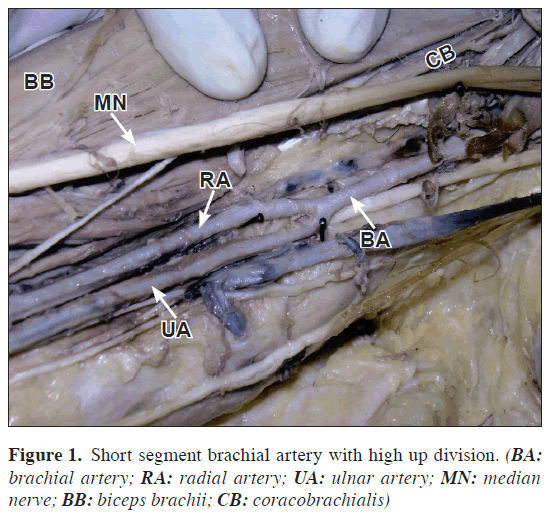
During routine dissection of a 40-year-old male cadaver in the Department of Anatomy, College of Medical Sciences, Bharatpur, Nepal, an unusually short segment brachial artery with high up division of brachial artery at the level of insertion of coracobrachialis in the middle of the right arm was observed. The unusually short-segment brachial artery was 11.5 cm in length and having slightly less caliber than usual. However it bifurcated normally into radial and ulnar arteries, both of same calibers. Further distribution of these two arteries was normal. Radial recurrent and common interosseous arteries arose normally from the radial and ulnar arteries, respectively. Profunda brachii and other branches of the brachial artery arose from it prior to its bifurcation. No other variation was found particularly in relation to the cords of the brachial plexus, their branches and their relations with axillary and brachial arteries. Dissection of the left upper limb revealed no unusual observations. Photographic documentation of the variation was made after thorough and meticulous dissection (Figure 1). Finally an attempt was made to explain the abnormality in the light of embryological development and to establish its clinical implications.
Discussion
Anomalies of the forelimb arterial tree are fairly common. This is mainly because of their multiple and plexiform sources, the temporal succession of emergence of principal arteries, anastomoses and periarticular networks and functional dominance followed by regression of some paths [2]. Several variations with regard to the termination of the brachial artery have been cited by many earlier researchers.
Occasionally the artery divides proximally into two trunks, which may reunite. Frequently it divides more proximally than usual, and this unusually short segment brachial artery may bifurcate as usual or it may trifurcate into radial, ulnar and common interosseous arteries. More often the radial branches arise proximally, leaving a common trunk for the ulnar and common interosseous; sometimes the ulnar artery arise proximally, the radial and common interosseous forming the other division; the common interosseous may also arise proximally [1].
Keen in 1961 mentioned high origin of radial artery from the brachial artery. He explained this variant on the basis of Arey’s observations regarding anomalous blood vessels [4,7]. In fact, Keen highlighted that there was persistence of the upper portion of the radial artery arising from the brachial artery proximal to the origin of ulnar artery followed by failure of development of the new connection of the radial artery with the brachial artery at the level of origin of ulnar artery [7].
Guha et al. observed high up division of brachial artery into radial and ulnar arteries in the middle of the arm associated with variant median nerve and absent musculocutaneous nerve [8].
The unusually short segment brachial artery with its high up division into radial and ulnar arteries as observed in the present study can be explained in the light of embryogenic development.
The early limb bud receives blood via intersegmental arteries, which contribute to a primitive capillary plexus. At the tip of the limb bud there is a terminal plexus that is constantly renewed in a distal direction as the limb grows. Later one main vessel supplies the limb and the terminal plexus; it is termed the axis artery.
The aforesaid terminal plexus at the tip of the limb bud is separated from the outer ectodermal sleeve of the limb by an avascular zone of mesenchyme. This avascular region contains an extracellular matrix consisting largely of hyaluronic acid. Removal of this hyaluronic acid by hyaluronidase results in vascularization of the tissue since partial degradation products of hyaluronic acid are angiogenic. Thus ectodermal-mesenchymal interactions and extracellular matrix components are controlling the initial patterning of blood vessels within the limb [3].
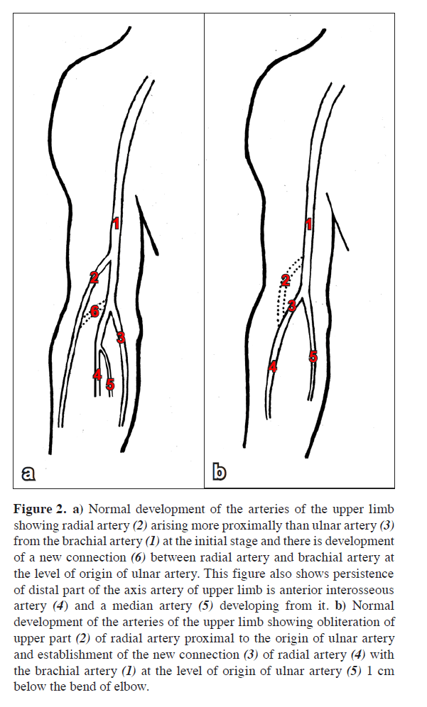
In the upper limb bud the axis artery is derived from the lateral branch of the seventh intersegmental artery (subclavian). The arterial trunk grows outwards along the ventral axial line and terminates in the deep plexus in the developing hand. Proximal part of the main trunk forms the axillary and brachial arteries and its distal part persists as the anterior interosseous artery and the deep palmar arch. The radial and ulnar arteries are the latest arteries to appear in the forearm from the axis artery (brachial). Initially the radial artery arises more proximally than the ulnar artery (Figure 2a). Later, it establishes a new connection with the main trunk at or near the level of origin of the ulnar artery and the upper portion of its original stem usually disappears to a large extent (Figure 2b).
Figure 2. a) Normal development of the arteries of the upper limb showing radial artery (2) arising more proximally than ulnar artery (3) from the brachial artery (1) at the initial stage and there is development of a new connection (6) between radial artery and brachial artery at the level of origin of ulnar artery. This figure also shows persistence of distal part of the axis artery of upper limb is anterior interosseous artery (4) and a median artery (5) developing from it. b) Normal development of the arteries of the upper limb showing obliteration of upper part (2) of radial artery proximal to the origin of ulnar artery and establishment of the new connection (3) of radial artery (4) with the brachial artery (1) at the level of origin of ulnar artery (5) 1 cm below the bend of elbow.
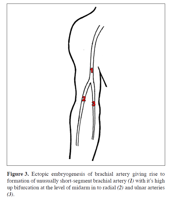
In the present case both the radial and ulnar arteries arose more proximally from the brachial artery leading to the formation of an unusually short segment and slightly narrow brachial artery which bifurcated into radial and ulnar arteries at a more proximal level near the middle of the arm (Figure 3). The profunda brachii artery took origin from the short segment brachial artery along with its other branches prior to its bifurcation. However, the subsequent distribution of the radial and ulnar arteries was normal.
It is pertinent to mention here that the normal vascular development including the patterning of the blood vessels is influenced greatly by local hemodynamic factors. Altered hemodynamic environment may give rise to variant patterning of blood vessels [5].
Campta highlighted diagnostic, interventional and surgical significance of such a variation. Diagnostically this type of variation may disturb the evaluation of angiographic images. Further knowledge of such variation has got clinical importance especially in the field of orthopedic, plastic and vascular surgeries [6].
Last but not the least, knowledge of this variation is important for the clinicians in day to day practice for measurement of blood pressure using sphygmomanometer cuff in the arm.
Conclusion
The short segment brachial artery and its variant termination in the form of high up bifurcation as noted in the present study are fairly common. The case can be explained in the light of embryological development. In addition, knowledge of such variation is important for carrying out surgical procedures in the arm.
References
- Johnson D, Ellis H, Collins P. Upper Arm. In: Standring S, Ellis H, Healy JC, Johnson D, Williams A, Collins P, eds. Gray’s Anatomy. 39th Ed. Philadelphia, Elsevier Churchill Livingstone. 2005; 856.
- Williams PL, Bannister LH, Berry MM, Collins P, Dyson M, Dussek JE, Ferguson MW, eds. Gray’s Anatomy. 38th Ed., London, Churchill Livingstone. 1999; 319, 1539.
- Feinberg RN. Vascular development in the embryonic limb bud. In: Feinberg RN, Sherer GK, Auerbach R, eds. The development of the vascular system. Basel, Karger (Issues Biomed). 1991; 14: 136–148.
- Arey LB. Developmental Anatomy. 6th Ed. Philadelphia, W.B. Saunders. 1957; 375–377.
- Rodriguez-Baeza A, Nebot J, Ferreira B, Reina F, Perez J, Sanudo JR, Roig M. An anatomical study and ontogenetic explanation of 23 cases with variations in the main pattern of the human brachio-antebrachial arteries. J Anat. 1995; 187: 473–479.
- Gonzalez-Compta X. Origin of the radial artery from the axillary artery and associated hand vascular anomalies. J Hand Surg Am. 1991; 16: 293–296.
- Keen JA. A study of the arterial variations in the limbs, with special reference to symmetry of vascular patterns. Am J Anat. 1961; 108: 245–261.
- Guha R, Palit S. A rare variation of anomalous median nerve with absent musculocutaneous nerve and high up division of brachial artery. J Interacad. 2005; 9: 398–403.
Namani Satyanarayana1*, P. Sunitha2, Munvar Miya Shaik3 and P Satya Vathi Devi4
1Departments of Anatomy, College of Medical Sciences, Bharatpur, Chitwan, Nepal
2Departments of Physiology, College of Medical Sciences, Bharatpur, Chitwan, Nepal
3Departments of Pharmacology, College of Medical Sciences, Bharatpur, Chitwan, Nepal
4Department of Anatomy, Prathima Institute of Medical Sciences Karimnagar, Andhra Pradesh, India
- *Corresponding Author:
- Dr. Namani Satyanarayana
Lecturer, Department of Anatomy, College of Medical Sciences, Bharatpur-23, Chitwan Dist., Nepal
Tel: +97 980 6809145
E-mail: satyam_n19@yahoo.com
Date of Received: August 22nd, 2009
Date of Accepted: February 10th, 2010
Published Online: April 23rd, 2010
© IJAV. 2010; 3: 56–58.
Abstract
Objectives: To document an unusually short segment brachial artery with its high up division into radial and ulnar arteries in the middle of the arm and to establish embryological and clinico-anatomical correlations of such variation. Methods: The finding was noted after thorough and meticulous dissection of the upper limbs of both sides (axilla, arm, cubital fossa, forearm and palm) of a 40-year-old adult male cadaver in the Department of Anatomy, College of Medical Sciences, Bharatpur, Nepal. Photographic documentation of the variation was also made. Results: An unusually short segment brachial artery was noted in the right arm. This short segment brachial artery bifurcated more proximally at the level of insertion of coracobrachialis in the middle of the right arm into radial and ulnar arteries both of same caliber. Further distribution of these two arteries was normal. No other variation was found particularly in relation to the cords of the brachial plexus and their branches. Dissection of the left upper limb revealed no unusual observations. Conclusion: The short segment brachial artery with high up bifurcation as noted in the present study is fairly common. The variation can be explained in the light of embryological development. In addition, knowledge of such variation is important for carrying out surgical procedures in the arm.
-Keywords
brachial artery, high-up division, radial artery, ulnar artery
Introduction
The brachial artery usually begins as a continuation of the axillary artery at the distal (inferior) border of the tendon of teres major and ends about a centimeter distal to the elbow joint (at the level of the neck of the radius) by dividing into radial and ulnar arteries [1].
Occasionally the artery divides proximally into two trunks, which may reunite. Frequently it divides more proximally than usual and this unusually short segment brachial artery may bifurcate as usual or it may trifurcate into radial, ulnar and common interosseous arteries [2]. Several other variations related to the termination of such a short segment brachial artery have been mentioned by some earlier workers.
Such variations can be explained on the basis of embryogenic development. According to Feinberg, ectodermal-mesenchymal interactions and extracellular matrix components within the developing limb bud are controlling the initial patterning of blood vessels [3].
Further, there is a view that some inductive factors from the limb mesenchyme cause the changes in the blood vessel pattern [2].
High up division of the brachial artery can also be explained on the basis of observations made by Arey in 1957 where he highlighted that, there may be persistence of vessels which normally obliterate and disappearance or failure of development of vessels which normally persist [4]. This reversal of the normal process of vascular significancedevelopment is largely due to altered local hemodynamic environment [5].
Finally, knowledge of such variations has got clinical importance especially in the field of orthopedic, vascular and plastic surgeries [6].
Case Report
During routine dissection of a 40-year-old male cadaver in the Department of Anatomy, College of Medical Sciences, Bharatpur, Nepal, an unusually short segment brachial artery with high up division of brachial artery at the level of insertion of coracobrachialis in the middle of the right arm was observed. The unusually short-segment brachial artery was 11.5 cm in length and having slightly less caliber than usual. However it bifurcated normally into radial and ulnar arteries, both of same calibers. Further distribution of these two arteries was normal. Radial recurrent and common interosseous arteries arose normally from the radial and ulnar arteries, respectively. Profunda brachii and other branches of the brachial artery arose from it prior to its bifurcation. No other variation was found particularly in relation to the cords of the brachial plexus, their branches and their relations with axillary and brachial arteries. Dissection of the left upper limb revealed no unusual observations. Photographic documentation of the variation was made after thorough and meticulous dissection (Figure 1). Finally an attempt was made to explain the abnormality in the light of embryological development and to establish its clinical implications.
Discussion
Anomalies of the forelimb arterial tree are fairly common. This is mainly because of their multiple and plexiform sources, the temporal succession of emergence of principal arteries, anastomoses and periarticular networks and functional dominance followed by regression of some paths [2]. Several variations with regard to the termination of the brachial artery have been cited by many earlier researchers.
Occasionally the artery divides proximally into two trunks, which may reunite. Frequently it divides more proximally than usual, and this unusually short segment brachial artery may bifurcate as usual or it may trifurcate into radial, ulnar and common interosseous arteries. More often the radial branches arise proximally, leaving a common trunk for the ulnar and common interosseous; sometimes the ulnar artery arise proximally, the radial and common interosseous forming the other division; the common interosseous may also arise proximally [1].
Keen in 1961 mentioned high origin of radial artery from the brachial artery. He explained this variant on the basis of Arey’s observations regarding anomalous blood vessels [4,7]. In fact, Keen highlighted that there was persistence of the upper portion of the radial artery arising from the brachial artery proximal to the origin of ulnar artery followed by failure of development of the new connection of the radial artery with the brachial artery at the level of origin of ulnar artery [7].
Guha et al. observed high up division of brachial artery into radial and ulnar arteries in the middle of the arm associated with variant median nerve and absent musculocutaneous nerve [8].
The unusually short segment brachial artery with its high up division into radial and ulnar arteries as observed in the present study can be explained in the light of embryogenic development.
The early limb bud receives blood via intersegmental arteries, which contribute to a primitive capillary plexus. At the tip of the limb bud there is a terminal plexus that is constantly renewed in a distal direction as the limb grows. Later one main vessel supplies the limb and the terminal plexus; it is termed the axis artery.
The aforesaid terminal plexus at the tip of the limb bud is separated from the outer ectodermal sleeve of the limb by an avascular zone of mesenchyme. This avascular region contains an extracellular matrix consisting largely of hyaluronic acid. Removal of this hyaluronic acid by hyaluronidase results in vascularization of the tissue since partial degradation products of hyaluronic acid are angiogenic. Thus ectodermal-mesenchymal interactions and extracellular matrix components are controlling the initial patterning of blood vessels within the limb [3].
In the upper limb bud the axis artery is derived from the lateral branch of the seventh intersegmental artery (subclavian). The arterial trunk grows outwards along the ventral axial line and terminates in the deep plexus in the developing hand. Proximal part of the main trunk forms the axillary and brachial arteries and its distal part persists as the anterior interosseous artery and the deep palmar arch. The radial and ulnar arteries are the latest arteries to appear in the forearm from the axis artery (brachial). Initially the radial artery arises more proximally than the ulnar artery (Figure 2a). Later, it establishes a new connection with the main trunk at or near the level of origin of the ulnar artery and the upper portion of its original stem usually disappears to a large extent (Figure 2b).
Figure 2. a) Normal development of the arteries of the upper limb showing radial artery (2) arising more proximally than ulnar artery (3) from the brachial artery (1) at the initial stage and there is development of a new connection (6) between radial artery and brachial artery at the level of origin of ulnar artery. This figure also shows persistence of distal part of the axis artery of upper limb is anterior interosseous artery (4) and a median artery (5) developing from it. b) Normal development of the arteries of the upper limb showing obliteration of upper part (2) of radial artery proximal to the origin of ulnar artery and establishment of the new connection (3) of radial artery (4) with the brachial artery (1) at the level of origin of ulnar artery (5) 1 cm below the bend of elbow.
In the present case both the radial and ulnar arteries arose more proximally from the brachial artery leading to the formation of an unusually short segment and slightly narrow brachial artery which bifurcated into radial and ulnar arteries at a more proximal level near the middle of the arm (Figure 3). The profunda brachii artery took origin from the short segment brachial artery along with its other branches prior to its bifurcation. However, the subsequent distribution of the radial and ulnar arteries was normal.
It is pertinent to mention here that the normal vascular development including the patterning of the blood vessels is influenced greatly by local hemodynamic factors. Altered hemodynamic environment may give rise to variant patterning of blood vessels [5].
Campta highlighted diagnostic, interventional and surgical significance of such a variation. Diagnostically this type of variation may disturb the evaluation of angiographic images. Further knowledge of such variation has got clinical importance especially in the field of orthopedic, plastic and vascular surgeries [6].
Last but not the least, knowledge of this variation is important for the clinicians in day to day practice for measurement of blood pressure using sphygmomanometer cuff in the arm.
Conclusion
The short segment brachial artery and its variant termination in the form of high up bifurcation as noted in the present study are fairly common. The case can be explained in the light of embryological development. In addition, knowledge of such variation is important for carrying out surgical procedures in the arm.
References
- Johnson D, Ellis H, Collins P. Upper Arm. In: Standring S, Ellis H, Healy JC, Johnson D, Williams A, Collins P, eds. Gray’s Anatomy. 39th Ed. Philadelphia, Elsevier Churchill Livingstone. 2005; 856.
- Williams PL, Bannister LH, Berry MM, Collins P, Dyson M, Dussek JE, Ferguson MW, eds. Gray’s Anatomy. 38th Ed., London, Churchill Livingstone. 1999; 319, 1539.
- Feinberg RN. Vascular development in the embryonic limb bud. In: Feinberg RN, Sherer GK, Auerbach R, eds. The development of the vascular system. Basel, Karger (Issues Biomed). 1991; 14: 136–148.
- Arey LB. Developmental Anatomy. 6th Ed. Philadelphia, W.B. Saunders. 1957; 375–377.
- Rodriguez-Baeza A, Nebot J, Ferreira B, Reina F, Perez J, Sanudo JR, Roig M. An anatomical study and ontogenetic explanation of 23 cases with variations in the main pattern of the human brachio-antebrachial arteries. J Anat. 1995; 187: 473–479.
- Gonzalez-Compta X. Origin of the radial artery from the axillary artery and associated hand vascular anomalies. J Hand Surg Am. 1991; 16: 293–296.
- Keen JA. A study of the arterial variations in the limbs, with special reference to symmetry of vascular patterns. Am J Anat. 1961; 108: 245–261.
- Guha R, Palit S. A rare variation of anomalous median nerve with absent musculocutaneous nerve and high up division of brachial artery. J Interacad. 2005; 9: 398–403.


