Case of an Ectopic Papillary Thyroid Carcinoma at the base of tongue
Received: 23-Apr-2018 Accepted Date: Apr 28, 2018; Published: 03-May-2018
Citation: Fang LLP. Case of an ectopic papillary thyroid carcinoma at the base of tongue. Surg Case Rep. 2018;2(1):14-15
This open-access article is distributed under the terms of the Creative Commons Attribution Non-Commercial License (CC BY-NC) (http://creativecommons.org/licenses/by-nc/4.0/), which permits reuse, distribution and reproduction of the article, provided that the original work is properly cited and the reuse is restricted to noncommercial purposes. For commercial reuse, contact reprints@pulsus.com
Abstract
Ectopic thyroid tissue is reported in 7-10% of adults common in females, especially in populations of Asian origin. Previous reports showed that ectopic thyroid tissue may present metastasis from thyroid carcinoma, and very rarely it may harbor a primary thyroid carcinoma. My patient is a young female patient who presented with submental and left neck swelling and on clinical examination showed that was a mass at the base of tongue and subsequent histopathological and CT scan finding showed that she had ectopic papillary thyroid carcinoma with metastasis to the cervical lymph node, in the absence of orthotopic thyroid. management of this disease.
Keywords
Orthotopic thyroid; Cervical Metastasis; Endocrine glands; Epithelial cells; Hypothyroidism; Therapy
Thyroid gland is the first body’s endocrine glands to develop, approximately on the 24th day of gestation. It formed from proliferation of endodermal epithelial cells, starts as a simple midline thickening and develops to form the thyroid diverticulum on the median surface of the developing pharyngeal floor. After the initial descent, the thyroid is still connected to the tongue via a tract, known later as thyroglossal duct, while the proximal segment retracts and subsequently obliterates entirely, leaving only the pit, known as foramen cecum at the posterior aspect of the tongue. A number of anomalies may occur during their migration. Here is a case of young female patient being diagnosed with Ectopic Papillary thyroid Carcinoma in which the ectopic tissue was located along the base of tongue to the left base of neck and subsequently treated [1,2].
Case Report
A 31-year-old lady, premorbid nil, presented with submental swelling for 6 months duration, which increasing in size and painless. She has no skin changes, no discharges noted and no fever. No symptoms of hyperthyroidism or hypothyroidism. No compressive symptoms and tolerating orally well.
On examination, there was a swelling felt at the submental region, with size of 2 × 2 cm and non-tender. On the left side of her neck, there was another swelling felt, which 3 × 3 cm in size, firm and non-tender. Scope finding showed there was a swelling at the base of tongue. Bilateral Fossa of Rossemuller, arythenoid, pyriform fossa, epiglottis and vellecula were clear.
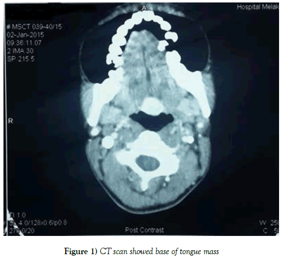
Subsequently fine neddle aspiration and cytology was carried out for the neck swelling and CT scan done. Histopathological findings were features keeping with metastatic papillary thyroid carcinoma. While the CT scan (Figure 1) showed quadruple ectopic thyroid, 3 superior lesions were in the midline along the base of tongue to hyoid bone and the distal most fourth lesion is on the left side of the base of neck, without normal located thyroid gland. Thyroid scan done showed 3 abnormal increased tracer uptakes seen at lingual region and thyroglossal region.
A Sistrunk procedure, excision of the left ectopic thyroid gland, excision of the right neck mass found intraoperatively (which later the HPE showed metastatic papillary carcinoma in the lymph node) and laser ablation over the base of tongue. Pre-operation her serum thyroglobulin level was >300ug/L. Radioactive isotope therapy was given twice since post-op. Serum thyroglobulin was reducing in trend. She was on L. thyroxine 100mcg daily.
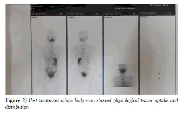
On subsequent follow up, 1 year since she was diagnosed with ectopic papillary thyroid carcinoma with cervical metastasis, she is currently well, remain asymptomatic. Latest diagnostic whole body scan (Figure 2) done showed physiological tracer uptake and distribution.
Discussion
Ectopic thyroid tissue is defined as thyroid tissue not located anterolaterally to the second to fourth tracheal cartilages. It has a reported prevalence of approximately 1 in 10,000 [3]. Lingual thyroid is the most common form of thyroid ectopy, accounting for 90% of the reported cases [4]. Other rarity include includes the mediastinum, esophagus, lung, heart, aorta and abdomen [4].
The thyroid gland formed from proliferation of endodermal epithelial cells, starts as a simple midline thickening and develops to form the thyroid diverticulum on the median surface of the developing pharyngeal floor, between the first and second pharyngeal pouches. This invagination and descends toward the anterior neck follow the primitive heart and passes the developing hyoid bone to reach it location in the anterior neck. The gland initially remains attached to the foramen cecum by the thyroglossal duct, which then begins to atrophy in the seventh week intrauterine. Failure or disturbance in the process of descent and the incomplete obliteration of its vertical tract lead to ectopic thyroid development.
Ectopic thyroid tissue can be subject to the same pathological processes as normal located thyroid tissue such as inflammation, hyperplasia, and tumorigenesis. Generally, malignant transformation in ectopic thyroid is rare [5]. Literature review appearing in the Journal of Clinical Endocrinology and Metabolism attempted to estimate the prevalence of such cases, ultimately finding just 400 reported cases of ectopic thyroid cancer. However, ectopic thyroid tissue alone, without any cancerous growth, is found in about one in 10,000 people.
Primary carcinoma in ectopic thyroid has been described in cases where an orthotopic thyroid exists. It can also arise in the absence of orthotopic thyroid, such as in our patient. Most tumors are papillary carcinomas. However, follicular, mixed follicular, papillary Hürthle cell, medullary carcinomas and anaplastic carcinoma have also been described. In the majority of reported cases of lingual thyroid carcinoma, the carcinoma did not extend beyond the tongue. Regional lymph node involvement has been documented in 21% of patients.
Management strategies, including performance of removal of the primary lesion, neck dissection, and treatment with radioiodine, should be based on individualized risk stratification. In the vast majority of reported cases, the tumors have been excised via the transoral route because this technique is both simpler and more cost. Treatment with 131-I alone is also an option for patients who are poor surgical candidates or who decline surgical intervention. All patients should receive levothyroxine therapy to suppress secretion of TSH.
Conclusion
Thyroid cancer arising from ectopic thyroid tissue in the neck occurs rarely and may represent either a de novo or metastatic process. Treatment of ectopic cervical thyroid cancer is based predominantly on the surgical excision of the malignant lesion and radioiodine therapy, as in our patient. Currently she is doing well, under proper observation and follow-up.
Conflict of Interest
None.
Patient Consent
None.
REFERENCES
- Sauk JJ, Ectopic lingual thyroid. J Pathol 102:239-245.
- Gopal RA, Acharya SV, Bandgar T, et al. Clinical profile of ectopic thyroid in Asian Indians: A single-center experience. Endocrine Practice 2009;15:322–325.
- Noyek AM, Friedberg J. Thyroglossal duct and ectopic thyroid disorders. Otolaryngol Clin North Am 14:187–201.
- Choi JY, Kim JH. A case of an ectopic thyroid gland at the lateral neck masquerading as a metastatic papillary thyroid carcinoma. J Korean Med Sci 2008;23(3):548-50.
- Shah BC, Ravichand CS, Juluri S, et al. Ectopic thyroid cancer. Ann Thorac Cardiovasc Surg 2007;13(2):122-4.


