Cerebral polymicrogyria: case report
Lazar Jelev1*, Alexandar Alexandrov2, Stanislav Hristov2 and Wladimir Ovtscharoff1
1Departments of Anatomy, Histology and Embryology, Medical University of Sofia, Sofia, Bulgaria
2Departments of Forensic Medicine and Deontology, Medical University of Sofia, Sofia, Bulgaria
- *Corresponding Author:
- Lazar Jelev, MD, PhD
Associate Professor, Department of Anatomy, Histology and Embryology Medical University of Sofia blvd. Sv. Georgi Sofiisky 1 BG-1431 Sofia, Bulgaria
Tel: +359 (2) 9172636
E-mail: ljelev@abv.bg
Date of Received: March 23rd, 2012
Date of Accepted: October 21st, 2012
Published Online: February 15th, 2013
© Int J Anat Var (IJAV). 2013; 6: 49–50.
[ft_below_content] =>Keywords
cerebral cortex, developmental anomaly, polymicrogyria, epilepsy
Introduction
Disruption of the major steps of the cerebral cortical development – cell proliferation/apoptosis, neuronal migration and cortical organization, result in a group of disorders called malformations of cortical development [1–4]. They are frequently associated with the presence of grossly unusual cortical gyri. With the improvement of modern imaging techniques this malformations have been increasingly recognized as a major cause of seizure disorders [3]. We herewith describe such a rare autopsy case.
Case Report
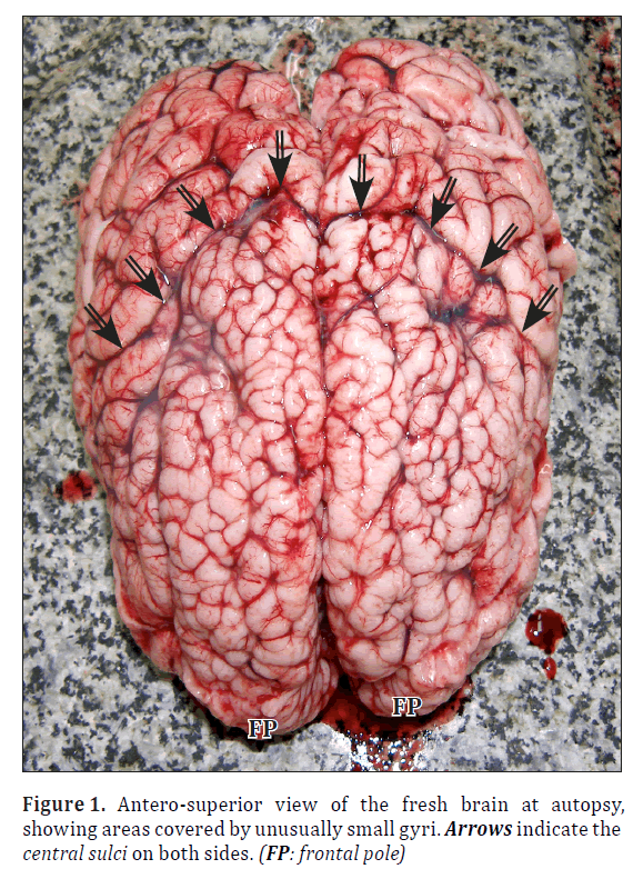
During routine forensic autopsy of the body of a 2-year-old boy, who died in status epilepticus, an unusual cortical formation was observed. On the superolateral surface of both hemispheres the size and location of the gyri differed considerably. In the anterior part of the cerebral area that includes the both left and right frontal lobes, there were numerous unusually small gyri. Careful examination of the brain surface, however, revealed that not the whole frontal lobes were affected, but the unusual gyri actually covered some regions corresponding to the normal superior and middle frontal gyri. On both sides the Sylvian sulcus was well developed. The central sulcus was well seen as a border between the unusual and normal cerebral surface. The gyri of the parietal and occipital lobes behind the central sulcus were relatively of normal size. The postcentral sulcus was better expressed than the precentral sulcus, which was possible to be recognized only in its lower part. The other parts of the cerebral cortex seemed normally developed.
Discussion
Based on the supposed embryonic developmental stage at witch the disruption is purported to cause the anomaly, the malformations of cortical development can be classified into three basic groups [2]. Group I (abnormal neuronal and glial proliferation or apoptosis) comprises microcephalies, megalencephalies, cortical dysgenesis with abnormal cell proliferation. Group II (abnormal neuronal migration) includes heterotopia, lissencephaly, subcortical heterotopia and sublobar dysplasia, cobblestone malformations. Group III (abnormal postmigrational development) includes polymicrogyria and schizencephaly, focal cortical dysplasia and postmigrational microcephaly.
The reported autopsy findings are consistent with a case of cerebral polymicrogyria. According to Friede the term “polymicrogyria” defines an excessive number of abnormally small gyri leading to an irregular cortical surface [5]. This condition can be localized to a single gyrus, may involve a part of the hemisphere, may be bilateral and asymmetrical, bilateral and symmetrical (as in our case) or diffuse throughout the cerebral cortex. According to the lobar topography established, various polymicrogyria syndromes have been described: bilateral perisylvian [6], bilateral parasagittal parietooccipital [7], bilateral frontal [8], bilateral frontoparietal [9] and unilateral perisylvian or multilobar polymicrogyria [10]. The main type is bilateral perisylvian polymicrogyria clinically characterized by a combination of pseudobulbar palsy, spastic tetraparesis, learning difficulties and epilepsy [1].
Polymicrogyria is associated with a wide number of patterns and syndromes and with mutation of several genes [2,4]. Its pathogenesis is still unknown. The brain pathology comprises at least two major histological types [1]: the most common four-layered cortex with simplified organization and the unlayered cortex. Because the most changes occur in the middle cerebral artery territory, probably in some cases hypoxic-ischemic insult may be related to the genesis of polymicrogyria. A congenital cytomegalovirus infection may also have an important role [1].
The described here case of frontal bilateral polymicrogyria is undoubtedly connected with the status epilepticus [8].
References
- Andrade CS, Leite Cda C. Malformations of cortical development: current concepts and advanced neuroimaging review. Arq Neuropsiquiatr. 2011; 69: 130–138.
- Barkovich AJ, Guerrini R, Kuzniecky RI, Jackson GD, Dobyns WB. A developmental and genetic classification for malformations of cortical development: update 2012. Brain. 2012; 135: 1348–1369.
- Colombo N, Salamon N, Raybaud C, Ozkara C, Barkovich AJ. Imaging of malformations of cortical development. Epileptic Disord. 2009; 11: 194–205.
- Guerrini R, Parrini E. Neuronal migration disorders. Neurobiol Dis. 2010; 38: 154–166.
- Friede RL. Developmental neuropathology. New York, Springer-Verlag. 1989; 577.
- Kuzniecky R, Andermann F, Guerrini R. Congenital bilateral perisylvian syndrome: study of 31 patients. The CBPS Multicenter Collaborative Study. Lancet. 1993; 341: 608–612.
- Guerrini R, Dubeau F, Dulac O, Barkovich AJ, Kuzniecky R, Fett C, Jones-Gotman M, Canapicchi R, Cross H, Fish D, Bonanni P, Jambaqué I, Andermann F. Bilateral parasagittal parietooccipital polymicrogyria and epilepsy. Ann Neurol. 1997; 41: 65–73.
- Guerrini R, Barkovich AJ, Sztriha L, Dobyns WB. Bilateral frontal polymicrogyria: a newly recognized brain malformation syndrome. Neurology. 2000; 54: 909–913.
- Piao X, Hill RS, Bodell A, Chang BS, Basel-Vanagaite L, Straussberg R, Dobyns WB, Qasrawi B, Winter RM, Innes AM, Voit T, Ross ME, Michaud JL, Déscarie JC, Barkovich AJ, Walsh CA. G protein-coupled receptor-dependent development of human frontal cortex. Science. 2004; 303: 2033–2036.
- Guerrini R, Genton P, Bureau M, Parmeggiani A, Salas-Puig X, Santucci M, Bonanni P, Ambrosetto G, Dravet C. Multilobar polymicrogyria, intractable drop attack seizures, and sleep-related electrical status epilepticus. Neurology. 1998; 51: 504–512.
Lazar Jelev1*, Alexandar Alexandrov2, Stanislav Hristov2 and Wladimir Ovtscharoff1
1Departments of Anatomy, Histology and Embryology, Medical University of Sofia, Sofia, Bulgaria
2Departments of Forensic Medicine and Deontology, Medical University of Sofia, Sofia, Bulgaria
- *Corresponding Author:
- Lazar Jelev, MD, PhD
Associate Professor, Department of Anatomy, Histology and Embryology Medical University of Sofia blvd. Sv. Georgi Sofiisky 1 BG-1431 Sofia, Bulgaria
Tel: +359 (2) 9172636
E-mail: ljelev@abv.bg
Date of Received: March 23rd, 2012
Date of Accepted: October 21st, 2012
Published Online: February 15th, 2013
© Int J Anat Var (IJAV). 2013; 6: 49–50.
Abstract
We describe a rare case of unusual development of the cerebral cortex in a 2-year-old boy, who died in status epilepticus. The brain regions, corresponding to the normal superior and middle frontal gyri, were covered with numerous unusually small gyri, a condition consisting with a case of polymicrogyria.
-Keywords
cerebral cortex, developmental anomaly, polymicrogyria, epilepsy
Introduction
Disruption of the major steps of the cerebral cortical development – cell proliferation/apoptosis, neuronal migration and cortical organization, result in a group of disorders called malformations of cortical development [1–4]. They are frequently associated with the presence of grossly unusual cortical gyri. With the improvement of modern imaging techniques this malformations have been increasingly recognized as a major cause of seizure disorders [3]. We herewith describe such a rare autopsy case.
Case Report
During routine forensic autopsy of the body of a 2-year-old boy, who died in status epilepticus, an unusual cortical formation was observed. On the superolateral surface of both hemispheres the size and location of the gyri differed considerably. In the anterior part of the cerebral area that includes the both left and right frontal lobes, there were numerous unusually small gyri. Careful examination of the brain surface, however, revealed that not the whole frontal lobes were affected, but the unusual gyri actually covered some regions corresponding to the normal superior and middle frontal gyri. On both sides the Sylvian sulcus was well developed. The central sulcus was well seen as a border between the unusual and normal cerebral surface. The gyri of the parietal and occipital lobes behind the central sulcus were relatively of normal size. The postcentral sulcus was better expressed than the precentral sulcus, which was possible to be recognized only in its lower part. The other parts of the cerebral cortex seemed normally developed.
Discussion
Based on the supposed embryonic developmental stage at witch the disruption is purported to cause the anomaly, the malformations of cortical development can be classified into three basic groups [2]. Group I (abnormal neuronal and glial proliferation or apoptosis) comprises microcephalies, megalencephalies, cortical dysgenesis with abnormal cell proliferation. Group II (abnormal neuronal migration) includes heterotopia, lissencephaly, subcortical heterotopia and sublobar dysplasia, cobblestone malformations. Group III (abnormal postmigrational development) includes polymicrogyria and schizencephaly, focal cortical dysplasia and postmigrational microcephaly.
Figure 1: Antero-superior view of the fresh brain at autopsy, showing areas covered by unusually small gyri. Arrows indicate the central sulci on both sides. (FP: frontal pole)
The reported autopsy findings are consistent with a case of cerebral polymicrogyria. According to Friede the term “polymicrogyria” defines an excessive number of abnormally small gyri leading to an irregular cortical surface [5]. This condition can be localized to a single gyrus, may involve a part of the hemisphere, may be bilateral and asymmetrical, bilateral and symmetrical (as in our case) or diffuse throughout the cerebral cortex. According to the lobar topography established, various polymicrogyria syndromes have been described: bilateral perisylvian [6], bilateral parasagittal parietooccipital [7], bilateral frontal [8], bilateral frontoparietal [9] and unilateral perisylvian or multilobar polymicrogyria [10]. The main type is bilateral perisylvian polymicrogyria clinically characterized by a combination of pseudobulbar palsy, spastic tetraparesis, learning difficulties and epilepsy [1].
Polymicrogyria is associated with a wide number of patterns and syndromes and with mutation of several genes [2,4]. Its pathogenesis is still unknown. The brain pathology comprises at least two major histological types [1]: the most common four-layered cortex with simplified organization and the unlayered cortex. Because the most changes occur in the middle cerebral artery territory, probably in some cases hypoxic-ischemic insult may be related to the genesis of polymicrogyria. A congenital cytomegalovirus infection may also have an important role [1].
The described here case of frontal bilateral polymicrogyria is undoubtedly connected with the status epilepticus [8].
References
- Andrade CS, Leite Cda C. Malformations of cortical development: current concepts and advanced neuroimaging review. Arq Neuropsiquiatr. 2011; 69: 130–138.
- Barkovich AJ, Guerrini R, Kuzniecky RI, Jackson GD, Dobyns WB. A developmental and genetic classification for malformations of cortical development: update 2012. Brain. 2012; 135: 1348–1369.
- Colombo N, Salamon N, Raybaud C, Ozkara C, Barkovich AJ. Imaging of malformations of cortical development. Epileptic Disord. 2009; 11: 194–205.
- Guerrini R, Parrini E. Neuronal migration disorders. Neurobiol Dis. 2010; 38: 154–166.
- Friede RL. Developmental neuropathology. New York, Springer-Verlag. 1989; 577.
- Kuzniecky R, Andermann F, Guerrini R. Congenital bilateral perisylvian syndrome: study of 31 patients. The CBPS Multicenter Collaborative Study. Lancet. 1993; 341: 608–612.
- Guerrini R, Dubeau F, Dulac O, Barkovich AJ, Kuzniecky R, Fett C, Jones-Gotman M, Canapicchi R, Cross H, Fish D, Bonanni P, Jambaqué I, Andermann F. Bilateral parasagittal parietooccipital polymicrogyria and epilepsy. Ann Neurol. 1997; 41: 65–73.
- Guerrini R, Barkovich AJ, Sztriha L, Dobyns WB. Bilateral frontal polymicrogyria: a newly recognized brain malformation syndrome. Neurology. 2000; 54: 909–913.
- Piao X, Hill RS, Bodell A, Chang BS, Basel-Vanagaite L, Straussberg R, Dobyns WB, Qasrawi B, Winter RM, Innes AM, Voit T, Ross ME, Michaud JL, Déscarie JC, Barkovich AJ, Walsh CA. G protein-coupled receptor-dependent development of human frontal cortex. Science. 2004; 303: 2033–2036.
- Guerrini R, Genton P, Bureau M, Parmeggiani A, Salas-Puig X, Santucci M, Bonanni P, Ambrosetto G, Dravet C. Multilobar polymicrogyria, intractable drop attack seizures, and sleep-related electrical status epilepticus. Neurology. 1998; 51: 504–512.


