Clinical Evaluation of a New Device to Measure Maximum Bite Force
#Equally contribution
Received: 28-Nov-2017 Accepted Date: Mar 22, 2018; Published: 03-Apr-2018
Citation: Amid R, Ebrahimi N, Kadkhodazadeh M, et al. New Device to Measurement of Maximum Bite Force. J Dent Oral Res 2018;2(1):4-7.
This open-access article is distributed under the terms of the Creative Commons Attribution Non-Commercial License (CC BY-NC) (http://creativecommons.org/licenses/by-nc/4.0/), which permits reuse, distribution and reproduction of the article, provided that the original work is properly cited and the reuse is restricted to noncommercial purposes. For commercial reuse, contact reprints@pulsus.com
Abstract
Background: Measurement of masticatory force has long been used to understand the biomechanics of chewing and assess the therapeutic effects of prostheses. This study aimed to design, fabricate and use a simple, portable and efficient electric circuit to reliably and reproducibly measure the masticatory load. Also, the results obtained by using this device were compared in a small population.
Methods: Using a standard pressure-sensitive sensor, an electrical circuit was designed and fabricated. After ensuring the function of device with minimum error, the masticatory loads of 100 subjects selected via non-random sampling, were measured and recorded at the right and left posterior regions, anterior regions of both jaws and the right and left quadrants of the maxilla and mandible. Data were analyzed using SPSS 16 and analytical and descriptive statistical tests.
Results: Significant correlations were found between 1. Males and females in terms of the mean masticatory force 2. Bilateral masticatory force and unilateral masticatory force (right and left) and 3. Masticatory load at the posterior regions and the masticatory force at the anterior regions of both jaws Comparison of the masticatory force among different groups in terms of craniofacial morphology revealed no significant differences.
Conclusion: The designed device had adequate accuracy. It has unique properties such as an external sensor, which is replaceable and can be disinfected and also a digital port. It has the ability to measure maximum and sub-maximum masticatory loads and the possibility to depict them in a graph.
Keywords
Dental occlusion; Mastication; Bite force
Introduction
The magnitude of masticatory force is an indicator of functional status of the masticatory system. The effect of chewing efficiency and bite force on oral health-related quality was evaluated in the field of geriatric and implant dentistry [1,2]. Measuring the masticatory force of individuals has been widely used to understand the biomechanical principles of masticatory muscles and outcomes of prosthodontics treatments [3]. Also, the masticatory force is very important for diagnosis and treatment of dysfunction and behavior of the stomatognathic system [4].
Masticatory force can be measured by two methods:
1. Directly, by placing a suitable load transfer tool in-between the two dental arches and
2. Indirectly, using physiological variables related to the masticatory force. Borelli in an experimental study in 1681 designed a gnathodynamometer for measuring masticatory forces for the first time. Since then, many scientists have investigated this topic and lever-manometer-springlever and micrometer devices have been designed for this purpose [5,6]. The strain-gage is the most commonly used recording device for masticatory forces [7,8]. This sensor is capable of measuring forces that bend it within the sensor’s range of flexibility from 446 to 1221 N [9]. Dental prescale is another measuring device made of a pressure sensitive foil (PSF) in the form of a horseshoe and a computerized monitoring system for data analysis. Supersensitive electronic devices are now used for this purpose. They are accurate enough to measure conventional forces. Previous studies have evaluated the effects of several factors such as craniofacial morphology [10-12], gender weight and height and pattern of occlusal contacts on masticatory loads. Gender differences are the most effective on masticatory loads. According to most researchers, masticatory forces are higher in males compared to females. This difference is probably due to the difference in muscle strength or size of teeth in males and females [13]. However, in the study by Abu Alhaija et al, in 2010 no difference was reported in the masticatory forces between males and females.
This study aimed to design and manufacture a simple, low-cost, applicable device with acceptable precision and quality for measurement of masticatory loads. Considering the lack of an applicable device for direct measurement of masticatory loads and also absence of any study on direct measurement of masticatory forces in our country,we designed and used adevice on a small Iranian population to record the mean value of masticatory load and analytically assess the correlation of masticatory load with physiological conditions.
Methods
The Flexiforcesensor A401 (Tekscan Inc., South Boston, USA) was used in this study. The dimensions of this sensor are shown in Table 1. This sensor is capable of measuring all types of loads and thus, it is considered as a strain-gage (for measuring sensor flexural loads) and also as a load cell (for measuring vertical loads applied to the sensor). Thus, it can absorb all types of loads applied from the teeth to sensor during mastication and displays interactions quantitatively in Newton (N) using a designed circuit (Table 1).
| Thickness | 0.208 mm (0.008 in.) |
|---|---|
| Length | 56.8 mm (2.24 in.) |
| Width | 31.8 mm (1.25 in.) |
| Sensing Area | 25.4 mm (1in.) |
| Pin Spacing | 2.54 mm (0.1 in.) |
Table 1 Dimensions of the sensor used in the designed device
Several methods are available to attach the Flexiforce sensor to the circuit. One way is to attach it to a load-voltage circuit according to the manufacturer’s instructions. If shear forces are required to be applied or the sensor needs to be placed on sharp edges, it must be covered with a flexible coat to prolong its service life. For this purpose, a flexible, compressible plastic shield with 1.5 mm thickness was used on both sides of the sensor. Thus, the device tip can be replaced or disinfected whenever required. Figure 1 shows the device components.
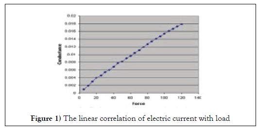
One important property of this device is its calibration ability. Calibration was done to signify the output as the measurement unit of our choice (N). For sensor calibration, the following steps were followed according to the manufacturer’s instructions: A specific mass was weighed using iBalance 500 (My Weigh Inc.,) digital scale. The respective mass was then placed on the sensor of the designed device in such way that its entire weight was applied to the sensor. The displayed output number was recorded. This process was repeated with other masses of different weights within the measuring limits of the sensor (at least 5 masses of different weights). Due to the linear function of the sensor, a suitable ratio was determined between the input and output loads of the device. By programming this ratio on a programmable IC, the figure displayed on the LCD of the device would be the equivalent of the applied load in N. Also, the diagram of the applied load can be plotted on the LCD to record the amount of load at each time point. The accuracy of the linear function of the sensor is shown in Diagram 1.
The level of reproducibility of the device was evaluated by using test-retest reliability that is measured of reliability obtained by administrating the same test over a period of time to a group of individuals. We calculated data from 10 persons repeated in 2 weeks. The level of agreement was 0.83 and p value for paired-T test was 0.75 that can be considered almost perfect.
To compare the masticatory forces between males and females, considering the mean difference of masticatory force between them reported by Koc et al, in 2011 [14] N1-N2=35.6-28.2=(7.4), the standard deviations (SD) of the two groups (S1=11.9, S2=10.6), power of 90% and 95% confidence interval (CI), the sample size was calculated to be 50 subjects in each group. A total of 100 subjects were selected among patients presenting to Shahid Beheshti School of Dentistry using non-random convenience sampling. These subjects had the following inclusion criteria: no history of a systemic disease, no specific asymmetry, no history of maxillofacial trauma or surgery, no temporomandibular joint (TMJ) problem, no history of bruxism, tooth mobility or orthodontic therapy, no anterior or posterior crossbite, permanent dentition and vital first molars without occlusal restorations or crowns. At the tested areas, patients did not have any removable denture or implant-supported prosthesis (fixed or removable). The mentioned criteria were ensured using a questionnaire filled by the subjects and also direct observation for evident confounders.
Method and objectives of the study were thoroughly explained to all participants and written informed consent was obtained. Age, gender, the dominant hand, facial height and Angle’s class of occlusion were recorded [14,15]. With the patient in a seated position and occlusal plane parallel to the horizontal plane, the sensor was placed in the patient’s mouth in-between the following teeth in an orderly fashion: 1. Occlusal surfaces of the maxillary right first molar and mandibular right first molar; 2. Occlusal surfaces of the maxillary left first molar and mandibular left first molar; 3. Occlusal surfaces of the maxillary and mandibular right and left first molars (by placing two sensors at both sides simultaneously); 4.Incisal edges of the maxillary and mandibular anterior teeth. The patients were asked to press their teeth in maximum intercuspation. By doing so, the maximum masticatory force in right and left molars, simultaneously at both sides and also in the anterior segment was measured and recorded. After every 10 measurements, the functions of the device and sensors were tested using a mass with a specific weight and they were exchanged if defective. The manufacturer claims that the sensor is capable of tolerating one million cycles of load measurement; however, high loads and non-standard conditions decrease its functional life. Eventually, 10 subjects were randomly selected and their masticatory forces were measured by the second examiner. Considering the high agreement between the test results, these results were generalized to the entire understudy population.
Data were analyzed using SPSS [16]. Descriptive statistics including the mean and SD values were used for reporting the data; and analytical statistics namely independent t-test, ANOVA and paired t-test were applied to assess the factors affecting the masticatory forces.
Results
This study was performed on 50 males and 50 females. The females had a mean age of 38.6 ± 11.4 years (range 20-58 years). The males had a mean age of 39.6 ± 11.8 years (range 22-59 years). The mean force measured at the right side was 636.5N in males and 484.8N in females. These values for the left side were 625.5N in males and 480.4N in females. The maximum masticatory forces in males and females were compared using t-test; which revealed a significant difference in this regard (P=0.000).
Table 2 shows the mean maximum masticatory force measured by the device in different areas of the mouth and in subjects with different craniofacial morphologies (normal, short, long).
| Mean maximum masticatory force (N) | |||||
|---|---|---|---|---|---|
| Areas of measurement of maximum masticatory force | Anterior segment | 268.64 | |||
| Left posterior | 555.17 | ||||
| Right posterior | 560.69 | ||||
| Bilateral posterior | 570.99 | ||||
| Anterior segment | Left posterior | Right posterior | Bilateral posterior | ||
| Craniofacial morphology | Long face | 271 | 577 | 564 | 575 |
| Normal | 270 | 557 | 563 | 569 | |
| Short face | 280 | 554 | 561 | 579 | |
Table 2 The maximum mean masticatory force measured by the device in different areas of the mouth and in subjects with different craniofacial morphologies.
Comparison of the masticatory forces among subjects in different craniofacial groups using ANOVA revealed that the forces at the mentioned areas were not significantly different among the three groups (P=0.630 for the anterior segment, P=0.192 for the right side, P=0.150 for the left side and P=0.189 for the bilateral posterior areas). Analysis of data with paired t-test showed that the bilateral masticatory force was significantly higher than the unilateral force at the right or left side (P=0.00).
Discussion
Considering the difference in tools for measuring masticatory forces in previous studies and absence of a gold standard for this purpose, this study aimed to design, manufacture and use a simple, portable and efficient electric circuit to reliably and reproducibly measure the masticatory force. Also, we compared the results in a small population.
The obtained results revealed that males had a significantly higher masticatory force than females, which confirms previous findings in this regard [17]. However, Abu Alhaija et al, in 2010 found no significant difference in maximum masticatory forces of men and women. It appears that the effect of gender on the difference in the masticatory forces is attributed to the higher muscular strength in men [18]; which per se is due to anatomical differences between the two sexes [19].
The more posterior the position of the converter in the mouth, the higher the masticatory force [20]. Our results showed that the masticatory force in the posterior areas was significantly higher than that in the anterior segment; which is in line with the results of previous studies [21]. Also, the bilateral masticatory force was significantly higher than the unilateral force, which is also in agreement with previous findings in this respect [22-25].
In the current study, no significant difference was noted in the load generated by the jaws and muscles among the three groups of normal, long and short facial height. Based on the results, the mean difference in loads measured in the posterior areas of both jaws in the three above mentioned groups was much higher than the mean load in the anterior regions of the jaws. The difference in jaw force in the posterior areas between the long face and short face individuals was greater than the difference in load in the anterior segments between the two mentioned groups. Pereira et al, in 2007 showed a reverse correlation between the masticatory force and angle of the mandible. Other studies have shown that long face individuals have lower masticatory force [26-28]. Researchers have stated that the masseter is thicker in short face compared to normal or long face individuals. It appears that short face individuals apply higher masticatory forces.
Significant difference in maximum masticatory load depends on several factors relating to the physiological and anatomical characteristics. Aside from these, the accuracy of the masticatory force is influenced by physiological characteristics of the load measuring device. Shinogaya et al, in 2000-2001 compared the occlusal force measured by a pressure sensitive film (PSF) and that measured by the conventional unilateral strain-gauge and reported that the maximum masticatory force recorded by the strain gauge was at 6-7 mm open bite on the mandibular models while in PSF with a thin foil diameter (0.97 mm), maximum masticatory force was recorded in maximum intercuspation. They stated that masticatory force measurement systems using a thin, pressure-sensitive film (0.1 mm thickness) are superior to those using a strain-gauge because in the former systems the masticatory force may be measured in a position close to intercuspation; which better simulates the clinical setting. On the other hand, these systems allow simultaneous study of load distribution in all areas of the dentition system. Tortopidis et al, in 1999 used acrylic appliances next to the metal surface of the strain gauge converters to minimize the risk of tooth fracture when applying maximum pressure to the converter [29]. Moreover, when the patient bites on the hard metal surface of the converter, some unusual movements are generated due to the neuromuscular reactions preventing the application of maximum force. Acrylic splints provide a softer surface and allow the generation of maximum masticatory force. In the current study, a sensor with 3.2mm thickness was used (0.2 mm original thickness of the sensor equal to the thickness of a paper sheet a plastic shield added for protection and infection control). This sensor was highly flexible and thus, there was no need to use an acrylic splint. On the other hand, due to the insignificant thickness of sensor, we may state that the measurements were close to the range of maximum intercuspation.
Floystrand et al, in 1982 introduced a small recorder using a semi-conductor in the form of a silicon sheet as a sensor, which was relatively reliable for measurement of loads between 10 and 1000N. The mechanism of action of all modern devices is measurement of masticatory force based on electrical resistance and most of them are capable of recording forces in the range of 50 to 800N with 10N accuracy and 80% precision. Ferrario et al, in 2004 and Kogawa et al, in 2006 measured the masticatory forces using a strain gauge converter measuring 4mm in height, 5mm in width and 7mm in length. Calibration of the device was done at room temperature (25 °C) between 0 and 3500N with ± 2% error. Most masticatory forces were measured to be in the range of 446N to 1221N. The device designed and manufactured in the current study is capable of measuring loads between 0 and 1200N with very high accuracy and low (3%) error rate. This device has a main body that analyzes the data sent from the sensor and also shows the load applied to the sensor on its touch display. Also, this device is capable of plotting the force/time diagram and maximum force (Figure 2). A software program was designed for this device to save the data in a computer and retrieve them whenever required.
Measurement of bite force can also be used for comparison of different prosthetic treatment modalities. Compared changes in bite force and masticatory efficiency between subjects with shortened dental arch and those rehabilitated with implant-supported restoration for first molar. They stated that there is significant differences between two groups at baseline and at 6 weeks after restoration. However the mean maximum bite force at 3 month were statistically insignificant [30,31]. Tripathi et.al measured the mean maximum bite force in 160 dentulous and edentulous individuals who had received treatment for missing teeth by fabrication of a complete denture. Mean maximum bite force were 41.3 ± 13.9 and 4.43 ± 2.4 Kg, respectively (p<0.001) in both groups, the bite force was significantly higher in males and subjects with square facial form [32]. Gonçalves et al assessed the influence of prosthetic type over the masticatory efficacy. They delivered 3 different prosthesis types (removable denture, implant-supported removable, and partial implant-borne fixed prosthesis) to each of twelve patients and calculated maximum bite force after each 2 months. Maximum bite force and masseter muscle thickness during chewing increased significantly after both implant-borne prosthesis.
The device manufactured in this study has the following applications:
1. Measuring, recording and saving the masticatory forces of patients presenting to dental clinics
2. Comparing the status of the masticatory system before and after orthodontic treatment
3. Comparing tooth-supported and implant-supported prosthetic treatments and describing the quality of each treatment by measuring the patient’s masticatory force
4. Help determine the proper site of implant placement
5. Help determine the number of implants required for edentulous areas
6. Determining the masticatory force in removable and fixed partial denture treatments
7. Determining the change in masticatory forces of individuals with increased age
8. Determining the masticatory forces of subjects with nocturnal bruxism and assessment of changes after its treatment
9. Determining and comparing the masticatory forces of subjects with rheumatoid arthritis at the time of diagnosis, disease progression or treatment
10. Determining and comparing the masticatory forces of subjects with muscular dystrophy at the time of diagnosis, disease progression or treatment
11. Determining the masticatory forces of patients before and after orthosurgery
12. Improving the current treatment plans
When measuring the masticatory forces, confounding factors such as bruxism also play a role and we cannot state with certainty that subjects apply the same masticatory force to the sensor during measurement as in actual mastication. Thus, this method is only capable of measuring maximum bite force. This issue indicates the need for further investigations to achieve the actual pattern of chewing in different individuals when measuring masticatory forces at the maximum and lower levels to obtain results specific for each subject. Analyzing these data can resolve many issues related to the diagnosis of masticatory conditions. Considering the absence of a standard device for measuring the maximum masticatory force, we used standard weights for initial calibration and ensuring the diagnostic potential of the device after clinical examinations. The results showed that the device maintained its calibration in different individuals.
Conclusion
The designed and manufactured device had adequate precision and functional life with unique properties such as having an external, replaceable and disinfectable sensor, digital outlet and the ability to measure the maximum and lower masticatory forces and plotting their diagram.
Despite the limited number of samples in this study, the results showed that bilateral masticatory forces in the posterior areas were higher than unilateral forces and those in the anterior regions. Different craniofacial patterns did not cause differences in maximum masticatory forces and men had a higher masticatory force than women.
Funding
This research did not receive any specific grant from funding agencies in the public, commercial, or not-for-profit sectors.
REFERENCES
- Abu Alhaija ES, Al Zo'ubi IA, Al Rousan ME, et al. Maximum occlusal bite forces in Jordanian individuals with different dentofacial vertical skeletal patterns. Eur J Orthod 2010;32:71-77.
- Alkan A, Keskiner I, Arici S, et al. The effect of periodontitis on biting abilities. J Periodontol 2006;77:1442-1445.
- Bakke M. Bite Force and Occlusion. Seminars in Orthodontics 2006;12:120-126.
- Bonakdarchian M, Askari N, Askari M. Effect of face form on maximal molar bite force with natural dentition. Arch Oral Biol 2009;54:201-204.
- Braun S, Bantleon HP, Hnat WP, et al. A study of bite force, part 1: Relationship to various cephalometric measurements. Angle Orthod 1995;65:367-372.
- Braun S, Bantleon HP, Hnat WP, et al. A study of bite force, part 2: Relationship to various cephalometric measurements. Angle Orthod 1995;65:373-377.
- Calderon PS, Kogawa EM, Lauris JR, et al. The influence of gender and bruxism on the human maximum bite force. J Appl Oral Sci 2006;14:448-453.
- Farella M, Bakke M, Michelotti A, et al. Masseter thickness, endurance and exercise-induced pain in subjects with different vertical craniofacial morphology. Eur J Oral Sci 2003;111:183-188.
- Ferrario VF, Sforza C, Serrao G, et al. Single tooth bite forces in healthy young adults. J Oral Rehabil 2004;31:18-22.
- Ferrario VF, Sforza C, Zanotti G, et al. Maximal bite forces in healthy young adults as predicted by surface electromyography. J Dent 2004;32:451-457.
- Floystrand F, Kleven E, Oilo G. A novel miniature bite force recorder and its clinical application. Acta Odontol Scand 1982;40:209-214.
- Fogle LL, Glaros AG. Contributions of facial morphology, age, and gender to EMG activity under biting and resting conditions: a canonical correlation analysis. J Dent Res 1995;74:1496-1500.
- Goncalves TM, Campos CH, Goncalves GM, et al. Mastication improvement after partial implant-supported prosthesis use. J Dent Res 2013;92:189s-194s.
- Koc D, Dogan A, Bek B. Effect of gender, facial dimensions, body mass index and type of functional occlusion on bite force. J Appl Oral Sci 2011;19:274-279.
- Jensen R, Rasmussen BK, Pedersen B, et al. Cephalic muscle tenderness and pressure pain threshold in a general population. Pain 1992;48:197-203.
- 16.Kikuchi M, Korioth TW, Hannam AG. The association among occlusal contacts, clenching effort, and bite force distribution in man. J Dent Res 1997;76:1316-1325.
- Ingervall B, Minder C. Correlation between maximum bite force and facial morphology in children. Angle Orthod 1997;67:415-422.
- Kogawa EM, Calderon PS, Lauris JR, et al. Evaluation of maximal bite force in temporomandibular disorders patients. J Oral Rehabil 2006;33:559-565.
- Mantyvaara J, Sjoholm T, Kirjavainen T, et al. Altered control of submaximal bite force during bruxism in humans. Eur J Appl Physiol Occup Physiol 1999;79:325-330.
- Meena A, Jain V, Singh N, et al. Effect of implant-supported prosthesis on the bite force and masticatory efficiency in subjects with shortened dental arches. J Oral Rehabil 2014;41:87-92.
- Ortug G. A new device for measuring mastication force (Gnathodynamometer). Ann Anat 2002;184:393-396.
- Raadsheer MC, van Eijden TM, van Ginkel FC, et al. Contribution of jaw muscle size and craniofacial morphology to human bite force magnitude. J Dent Res 1999;78:31-42.
- Pizolato RA, Gaviao MB, Berretin-Felix G, et al. Maximal bite force in young adults with temporomandibular disorders and bruxism. Braz Oral Res 2007;21:278-283.
- Proffit WR, Fields HW, Nixon WL. Occlusal forces in normal- and long-face adults. J Dent Res 1983;62:566-570.
- Pereira-Cenci T, Pereira LJ, Cenci MS, et al. Maximal bite force and its association with temporomandibular disorders. Braz Dent J 2007;18:65-68.
- Regalo SC, Santos CM, Vitti M, et al. Evaluation of molar and incisor bite force in indigenous compared with white population in Brazil. Arch Oral Biol 2008;53:282-286.
- Shinogaya T, Bakke M, Thomsen CE, et al. Bite force and occlusal load in healthy young subjects-a methodological study. Eur J Prosthodont Restor Dent 2000;8:11-15.
- Shinogaya T, Bakke M, Thomsen CE, et al. Effects of ethnicity, gender and age on clenching force and load distribution. Clin Oral Investig, 2001;5:63-68.
- Tortopidis D, Lyons MF, Baxendale RH. Bite force, endurance and masseter muscle fatigue in healthy edentulous subjects and those with TMD. J Oral Rehabil 1999;26:321-328.
- Takahashi M, Yamaguchi S, Fujii T, et al. Contribution of each masticatory muscle to the bite force determined by MRI using a novel metal-free bite force gauge and an index of total muscle activity. J Magn Reson Imaging 2016;44:804-813.
- Tripathi GA, Rajwadha AP, Chhaparia N, et al. Comparative evaluation of maximum bite force in dentulous and edentulous individuals with different facial forms. J Clin Diagn Res 2014;8:Zc37-40.
- Tortopidis D, Lyons MF, Baxendale RH, et al. The variability of bite force measurement between sessions, in different positions within the dental arch. J Oral Rehabil 1998;25:681-686.


