Complete aberrance of all major visceral branches of abdominal aorta
Received: 03-Oct-2022, Manuscript No. ijav-22-5505 ; Editor assigned: 05-Oct-2022, Pre QC No. ijav-22-5505 (PQ); Accepted Date: Oct 24, 2022; Reviewed: 19-Oct-2022 QC No. ijav-22-5505; Revised: 24-Oct-2022, Manuscript No. ijav-22-5505 (R); Published: 31-Oct-2022, DOI: 10.37532/1308-4038.15(10).221
Citation: Tessema CB. Complete Aberrance of All Major Visceral Branches of Abdominal Aorta. Int J Anat Var. 2022;15(10):225-228.
This open-access article is distributed under the terms of the Creative Commons Attribution Non-Commercial License (CC BY-NC) (http://creativecommons.org/licenses/by-nc/4.0/), which permits reuse, distribution and reproduction of the article, provided that the original work is properly cited and the reuse is restricted to noncommercial purposes. For commercial reuse, contact reprints@pulsus.com
Abstract
Dissections of the peritoneal cavity and retroperitoneal space of an 80-year-old male cadaver revealed co-exiting complex variations of the major unpaired and paired visceral branches of the abdominal aorta. The celiac trunk appeared as Gastro-phrenico-splenic trunk; bifurcated into gastro-phrenic trunk and the splenic artery. The common hepatic and superior mesenteric arteries originated from a hepato-mesenteric trunk. The renal arteries were bilaterally duplicated, where the right accessory renal artery ran between the inferior vena cava and the right testicular vein and those on the left shared a short common trunk arising from the aorta. The left testicular artery arched around the left renal vein. Such intricate variations could be causes of various low- or high-flow abdominal vascular syndromes, compressive syndromes and damages causing blood loss during surgery. Therefore, to be aware of these variations is very important during gastro-esophageal, hepato-biliary, pancreatic, splenic, intestinal, nephron-urologic and vascular surgeries and during imaging procedures.
Keywords
Abdominal aorta; Gastro-phrenico-splenic trunk; Gastro-phrenic trunk; Hepato-mesenteric trunk; Accessory renal artery; Arched testicular artery
INTRODUCTION
The abdominal aorta extends on the ventral surface of the bodies of the vertebrae from T12 to L4 slightly to the left of the midline, which can be represented on the body surface between a point about 3 cm. above the transpyloric plane and the supracristal plane below. It gives off four sets of branches: 1) Unpaired anterior visceral branches to the digestive tract and associated glands (celiac trunk, superior mesenteric artery and inferior mesenteric artery), 2) Paired lateral visceral branches to the thoracic diaphragm and organs derived from the intermediate mesoderm (inferior phrenic, middle suprarenal, renal and gonadal arteries), 3) Dorsal branches to the body wall, spinal canal and its contents (paired lumbar and unpaired median sacral) and 4) Terminal branches (common iliac arteries) [1]). The celiac trunk originates from the abdominal aorta at the level of T12, while the superior mesenteric and inferior mesenteric arteries originate at of L1 and L3 levels respectively [2]. The celiac trunk divides in a classic trifurcation pattern into three branches: the left gastric, common hepatic and splenic arteries that supply derivatives of the abdominal part of the foregut and the spleen. However, there are variations in the origin and branching patterns, where the left gastric or splenic artery my not arise from the celiac trunk. If the left gastric artery arises from the abdominal aorta, the celiac trunk appears as hepato-splenic trunk that branches into common hepatic and splenic arteries, whereas, if the splenic artery arises directly from the abdominal aorta a hepato-gastric trunk that divides into common hepatic and left gastric arteries will be formed [2]. The celiac trunk could also originate from the abdominal aorta via a common trunk with the superior mesenteric artery as celiacomesenteric trunk [2]. It was also described that the most common celiac trunk variation is the formation of hepato-splenic trunk even though gastrosplenic trunk and hepato-gastric trunks could also be observed [3]. A previous case report on two dissection cadavers noted two important variations in the area of origin of the celiac trunk: 1) a gastro-splenic trunk originated directly from the abdominal aorta and divided into left gastric and splenic arteries, while the common hepatic artery arose from the abdominal aorta as separate independent branch and 2) a gastro-phrenic trunk that divided into left gastric and left phrenic arteries, also originated from the abdominal aorta [4]). A number of other studies conducted on the variations of the two inferior phrenic arteries also found that these arteries can arise independently, or by a common trunk either from the abdominal aorta or the celiac trunk [5-6]. The case report by Potgieter RE in 2018 documented a combined variations of the different branches of the abdominal aorta, including a celiaco-mesenteric trunk, bilaterally duplicated renal arteries, and right and left testicular arteries arising from the inferior renal artery of the respective side [7]. Unilateral and bilateral duplications of renal arteries with prehilar trifurcation and bifurcation division patterns were also recently reported [8].
The testicular arteries, one of the paired lateral branches of the abdominal aorta, arise at the level of L2, usually inferior to the renal arteries, but this arteries can have variable origins, numbers and courses [9]. The review study by Nallikuzhy et al generally showed that testicular arterial variations, including arching of testicular artery and testicular artery passing through a bifid renal vein, are more common on the left side than on the right side [9]. A high origin of testicular artery deep to the left renal vein and its course through a vascular hiatus in the renal vein to descend anterior to the left renal vein was also reported [10].
It is well a known fact; that variations of the branches of the abdominal aorta are common, and can be causes of various abdominal vascular syndromes that can result in hemorrhagic or compressive complications including celiac artery compression syndrome, right hepatic artery syndrome, nutcracker syndrome, superior mesenteric artery syndrome, May-Thunder syndrome and etc. that might require the application of major interventional procedures [11-12]).
MATERIALS AND METHODS
During the dissection of the peritoneal cavity and retroperitoneal space of an 81-year-old male cadaver multiple variations of the unpaired anterior visceral branches and paired lateral visceral branches of the abdominal aorta were found. All the visceral branches were carefully dissected cleaned, and the variant arteries were painted red and designated with unusual name for the purpose of description. Multiple pictures were taken for illustration.
CASE REPORT
A. Observed variations of the unpaired anterior visceral branches
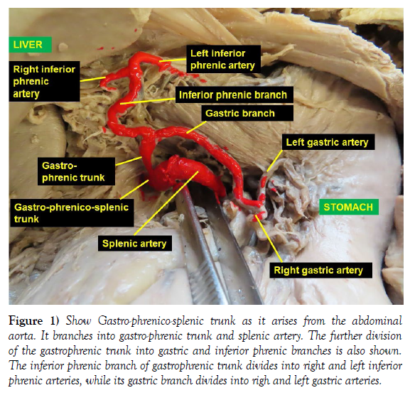
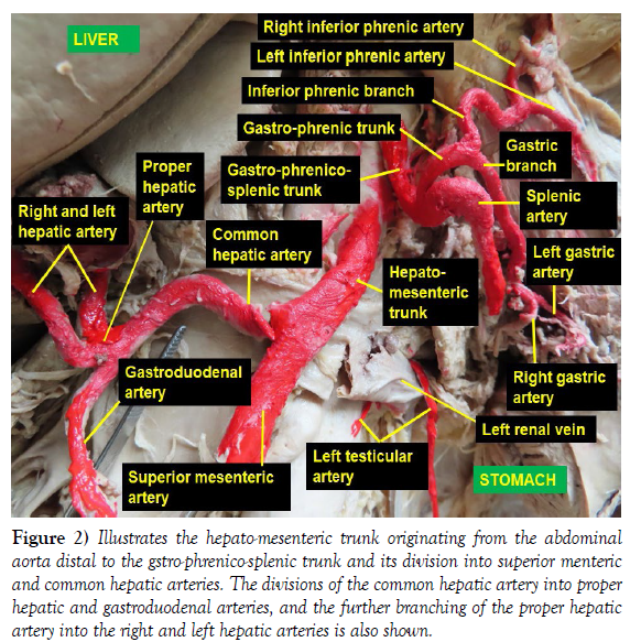
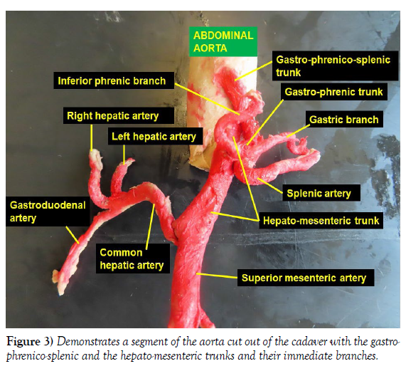
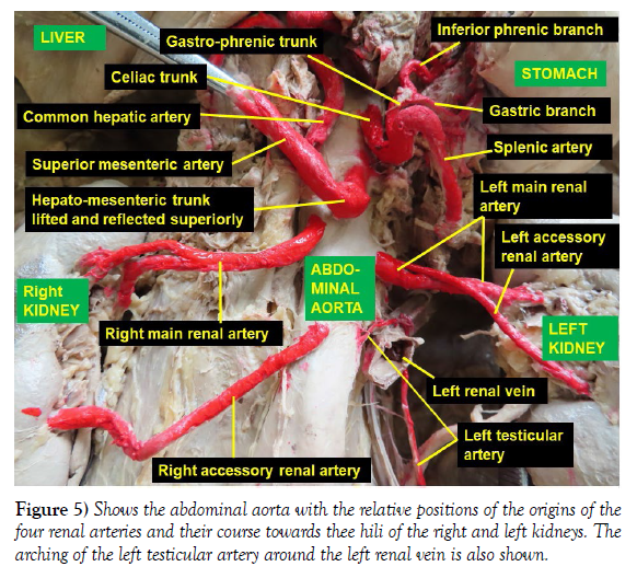
1. In the place of the celiac trunk a gastro-phrenico-splenic trunk originated from the abdominal aorta and gave branch to two trunks: gastrophrenic trunk and the splenic artery. The gastro-phrenic trunk further divided into inferior phrenic and gastric branches. The inferior phrenic branch ascended slightly along the right side of the abdominal part of the esophagus and divided into the right and left inferior phrenic arteries. Both inferior phrenic arteries entered the inferior surface of the thoracic diaphragm on the respective side but the left inferior phrenic artery crossed to the left ventral the abdominal part of the esophagus before entering the diaphragm. The gastric branch of the gastro-phrenic trunk descended towards the lesser curvature of the stomach and divided into an ascending left gastric artery and a descending right gastric artery. The splenic artery was found to run dorsal to the stomach towards the spleen as usual (Figure 1-3).
Figure 1: Show Gastro-phrenico-splenic trunk as it arises from the abdominal aorta. It branches into gastro-phrenic trunk and splenic artery. The further division of the gastrophrenic trunk into gastric and inferior phrenic branches is also shown. The inferior phrenic branch of gastrophrenic trunk divides into right and left inferior phrenic arteries, while its gastric branch divides into righ and left gastric arteries.
Figure 2: Illustrates the hepato-mesenteric trunk originating from the abdominal aorta distal to the gstro-phrenico-splenic trunk and its division into superior menteric and common hepatic arteries. The divisions of the common hepatic artery into proper hepatic and gastroduodenal arteries, and the further branching of the proper hepatic artery into the right and left hepatic arteries is also shown.
2. At the point of origin of the superior mesenteric artery from the abdominal aorta, a hepato-mesenteric trunk arose just distal to the origin of the gastro-phrenico-splenic trunk, and branched into common hepatic and superior mesenteric arteries. The common hepatic artery ascended to the right dorsal to the duodenum and pancreas to enter the hepatoduodenal ligament. Then it divided into gastroduodenal artery that descended dorsal to the first part of the duodenum and the proper hepatic artery that ascended in the hepatoduodenal ligament and further divided into right and left hepatic arteries, which entered the porta hepatis (Figure2-3). No variation in the division and further course of superior mesenteric artery was observed.
B. Observed variations of the paired lateral visceral branches
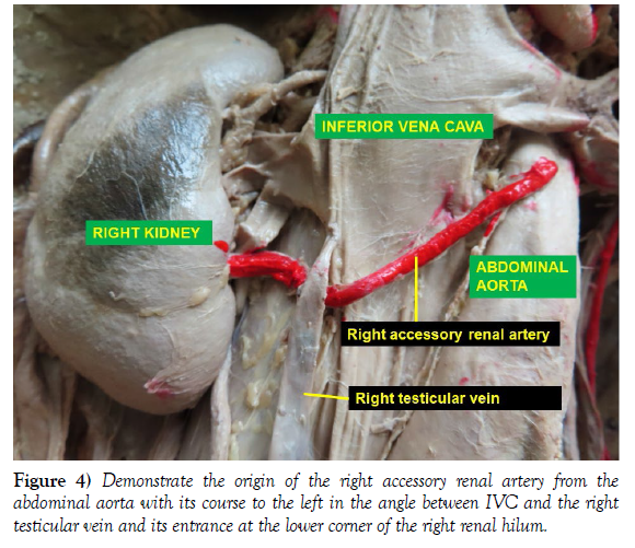
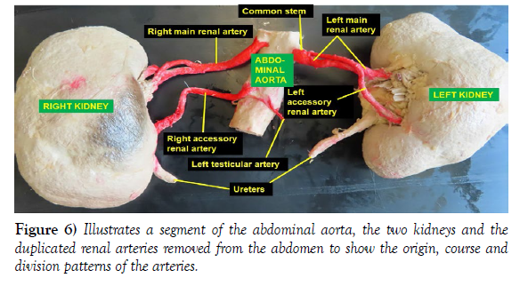
1. Bilateral double renal arteries (one main and one accessory) originating from the abdominal aorta were observed. The right accessory renal artery arose from the ventral surface of the abdominal aorta and ran to the right in the tight angle between the inferior vena cava and the right testicular vein and entered the lower corner of the right renal hilum. The right main renal artery arose from the abdominal aorta about 2 cm distal to the level of the origin of the hepato-mesenteric trunk, ran dorsal to the inferior vena cava, and divided into three prehilar (extrahilar) branches that entered the upper corner of the right renal hilum (Figure 4-6). On the left side, the main and accessory renal arteries shared a very short common trunk that arose from the abdominal aorta at a level half way between the origins of the right main and accessory renal arteries (Figure 5-7). This short common trunk then divided into a smaller upper branch (accessory renal artery) and a larger lower branch (main renal artery). As it courses to the left, the small upper branch crossed and descended ventral to the larger inferior branch and all structure of the renal pedicle, and entered the lower corner of the left renal hilum, while the larger lower branch entered the upper corner of the left renal hilum (Figure5- 6).
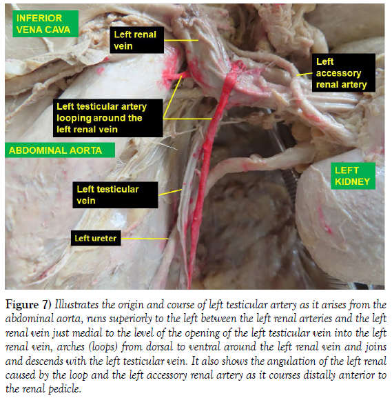
The left testicular artery arose from the left side of the abdominal aorta at the level of the right accessory renal artery, ascended to the left dorsal to the left renal vein and wrapped around it ventrally a the level slightly medial to the opening of the left testicular vein into the left renal vein and descended with the left testicular vein (Figure 5-7). As it runs around the left testicular vein, it caused a slight angulation of the left renal vein.
Figure 7: Illustrates the origin and course of left testicular artery as it arises from the abdominal aorta, runs superiorly to the left between the left renal arteries and the left renal vein just medial to the level of the opening of the left testicular vein into the left renal vein, arches (loops) from dorsal to ventral around the left renal vein and joins and descends with the left testicular vein. It also shows the angulation of the left renal caused by the loop and the left accessory renal artery as it courses distally anterior to the renal pedicle.
DISCUSSION
The variations of the anterior and lateral visceral branches of the abdominal aorta are widely documented. A computed tomographic study done on 102 patients revealed that there are different variations in the origin, number, course and branching patterns of anterior visceral branches of the abdominal aorta. These variations could be in such a way that the left gastric artery may arise from the celiac trunk resulting in the formation of hepato-splenic trunk, or splenic artery may fail to arise from the celiac trunk resulting in the formation of hepato-gastric trunk, which are due to errors that occur during the process of development [2]. According to this study, in about 0.98% of the case, a common origin of celiac artery and superior mesenteric artery as celiaco-mesenteric trunk and a similar percentage of splenic artery arising directly from the abdominal aorta were observed [2]. As it was documented in CT angiographic study of 110 patients, the most common celiac trunk variation was the presence of hepato-splenic trunk (60%), while gastrosplenic and hepato-gastric trunks were also found in 4.55% and 1.82% of the cases respectively. Gastro-splenic trunk along with hepato-mesenteric trunk was also observed in 1.82% of the cases. This study also described that both inferior phrenic arteries originated from the left gastric artery in 0.98% of the cases [3]. As noted by a case report, the splenic and left gastric arteries were found to spring from the abdominal aorta as gastro-splenic trunk, while the common hepatic artery arose as separate independent branch. In the same report but a different cadaver a gastro-phrenic trunk originating from the abdominal aorta and dividing into left gastric and left phrenic arteries was also noted [4]). Though, one of the observations in this current report related to celiac trunk appears to have some similarity with some of the previous findings, it is different in that, what originated from the abdominal aorta in the place of the celiac trunk is a gastro-phrenico-splenic trunk that directly or indirectly provided the two inferior phrenic arteries to the diaphragm, right and left gastric arteries to the lesser curvature of the stomach and the splenic artery to spleen. Other studies conducted on the variations of the origins of the two inferior phrenic arteries found that these arteries can arise independently directly from the abdominal aorta, or by a common trunk either from the abdominal aorta or the celiac trunk [5, 6]). However, the origin of the inferior phrenic arteries in the current case report is completely different in that they arose from the gastro-phrenic trunk which is a division of the gastro-phrenico-splenic trunk but not from the left gastric, abdominal aorta or the celiac trunk.
A combined variation of the branches of abdominal aorta, including celiacomesenteric trunk, bilaterally duplicated renal arteries, and right and left testicular arteries arising from the inferior renal arteries (accessory renal arteries) were also reported [7]. The report further elucidated that the celiacomesenteric trunk arose from the abdominal aorta and divides into hepatogastric trunk that gave branch to common hepatic and left gastric arteries, superior mesenteric artery, splenic artery and right gastric artery. The same report also showed bilateral double renal arteries with retroaortic left renal vein and retrocaval right testicular artery. A previous case report by Tessema, CB on bilaterally duplicated renal arteries arising from the abdominal aorta demonstrated that the arteries showed prehilar trifurcation on the right side and bifurcation on the left [8]. Even though, bilateral duplication of renal arteries were also observed in present case, the origin and course of the renal arteries are different from those previous reports in that the right accessory renal artery coursed between the inferior vena cava and the left testicular vein and on the left side the main and accessory renal arteries took a short common origin from the abdominal aorta (which can also be considered as early division of the left renal artery into two branches). However, the prehilar trifurcated division pattern of the right main renal artery is similar to the recent report by Tessema [8]. What is specifically off not on the left side of this case was that the accessory renal artery descended anterior to all the structures in the renal pedicle and entered the left kidney.
The testicular arteries can have variable origins, numbers and courses [9]. The review study by Nallikuzhy et al generally showed that testicular arterial variations are more common on left side than on the right side and therefore, arching of testicular artery and testicular artery passing through a bifid renal vein are also more common on the left side [9]. This is supported by a previous report that documented a unique case of high testicular artery origin from the abdominal aorta with a course through a hiatus in the left renal vein [10]. The observation of arching left testicular artery in this current case report seems to be consistent with the previous observations in being on the left side and in its arching course around the left renal vein. I believe, this course of testicular artery can potentially affect arterial blood flow to the left testis and venous drainage of both the left kidney and the left testis. This could happen in such a way that, kicking of the artery can occur as it loops around the renal vein resulting in testicular ischemia. On the other hand, as it loops around the renal vein, medial to the level of the opening of the left testicular vein into the left renal vein, it can cause partial constriction of the renal vein at the level of the slight angulation, and increases renal venous pressure, leading to retrograde flow of blood in left renal and testicular veins causing interstitial edema of the kidney and venous congestion of the testis with consequent AKI and varicocele in the testis. The testicular ischemia and varicocele, if chronic, can result in impaired fertility.
According to Cardarelli-Lite et al and Gozzo C et al, such variations of branches of the abdominal aorta, in general, can be causes of various abdominal vascular syndromes that can result in hemorrhagic or compressive complications including celiac artery compression syndrome, nutcracker syndrome, and superior mesenteric artery syndrome that might necessitate major interventional procedures [11, 12].
CONCLUSION
Variations of individual visceral branches of the abdominal aorta are common but complete variations of almost all visceral branches like in this case are very rare but very relevant in the practice of medicine, radiology and surgery. The unusual findings in this report, like uncommon trunk formations, variable branches and branching patterns, combination of cross over and arching are very relevant findings to be taken into consideration. Such intricate variations could be causes of various low- or high-flow abdominal vascular syndromes, compressive syndromes and damages resulting in blood loss during surgery. Therefore, lack of awareness of such variations can be very consequential during hepatobiliary, gastro-esophageal, pancreatic, splenic, intestinal, nephron-urologic and vascular surgeries, and imaging procedures.
ACKNOWLEDGEMENT
I am thankful to the donor and his families for their invaluable donation and consent for education, research and publication. I would also like to express my gratitude to the department of biomedical sciences for the encouragement and uninterrupted support. As always, I am also grateful to Denelle Kees and John Opland for their immense assistance during the dissection of this cadaver in the gross anatomy lab.
REFERENCES
- Rosse C, Gaddum-Rosse P, Hollinshead W. Hollinshead’s textbook of anatomy. Medicine. 1997; 600-602.
- Arudchelvam J. Study on variations of the ventral abdominal aortic branches: A computed tomography based study. SLJS. 2021; 39(1):26-29.
- Malviya KK, Verma A, Nayak AK, Mishra A, More RS, et al. Unraveling variation in celiac trunk and hepatic artery by CT angiography to aid in surgeries of upper abdominal region. Diagnositics. 2021; 11:22-62.
- Parasanna LC, Alva R, Senha GK, Bhat KMR, et al. Rare variation in the origin, branching pattern and course of celiac trunk: report of two cases. Malays J Med Sci. 2016; 20(1):77-81.
- Chandrachari JK, Jayaramu PA, Shetty S. A study of variations in the origin of inferior phrenic artery in adult human cadavers with clinical and embryological significance. Int J Anat Radiol Surg. 2016.
- Aslaner R, Pekcevik Y, Sahin H, Toka O, et al. Variations in the origin of inferior phrenic arteries and their relationship to celiac axis variations on CT angiography. Korean J radiol. 2017; 18(2): 336-344.
- Potgieter RE, Taylor AM, Wessels Q. A rare combined variation of the celiac trunk, renal and testicular vasculature. Anat Cell Biol. 2018; 51(1):62-65.
- Tessema CB. Duplication of the renal arteries with unusual extra renal branching patterns and positional relationship to hilar structures. Int J Anat Var 2022; 15 (4):166-168.
- Nallikuzhy TJ, Rajasekhar SSSN, Malik S, TAmagire DW, Johnson P, et al. Variations of testicular artery and vein: A meta-analysis with proposed classification. Clin Anat. 2018; 31:854-869.
- Padur AA, Kumar N. Unique variation of left testicular artery passing through a vascular hiatus in left renal vein. Anat cell Biol. 2019; 52:105-107.
- Cardarelli-leite L, Valloni FG, Salvadori PS, Lemos MD, D’lppolito G, et al. Abdominal vascular syndromes: characteristic imaging findings. Radiol Bras. 2016; 49(4):257-263.
- Gozzo C, Giambelluca D, Cannella R, Caruana G, Jukan A, et al. CT imaging findings of abdominopelvic vascular compression syndromes: what the radiologist needs to know. Insights Imaging. 2020; 11:48.
Indexed at, Google Scholar, Crossref
Indexed at, Google Scholar, Crossref
Indexed at, Google Scholar, Crossref
Indexed at, Google Scholar, Crossref
Indexed at, Google Scholar, Crossref
Indexed at, Google Scholar, Crossref
Indexed at, Google Scholar, Crossref
Indexed at, Google Scholar, Crossref


