Complete lateral discoid meniscus in a South Indian fetus: a case report and review of literatureThe medial menisci in both the knee joints of the fetus were having
BV Murlimanju1*, Narga Nair1 and Vishal Kumar2
1Department of Anatomy, Centre for Basic sciences, Kasturba Medical College, Manipal University, Mangalore, India
2Department of Anatomy, K.S. Hegde Medical Academy, Mangalore, India
- *Corresponding Author:
- BV Murlimanju, MD
Assistant Professor, Department of Anatomy, Kasturba Medical College, Manipal University Mangalore, 575004, India
Tel: +91 824 2211746
E-mail: murali.manju@manipal.edu
Date of Received: December 3rd, 2009
Date of Accepted: July 10th, 2010
Published Online: August 9th, 2010
© Int J Anat Var (IJAV). 2010; 3: 110–111.
[ft_below_content] =>Keywords
development, discoid, fetus, lateral meniscus, menisci
Introduction
The discoid lateral meniscus (DLM) was first described by Young in 1889 in a dissected cadaver specimen [1], but it was not until 1910 that Kroiss [2] attributed the snapping knee syndrome to the anomaly. The discoid menisci have been the object of many studies, because they are frequently the source of symptoms [3,4]. The incidence is estimated to be 3% to 5% in the general population and slightly higher in Asian populations [5]. The lesion initially was considered to be atavistic [3], but most authors currently believe that the discoid meniscus must be of congenital origin [6] and it might be symptomatic or not. In this study we report a DLM, which was found in a female fetus of South Indian origin. Only a very few reports of this were found in the literature. The objectives of this report is to discuss the embryological basis, anatomical features, etiological factors and clinical implications of this variant.
Case Report
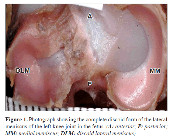
During the dissections of embalmed fetuses done for the morphometric study of the knee joints, this variant was observed at the Anatomy Laboratory, Kasturba Medical College, Manipal, Manipal University in the year 2009. The complete type of DLM was found on the left knee joint of a female fetal cadaver (Figure 1). It was a disc-shaped meniscus and occupied almost more than 90% of the tibial plateau area. At the middle one-third region its width measured 5.2 mm and the thickness was 2.8 mm. The lateral meniscus on the right side was found normal. literatureThe medial menisci in both the knee joints of the fetus were having normal shape.
Discussion
The discoid meniscus is the most common abnormal meniscal variant in children [7]. The concept of a clinical syndrome of a snapping knee that is caused by this type of DLM is widely accepted in the pediatric orthopedic literature. The classically described clinical presentation of the symptomatic DLM is a snapping or popping knee [7]. Cadaver studies have reported the prevalence of lateral discoid menisci to be between 0 and 7%, whereas arthroscopic studies have demonstrated ranges from 0.4 to 16.6% [7]. But racial differences do exist.
In this study, we report a complete type of DLM which was found on the left knee joint of an embalmed female fetal cadaver (Figure 1). The lateral meniscus on right knee joint and both sides medial menisci were found normal in shape. Le Minor [8] reported that the DLM was usually unilateral, however bilateral observations were reported in the literature. Hereditary transmission of the discoid lateral menisci is shown in some cases [9]. The discoid lateral meniscus is more common in females. The female preponderance of DLM (7:5) was observed earlier by Rao and Rao [10].
The DLM is a mega meniscus. It is disc shaped and covers the tibial plateau surface and prevents any contact between the femoral and tibial hyaline cartilages. In the present case the meniscus occupied almost more than 90% of the tibial plateau area. At the middle one-third region its width measured 5.2 mm and the thickness was 2.8 mm. The measurement was taken with a vernier caliper of accuracy 0.02 mm.
Since the publication of Watanabe’s Atlas [2], three types of lateral meniscal abnormalities are generally accepted. Their classification combined elements of both Smillie’s [3] and Kaplan’s [4] descriptions. The most commonly used classification system for discoid lateral menisci, by Watanabe, is based upon their shape and tibial attachments. This classification was developed from arthroscopic observations, divides discoid lateral menisci into three types, complete, incomplete, and Wrisberg ligament. If the meniscus occupies more than 80% of tibial plateau it is considered as complete type and less than 80% which is wider than usual is called as incomplete type [5].
In 1948, Smillie [3] proposed that there are three types of discoid lateral meniscus – primitive, intermediate, and infantile. He believed that there was a failure of resorption of the central area of the cartilage plate during the fetal stages of normal development, proposed the first theory on the development of the discoid meniscus. In his article, he states that the meniscus exists as a cartilaginous disc at an early stage of development, and that a congenital discoid meniscus is caused by an occasional persistence of the fetal state.
This theory was refuted by Kaplan in 1957 [4], when dissections of embryonic specimens of humans and animals failed to demonstrate the meniscus as a cartilaginous disk at any stage of normal development. They also reported that the lateral meniscus was having its adult crescent shape from its inception.
Kaplan [4] proposed a developmental theory, hypothesizing that the discoid meniscus developed because of the absence of the posterior tibial attachments of a normal meniscus. He noted that in his series of six patients with discoid menisci, at surgery, the only posterior attachment was the posterior meniscofemoral ligament (ligament of Wrisberg). Because of the lack of sufficient posterior attachments, he realized that the meniscus abnormally subluxates into the notch during extension and is pulled laterally by the coronary ligaments and the popliteal tendon with flexion. He believed that this abnormal motion resulted in repetitive microtrauma and produced the increased size and shape of the discoid meniscus. This theory does not explain the development of the more common discoid meniscus with normal posterior attachments (found 76-100% of the time).
Conclusions
Most authors now believe that the discoid meniscus must be of congenital origin. Further support for a congenital theory comes from evidence of familial transmission and reports of occurrence in twins. We believe that this report discusses the detailed anatomy, embryological basis, etiological factors and clinical implications of the DLM with relevant review of literature. This report discusses the surgical anatomy of the DLM which is not only important for the arthroscopic surgeons, clinicians and anatomists but also for the morphologists and embryologists.
Acknowledgements
The authors sincerely thank Professor Shakuntala R. Pai, Head of Department of Anatomy and Associate Dean, Kasturba Medical College, Manipal for her help and support for this study.
References
- Young R. The external semilunar cartilage as a complete disc. In: Memoirs and Memoranda in Anatomy. Cleland J, Mackay J, Young R, eds. London, Williams and Norgate. 1889; 179.
- Kroiss F. Die Verletzungen der Kniegelenkoszwischenknorpel und ihrer Verbindungen. Beitr Klin Chir. 1910; 66: 598?801. (German)
- Smillie IS. The congenital discoid meniscus. J Bone Joint Surg Am. 1948; 30B: 671?682.
- Kaplan EB. Discoid lateral meniscus of the knee joint; nature, mechanism, and operative treatment. J Bone Joint Surg Am. 1957; 39-A: 77?87.
- Kocher MS, Klingele K, Rassman SO. Meniscal disorders: normal, discoid, and cysts. Orthop Clin North Am. 2003; 34: 329?340.
- Woods GW, Whelan JM. Discoid meniscus. Clin Sports Med. 1990; 9: 695?706.
- Kelly BT, Green DW. Discoid lateral meniscus in children. Curr Opin Pediatr. 2002; 14: 54?61.
- Le Minor JM. Comparative morphology of the lateral meniscus of the knee in primates. J Anat. 1990; 170: 161?171.
- Dashefsky JH. Discoid lateral meniscus in three members of a family. Case reports. J Bone Joint Surg Am. 1971; 53: 1208?1210.
- Rao SK, Sripathi Rao P. Clinical, radiologic and arthroscopic assessment and treatment of bilateral discoid lateral meniscus. Knee Surg Sports Traumatol Arthrosc. 2007; 15: 597?601.
BV Murlimanju1*, Narga Nair1 and Vishal Kumar2
1Department of Anatomy, Centre for Basic sciences, Kasturba Medical College, Manipal University, Mangalore, India
2Department of Anatomy, K.S. Hegde Medical Academy, Mangalore, India
- *Corresponding Author:
- BV Murlimanju, MD
Assistant Professor, Department of Anatomy, Kasturba Medical College, Manipal University Mangalore, 575004, India
Tel: +91 824 2211746
E-mail: murali.manju@manipal.edu
Date of Received: December 3rd, 2009
Date of Accepted: July 10th, 2010
Published Online: August 9th, 2010
© Int J Anat Var (IJAV). 2010; 3: 110–111.
Abstract
The discoid meniscus is a relatively rare abnormality of the knee joint. Although both menisci have been reported to have discoid shape, lateral tends to be more common than the medial meniscus. In this study, we report a complete type of discoid lateral meniscus, which was found on the left knee joint of an embalmed female fetal cadaver. This report discusses the anatomy, etiology, embryological basis, clinical features and evaluation of this abnormality with relevant review of literature. Importance of the knowledge of this abnormal shaped meniscus in clinical diagnosis and therapeutic implications are discussed.
-Keywords
development, discoid, fetus, lateral meniscus, menisci
Introduction
The discoid lateral meniscus (DLM) was first described by Young in 1889 in a dissected cadaver specimen [1], but it was not until 1910 that Kroiss [2] attributed the snapping knee syndrome to the anomaly. The discoid menisci have been the object of many studies, because they are frequently the source of symptoms [3,4]. The incidence is estimated to be 3% to 5% in the general population and slightly higher in Asian populations [5]. The lesion initially was considered to be atavistic [3], but most authors currently believe that the discoid meniscus must be of congenital origin [6] and it might be symptomatic or not. In this study we report a DLM, which was found in a female fetus of South Indian origin. Only a very few reports of this were found in the literature. The objectives of this report is to discuss the embryological basis, anatomical features, etiological factors and clinical implications of this variant.
Case Report
During the dissections of embalmed fetuses done for the morphometric study of the knee joints, this variant was observed at the Anatomy Laboratory, Kasturba Medical College, Manipal, Manipal University in the year 2009. The complete type of DLM was found on the left knee joint of a female fetal cadaver (Figure 1). It was a disc-shaped meniscus and occupied almost more than 90% of the tibial plateau area. At the middle one-third region its width measured 5.2 mm and the thickness was 2.8 mm. The lateral meniscus on the right side was found normal. literatureThe medial menisci in both the knee joints of the fetus were having normal shape.
Discussion
The discoid meniscus is the most common abnormal meniscal variant in children [7]. The concept of a clinical syndrome of a snapping knee that is caused by this type of DLM is widely accepted in the pediatric orthopedic literature. The classically described clinical presentation of the symptomatic DLM is a snapping or popping knee [7]. Cadaver studies have reported the prevalence of lateral discoid menisci to be between 0 and 7%, whereas arthroscopic studies have demonstrated ranges from 0.4 to 16.6% [7]. But racial differences do exist.
In this study, we report a complete type of DLM which was found on the left knee joint of an embalmed female fetal cadaver (Figure 1). The lateral meniscus on right knee joint and both sides medial menisci were found normal in shape. Le Minor [8] reported that the DLM was usually unilateral, however bilateral observations were reported in the literature. Hereditary transmission of the discoid lateral menisci is shown in some cases [9]. The discoid lateral meniscus is more common in females. The female preponderance of DLM (7:5) was observed earlier by Rao and Rao [10].
The DLM is a mega meniscus. It is disc shaped and covers the tibial plateau surface and prevents any contact between the femoral and tibial hyaline cartilages. In the present case the meniscus occupied almost more than 90% of the tibial plateau area. At the middle one-third region its width measured 5.2 mm and the thickness was 2.8 mm. The measurement was taken with a vernier caliper of accuracy 0.02 mm.
Since the publication of Watanabe’s Atlas [2], three types of lateral meniscal abnormalities are generally accepted. Their classification combined elements of both Smillie’s [3] and Kaplan’s [4] descriptions. The most commonly used classification system for discoid lateral menisci, by Watanabe, is based upon their shape and tibial attachments. This classification was developed from arthroscopic observations, divides discoid lateral menisci into three types, complete, incomplete, and Wrisberg ligament. If the meniscus occupies more than 80% of tibial plateau it is considered as complete type and less than 80% which is wider than usual is called as incomplete type [5].
In 1948, Smillie [3] proposed that there are three types of discoid lateral meniscus – primitive, intermediate, and infantile. He believed that there was a failure of resorption of the central area of the cartilage plate during the fetal stages of normal development, proposed the first theory on the development of the discoid meniscus. In his article, he states that the meniscus exists as a cartilaginous disc at an early stage of development, and that a congenital discoid meniscus is caused by an occasional persistence of the fetal state.
This theory was refuted by Kaplan in 1957 [4], when dissections of embryonic specimens of humans and animals failed to demonstrate the meniscus as a cartilaginous disk at any stage of normal development. They also reported that the lateral meniscus was having its adult crescent shape from its inception.
Kaplan [4] proposed a developmental theory, hypothesizing that the discoid meniscus developed because of the absence of the posterior tibial attachments of a normal meniscus. He noted that in his series of six patients with discoid menisci, at surgery, the only posterior attachment was the posterior meniscofemoral ligament (ligament of Wrisberg). Because of the lack of sufficient posterior attachments, he realized that the meniscus abnormally subluxates into the notch during extension and is pulled laterally by the coronary ligaments and the popliteal tendon with flexion. He believed that this abnormal motion resulted in repetitive microtrauma and produced the increased size and shape of the discoid meniscus. This theory does not explain the development of the more common discoid meniscus with normal posterior attachments (found 76-100% of the time).
Conclusions
Most authors now believe that the discoid meniscus must be of congenital origin. Further support for a congenital theory comes from evidence of familial transmission and reports of occurrence in twins. We believe that this report discusses the detailed anatomy, embryological basis, etiological factors and clinical implications of the DLM with relevant review of literature. This report discusses the surgical anatomy of the DLM which is not only important for the arthroscopic surgeons, clinicians and anatomists but also for the morphologists and embryologists.
Acknowledgements
The authors sincerely thank Professor Shakuntala R. Pai, Head of Department of Anatomy and Associate Dean, Kasturba Medical College, Manipal for her help and support for this study.
References
- Young R. The external semilunar cartilage as a complete disc. In: Memoirs and Memoranda in Anatomy. Cleland J, Mackay J, Young R, eds. London, Williams and Norgate. 1889; 179.
- Kroiss F. Die Verletzungen der Kniegelenkoszwischenknorpel und ihrer Verbindungen. Beitr Klin Chir. 1910; 66: 598?801. (German)
- Smillie IS. The congenital discoid meniscus. J Bone Joint Surg Am. 1948; 30B: 671?682.
- Kaplan EB. Discoid lateral meniscus of the knee joint; nature, mechanism, and operative treatment. J Bone Joint Surg Am. 1957; 39-A: 77?87.
- Kocher MS, Klingele K, Rassman SO. Meniscal disorders: normal, discoid, and cysts. Orthop Clin North Am. 2003; 34: 329?340.
- Woods GW, Whelan JM. Discoid meniscus. Clin Sports Med. 1990; 9: 695?706.
- Kelly BT, Green DW. Discoid lateral meniscus in children. Curr Opin Pediatr. 2002; 14: 54?61.
- Le Minor JM. Comparative morphology of the lateral meniscus of the knee in primates. J Anat. 1990; 170: 161?171.
- Dashefsky JH. Discoid lateral meniscus in three members of a family. Case reports. J Bone Joint Surg Am. 1971; 53: 1208?1210.
- Rao SK, Sripathi Rao P. Clinical, radiologic and arthroscopic assessment and treatment of bilateral discoid lateral meniscus. Knee Surg Sports Traumatol Arthrosc. 2007; 15: 597?601.


