Congenital absence of the right coronary artery with a unique origin of the artery to the sinu-atrial node: A case study
Gail Elizabeth Elliott and Alexander Hamilton Martin*
Ross University School of Medicine, Portsmouth, Commonwealth of Dominica, West Indies
- *Corresponding Author:
- Dr. Alexander H. Martin
Professor of Anatomy, Ross University, School of Medicine, P.O. Box 266 Portsmouth, Commonwealth of Dominica 00152, West Indies
Tel: +1 (767) 255-6312
E-mail: AMartin@Rossmed.edu.dm
Date of Received: November 9th, 2011
Date of Accepted: September 3rd, 2012
Published Online: December 20th, 2012
© Int J Anat Var (IJAV). 2012; 5: 120–125.
[ft_below_content] =>Keywords
coronary, circulation, sinu-atrial
Introduction
Literature regarding the anatomy of the coronary circulation and its variations has grown exponentially with the increased application of imaging techniques such as coronary angiography and coronary computer tomography, higher numbers of medical schools carrying out gross anatomical dissection, methodical documentation of autopsy instances of variations, and advancing methods of cardiac surgery [1]. With all of these different means of gaining information, the major influence over the expansion in the literature has been the installment of extensive preoperative procedures to document the coronary artery tree in order to prevent accidental ligation during the procedure [1]. The increased instances of autopsy have also revealed a number of cases of variations which may not display any symptoms and are only discovered when an autopsy is performed. While some of the variations are clinically silent, some may alter the interpretations of the clinical symptoms expressed. Given the numerous instances of cardiac surgeries and the awareness of coronary diseases, it is essential to document unique variations to raise the awareness of possible anomalies in the event they present on the table during surgery.
The coronary arteries begin to develop in utero at the start of the third week of embryogenesis. The initiation of their development is considered to be the thickening of the myocardium in the ventricles and the subsequent need for increased vascularization of the growing heart [2,3]. The arteries pass through several complex steps during their development in utero, some of which are unique to the development of the arteries in the coronary circulation [2]. The steps include: vasculogenesis, angiogenesis, arteriogenesis and remodeling [3], which create the opportunity for variation to occur within the general arterial pattern although the basic coronary tree is present at the end of the vasculogenesis event [2]. Research indicates that the arteries are apparent within the aortic sinuses before the openings of the coronary arteries become patent in the aorta, giving weight to the concept of the coronary arteries growing into the aorta, rather than forming as an out pouching from it [2]. As the arteries grow towards the aorta, they do so as several vessels but only a single vessel on each side will penetrate through the aorta to become continuous with its lumen as a coronary artery [2].
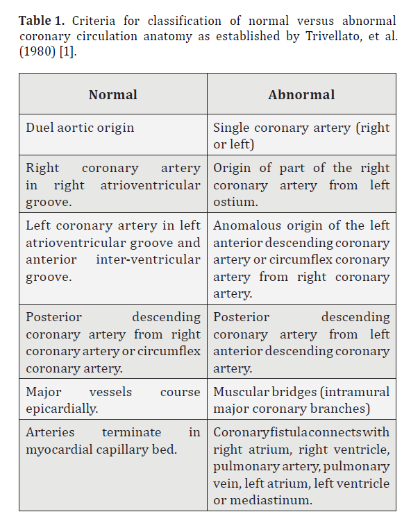
Due to the complex nature by which the coronary arteries develop in utero, variation is a common characteristic of the coronary anatomy [1]. Trivellato, et al. discuss the dissimilarities in the coronary arteries and divide the variations into two groups – ‘normal’ and ‘abnormal’ according to the nature of the variations present, whilst laying down a baseline criteria for a ‘normal’ coronary circulation anatomy categorization (Table 1). The ‘normal’ anatomy classified by these authors [1] displays minor variations including the diameter of the vessels and the length of the branches. The ‘abnormal’ circulation contains acute variations such as the absence of a coronary artery. Even with the basic criteria established, however, Trivellato, et al. found that each coronary system, even if considered ‘normal’ is individualistic due to the minimal variations present [1]. The authors also go on to acknowledge that statistical measures would have to be employed to establish the minimum number of features present for a categorization of ‘normal’. Furthermore, empirical procedures would be necessary to determine the line of separation between the classification of ‘normal’ and ‘abnormal’ [1].
Table 1. Criteria for classification of normal versus abnormal coronary circulation anatomy as established by Trivellato, et al. (1980) [1].
The Single Coronary Artery
Table 1 divides the spectrum of coronary variation into two specific groups which permits a general exploration into the expression, appearance, and clinical significances of ‘abnormal’ variations. Congenital abnormalities are essential to take not of due to the higher risk they pose through their potential clinical significance and the serious surgical problems they may pose due to their deviations from ‘normal’ and expected pathways. Coronary artery variations are congenital with the frequency of their appearance apparently unaffected by sex or race [4,5]. Abnormal variations are also rare and only present in 0.2–1.3% of the population [6]. The literature, however, is controversial regarding the topic of the clinical significance of coronary abnormalities. Yamanaka and Hobbs, indicate that coronary abnormalities generally do not cause clinical symptoms and usually go undiscovered until the time of autopsy. The authors also found no significant correlation between coronary heart disease and an abnormal coronary circulation [4]. They do, however, suggest that a young person showing signs of cardiac distress such as exertional synocope or myocardial infarction will most likely have an “abnormal” coronary circulation [4]. Taimur, et al., indicate that congenital coronary abnormalities are one of the leading causes of death in young athletes, only second to hypertropic cardiomyopathy [6].
A single coronary artery is one of the major variations possible in an “abnormal” coronary circulation and is present in 0.29% of the population [6]. It is suggested that the reason for the single artery to form is because of the underdevelopment of the proximal portion of the coronary artery which is expected to grow into the aorta during the final stages of the coronary artery formation [2,7]. In some instances, although this portion of the artery may be absent, the distal part of the artery will remain patent and be present in its expected epicardial locations – the atrioventricular and interventricular sulci [7]. Because of the failure of the proximal part of the artery to penetrate through the tunica media and the endothelial lining of the aorta, the coronary orifice is often not present or patent for the absent coronary artery [2]. Hyrtle was the first person to document and define a single coronary artery in 1841 on the heart of a seven month old fetus. He then went on to define a single coronary artery circulation occurring when, “one artery supplies the entire heart” [7]. He also further narrows the definition by adding that this variation should not arise because of, “a common aortic orifice of the two arteries or an unusual origin of the missing one” [7].
In regard to the clinical significance of a single coronary artery, the literature is divided again. Trivellato et al., mention that in cases of single coronary arteries with no atherosclerotic obstruction, myocardial ischemia and exertional angina have been documented. The authors suggest this may be attributed to the increased systemic and pulmonic pressures, causing the enlargement of the great vessels and the narrowing of the single coronary artery as a result [1]. They suggest, therefore, that -the single coronary artery is a potential clinical symptom waiting to occur. Eckart et al. completed a study of individuals in the military service dying sudden non-trauma related deaths [8]. The study encompassed over six million individuals and within that study, 126 cases of deaths of this nature were reported. Of these, 64 cases displayed extreme coronary anatomy variations and it was concluded that these variations were the major cause of death. They also found that in 21 cases of the abnormal coronary anatomy, there was a single coronary artery system present. In all instances, these single arteries were the left coronary artery which arose from over the right cusp of the aorta. Fifty percent of these 21 cases also presented pre-mortem reports of symptoms such as chest pain and synocope [8].
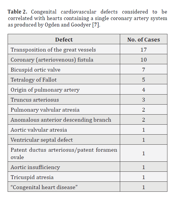
Conversely, Ogden and Goodyer indicate that deaths of young individuals with single coronary arteries have been as a result of other cardiac complications rather than the single artery itself while suggesting that in most cases the artery is clinically silent and only discovered during autopsy. Additionally, they mention that in some cases it may be the cause of myocardial ischemia [7]. There is also a correlation between a single coronary artery and additional congenital cardiovascular variations as shown below in Table 2 produced by Ogden and Goodyer [7].
Table 2. Congenital cardiovascular defects considered to be correlated with hearts containing a single coronary artery system as produced by Ogden and Goodyer [7].
Variations in origins of the arterial supply to the sinu-atrial node
The arterial supply to the atria is relatively poorly documented in the expanse of coronary circulation literature. This is surprising, given the knowledge of the atrial arterial supply and its influence over the functions of the heart’s pacemakers especially, regarding cardiac arrhythmia and subsequent clinical interventions [9]. In textbooks, the arterial supply to the atria is also generally overlooked by a brief mention of the atria being supplied by branches of the coronary arteries on the respective sides of the heart but offer no further additional information [9]. Knowledge of the arterial supply to the atria and its clinical significance in regard to the sinu-atrial node and the atrial aspect of the conduction system did increase during the period of discovery of the arteries to the sinu-atrial and the atrioventricular nodes but is still minimal in its coverage of the arterial variations in this region [9].
The right anterior atrial artery, the largest of the branches involved in the arterial supply to the atria supplies the area of the heart near the entrance of the superior vena cava by surrounding the area, and most frequently (50% of the time) supplies the artery to the sinu-atrial node [5,9]. This particular artery is given a number of names in literature. On occasion, it is referred to as the right anterior atrial branch, classified by the position in which the arterial branch arises from the right coronary artery as established by Spalteholz [9]. However, if this artery arises from the left coronary artery then it is considered to be the left anterior atrial artery which causes confusion and conflict during literature revision. Gross refers to this artery as the ramus ostii cavae superioris regardless of where it arises in the coronary circulation [9]. This artery does vary in its origin, although described most often as arising from proximal aspect of the right coronary artery and is regarded as being as constant as any of the other significant arteries in the human body [5,9]. In cases when this artery arises from the left coronary artery, it will arise from the circumflex branch (30%) more frequently than it does directly from the proximal region (12%) and is considered to give rise to the artery to the sinu-atrial node in 25% of cases [9]. When this artery does arise from the more medial aspect of the left coronary, it may also give rise to two smaller arteries instead of a single larger artery. The clinical importance of this artery is the spontaneous atrial fibrillation in elderly patients due to atherosclerotic disease of the ramus ostii cavae superioris [9].
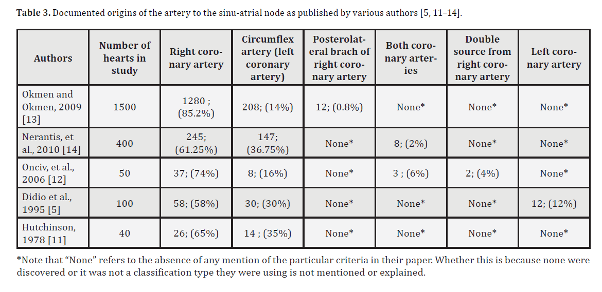
The arterial supply to the sinu-atrial node is highly variable but very well researched as it is of important clinical significance. The abnormal pathways of the artery to the sinu-atrial node affect the ways that the ischemia is expressed clinically and several papers are available researching the frequency of the various origins of this vessel [9]. It is also essential that a coronary angiography or coronary computer tomography be carried out prior to cardiac surgery in order to identify an anomalous course of this vessel, to map its path and to prevent its ligation [10,11]. As mentioned earlier, there are a number of publications researching the variations in the origins of this artery and a summary of the results found are presented in Table 3 [5,11–14]. The artery arises from the right coronary artery in most instances with varying percentages as displayed in table 3 and in 50% of these instances, it arises as a branch of the right anterior atrial artery [12] or it may arise directly from the proximal part of the right coronary artery [10,11]. Sex and ancestry do not affect the variations of the origin of the artery to the sinu-atrial node [5].
Case Report
In the case study being presented, a donor arrived in the Gross Anatomy Laboratory at Ross University School of Medicine during the period of May to August 2011. The donor was a 67-year-old female, who died from aspiration pneumonia. The coronary artery anatomy would be classified as “abnormal” if applying the criteria as described by Trivellato, et al. (Table 1) [1]. The classification is due notably but not exclusively to the absence of the right coronary artery and the unique origins of the right anterior atrial artery and the artery to the sinu-atrial node. Due to the nature of the donor program, donor medical records are not made available to the university; therefore, it is impossible to say whether or not the variations presenting in this individual resulted in clinical symptoms for which they may have sought out a cardiologist. There were no visible signs of a surgical intervention on the heart but it is impossible to say whether or not the abnormalities were clinically silent. The heart was also fairly small which may indicate that relatively little stress was placed upon it and it is possible that under more exertional conditions, cardiac symptoms may have arisen.
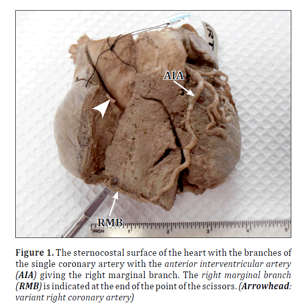
The single coronary artery in this instance arose from the left aortic cusp in the normal location of the left coronary artery and displayed the major characteristics of the left coronary artery (Figure 1). After it has arisen from the left cusp, the artery immediately divided into the left anterior descending artery or anterior interventricular branch and the circumflex artery in the expected pattern of branching in a “normal” left coronary artery. The left anterior interventricular branch continued in the anterior inter-ventricular sulcus on the sternocostal surface of the heart towards the apex of the heart, as is characteristic of this branch. At the apex, the terminal aspect of the left anterior descending branch continued onto the right margin as the right marginal artery. The circumflex branch of the left coronary artery continued onto the diaphragmatic surface of the heart giving off the larger left marginal branch and then terminated as the posterior interventricular artery in the posterior interventricular sulcus (Figure 2). In a normal coronary circulation in which both coronary arteries are present, the artery which gives rise to the posterior interventricular artery is said to have dominance in the heart because the diaphragmatic surface of the heart is largest and the posterior interventricular branch would supply this region. In this particular instance, the left coronary artery is dominant because of the absence of the right coronary artery, which again is a fairly rare occurrence, even in a “normal” coronary circulation where both coronary arteries are present, but is not unusual.
Figure 1. The sternocostal surface of the heart with the branches of the single coronary artery with the anterior interventricular artery (AIA) giving the right marginal branch. The right marginal branch (RMB) is indicated at the end of the point of the scissors. (Arrowhead: variant right coronary artery)
In a manner uncharacteristic of the single coronary artery circulation, the coronary orifice for the absent right coronary artery was patent [2]. Arising from this opening was the right anterior atrial artery which passed into the region between the superior vena cava and the aorta to supply the right atrium and the artery to the sinu-atrial node (Figure 3). It is important to note here that at first glance, the right anterior atrial artery was thought to be the right coronary artery by two highly experienced anatomists due to the relatively small size of the heart and the smaller diameter of the coronary vessels. However, upon further dissection, it was discovered that in fact that the initial diagnosis was mistaken and the vessel was actually the right anterior atrial artery.
Figure 3. Figure showing the the sternocostal surface of the heart on the right hand side. The orifice for the right anterior atrial artery (ORAA) can also be clearly seen where the artery would directly arise from the aorta. The figure also shows the atrioventricular sulcus (arrowheads) where the right coronary artery would be if it were present. (RAA: right anterior atrial artery; AIA: anterior interventricular artery)
Discussion
Information regarding the coronary circulation has grown with the increased frequency of cardiac surgeries, the use of non-invasive techniques to explore the coronary tree for pre-operative purposes or to investigate clinical symptoms, as well as the growing numbers of educational dissections of the heart. However, the information regarding the atrial arterial supply is relatively isolated towards the origins of the arteries to the sinu-atrial and atrioventricular nodes. An extensive search into the arterial supply to the atria has indicated that the artery to the sinu-atrial node arises from a number of origins as previously discussed, but to the best of our knowledge, the origin being presented in this study has not been mentioned in literature before. The variant which presented in this case study arose over the right cusp of the aorta where the right coronary artery would normally arise. During the initial inspection, it was thought, without further dissection, that the artery was in fact the right coronary artery. However, upon further examination and dissection, it was discovered that the mentioned variation was present instead. An extensive search of the literature regarding arterial supply to the atria reveals that a variation of this nature has not yet been documented. It is important to raise awareness of this unusual presentation to prepare the surgeon in the event that such a variation appears on the operating table.
References
- Trivellato M, Angelini P, Leachman RD. Variations in coronary artery anatomy: normal versus abnormal. Cardiovasc Dis. 1980; 7: 357–370.
- De Oliviera Silva-Junior G, Wilson da Silva Miranda S, Mandarim-de-Lacerda CA. Origin and development of the coronary arteries. Int J Morphol. 2009; 27: 891–898.
- Sadler TW. Medical Embryology. 11th Ed., London, Lippincott, Williams and Wilkins. 2009; 183–190.
- Yamanaka O, Hobbs RE. Coronary artery anomalies in 126,595 patients undergoing coronary arteriography. Cathet Cardiovasc Diagn. 1990; 21: 28–40.
- DiDo LJ, Lopes AC, Caetano AC, Prates JC. Variations of the origin of the artery of the sinoatrial node in normal human hearts. Surg Radiol Anat. 1995; 17: 19–26.
- Taimur SD, Khan SR, Haq MM, Mansur M, Haque H, Sultan AU. Congenital absence of right coronary artery without any other associated anomalies – a case report. University Heart Journal. 2010; 6: 45–47.
- Ogden JA, Goodyer AVN. Patterns of distribution of the single coronary artery. Yale J Biol Med. 1970; 43: 11-21.
- Eckart RE, Scoville SL, Campbell CL, Shry EA, Stajduhar KC, Potter RN, Pearse LA, Virmani R. Sudden death in young adults: a 25-year review of autopsies in military recruits. Ann Intern Med. 2004; 141: 829–834.
- James TN, Burch GE. The atrial coronary arteries in man. Circulation. 1958; 17: 90–98.
- Gavrielatos G, Nerantzis CE. Post mortem coronary angiographic visualization of a rare sinus node artery anatomical variant. Hellenic J Cardiol. 2011; 52: 84–85.
- Hutchinson MC. A study of the atrial arteries in man. J Anat. 1978; 125: 39–54.
- Onciu M, Tuta LA, Baz R, Leonte T. Specifics of the blood supply of the sinoatrial node. Rev Med Chir Soc Med Nat Iasi. 2006; 110: 667–673. (Romanian)
- Okmen AS, Okmen E. Sinoatrial node artery arising from posterolateral branch of right coronary artery: definition by screening consecutive 1500 coronary angiographies. Anadolu Karadiyol Derg. 2009; 9: 481–485.
- Nerantzis CE, Anninos H, Kouysaffis PN. Variation in the blood supply of the sinus node. Surg Radiol Anat. 2010; 32: 983–984.
Gail Elizabeth Elliott and Alexander Hamilton Martin*
Ross University School of Medicine, Portsmouth, Commonwealth of Dominica, West Indies
- *Corresponding Author:
- Dr. Alexander H. Martin
Professor of Anatomy, Ross University, School of Medicine, P.O. Box 266 Portsmouth, Commonwealth of Dominica 00152, West Indies
Tel: +1 (767) 255-6312
E-mail: AMartin@Rossmed.edu.dm
Date of Received: November 9th, 2011
Date of Accepted: September 3rd, 2012
Published Online: December 20th, 2012
© Int J Anat Var (IJAV). 2012; 5: 120–125.
Abstract
A 67 year-old female, presented with an absent right coronary artery during dissection in the Gross Anatomy Laboratory, Ross University School of Medicine. This is a previously undocumented origin of the right anterior atrial artery and a unique origin of the sinu-atrial node artery. Although the absence of the right coronary artery is rare, with an autopsy occurrence rate of 0.29%, it is extensively documented and not the novelty in this case. The more significant variation is the unusual origin of the right anterior atrial artery and the artery to the sinu-atrial node. Previous studies indicate the artery to the sinu-atrial node arises from the right coronary artery in 54% of the population. In this instance, however, the artery to the sinu-atrial node originates from the right anterior atrial artery which arises directly from the aorta over the right cusp, an origin, which has not been previously documented. Knowledge of the coronary anatomy is important in the understanding of clinical symptoms and in order for successful cardiac surgery to be performed. It is essential, therefore, for this previously undocumented variant to be put forward to raise knowledge of this very unique variation.
-Keywords
coronary, circulation, sinu-atrial
Introduction
Literature regarding the anatomy of the coronary circulation and its variations has grown exponentially with the increased application of imaging techniques such as coronary angiography and coronary computer tomography, higher numbers of medical schools carrying out gross anatomical dissection, methodical documentation of autopsy instances of variations, and advancing methods of cardiac surgery [1]. With all of these different means of gaining information, the major influence over the expansion in the literature has been the installment of extensive preoperative procedures to document the coronary artery tree in order to prevent accidental ligation during the procedure [1]. The increased instances of autopsy have also revealed a number of cases of variations which may not display any symptoms and are only discovered when an autopsy is performed. While some of the variations are clinically silent, some may alter the interpretations of the clinical symptoms expressed. Given the numerous instances of cardiac surgeries and the awareness of coronary diseases, it is essential to document unique variations to raise the awareness of possible anomalies in the event they present on the table during surgery.
The coronary arteries begin to develop in utero at the start of the third week of embryogenesis. The initiation of their development is considered to be the thickening of the myocardium in the ventricles and the subsequent need for increased vascularization of the growing heart [2,3]. The arteries pass through several complex steps during their development in utero, some of which are unique to the development of the arteries in the coronary circulation [2]. The steps include: vasculogenesis, angiogenesis, arteriogenesis and remodeling [3], which create the opportunity for variation to occur within the general arterial pattern although the basic coronary tree is present at the end of the vasculogenesis event [2]. Research indicates that the arteries are apparent within the aortic sinuses before the openings of the coronary arteries become patent in the aorta, giving weight to the concept of the coronary arteries growing into the aorta, rather than forming as an out pouching from it [2]. As the arteries grow towards the aorta, they do so as several vessels but only a single vessel on each side will penetrate through the aorta to become continuous with its lumen as a coronary artery [2].
Due to the complex nature by which the coronary arteries develop in utero, variation is a common characteristic of the coronary anatomy [1]. Trivellato, et al. discuss the dissimilarities in the coronary arteries and divide the variations into two groups – ‘normal’ and ‘abnormal’ according to the nature of the variations present, whilst laying down a baseline criteria for a ‘normal’ coronary circulation anatomy categorization (Table 1). The ‘normal’ anatomy classified by these authors [1] displays minor variations including the diameter of the vessels and the length of the branches. The ‘abnormal’ circulation contains acute variations such as the absence of a coronary artery. Even with the basic criteria established, however, Trivellato, et al. found that each coronary system, even if considered ‘normal’ is individualistic due to the minimal variations present [1]. The authors also go on to acknowledge that statistical measures would have to be employed to establish the minimum number of features present for a categorization of ‘normal’. Furthermore, empirical procedures would be necessary to determine the line of separation between the classification of ‘normal’ and ‘abnormal’ [1].
Table 1. Criteria for classification of normal versus abnormal coronary circulation anatomy as established by Trivellato, et al. (1980) [1].
The Single Coronary Artery
Table 1 divides the spectrum of coronary variation into two specific groups which permits a general exploration into the expression, appearance, and clinical significances of ‘abnormal’ variations. Congenital abnormalities are essential to take not of due to the higher risk they pose through their potential clinical significance and the serious surgical problems they may pose due to their deviations from ‘normal’ and expected pathways. Coronary artery variations are congenital with the frequency of their appearance apparently unaffected by sex or race [4,5]. Abnormal variations are also rare and only present in 0.2–1.3% of the population [6]. The literature, however, is controversial regarding the topic of the clinical significance of coronary abnormalities. Yamanaka and Hobbs, indicate that coronary abnormalities generally do not cause clinical symptoms and usually go undiscovered until the time of autopsy. The authors also found no significant correlation between coronary heart disease and an abnormal coronary circulation [4]. They do, however, suggest that a young person showing signs of cardiac distress such as exertional synocope or myocardial infarction will most likely have an “abnormal” coronary circulation [4]. Taimur, et al., indicate that congenital coronary abnormalities are one of the leading causes of death in young athletes, only second to hypertropic cardiomyopathy [6].
A single coronary artery is one of the major variations possible in an “abnormal” coronary circulation and is present in 0.29% of the population [6]. It is suggested that the reason for the single artery to form is because of the underdevelopment of the proximal portion of the coronary artery which is expected to grow into the aorta during the final stages of the coronary artery formation [2,7]. In some instances, although this portion of the artery may be absent, the distal part of the artery will remain patent and be present in its expected epicardial locations – the atrioventricular and interventricular sulci [7]. Because of the failure of the proximal part of the artery to penetrate through the tunica media and the endothelial lining of the aorta, the coronary orifice is often not present or patent for the absent coronary artery [2]. Hyrtle was the first person to document and define a single coronary artery in 1841 on the heart of a seven month old fetus. He then went on to define a single coronary artery circulation occurring when, “one artery supplies the entire heart” [7]. He also further narrows the definition by adding that this variation should not arise because of, “a common aortic orifice of the two arteries or an unusual origin of the missing one” [7].
In regard to the clinical significance of a single coronary artery, the literature is divided again. Trivellato et al., mention that in cases of single coronary arteries with no atherosclerotic obstruction, myocardial ischemia and exertional angina have been documented. The authors suggest this may be attributed to the increased systemic and pulmonic pressures, causing the enlargement of the great vessels and the narrowing of the single coronary artery as a result [1]. They suggest, therefore, that -the single coronary artery is a potential clinical symptom waiting to occur. Eckart et al. completed a study of individuals in the military service dying sudden non-trauma related deaths [8]. The study encompassed over six million individuals and within that study, 126 cases of deaths of this nature were reported. Of these, 64 cases displayed extreme coronary anatomy variations and it was concluded that these variations were the major cause of death. They also found that in 21 cases of the abnormal coronary anatomy, there was a single coronary artery system present. In all instances, these single arteries were the left coronary artery which arose from over the right cusp of the aorta. Fifty percent of these 21 cases also presented pre-mortem reports of symptoms such as chest pain and synocope [8].
Conversely, Ogden and Goodyer indicate that deaths of young individuals with single coronary arteries have been as a result of other cardiac complications rather than the single artery itself while suggesting that in most cases the artery is clinically silent and only discovered during autopsy. Additionally, they mention that in some cases it may be the cause of myocardial ischemia [7]. There is also a correlation between a single coronary artery and additional congenital cardiovascular variations as shown below in Table 2 produced by Ogden and Goodyer [7].
Table 2. Congenital cardiovascular defects considered to be correlated with hearts containing a single coronary artery system as produced by Ogden and Goodyer [7].
Variations in origins of the arterial supply to the sinu-atrial node
The arterial supply to the atria is relatively poorly documented in the expanse of coronary circulation literature. This is surprising, given the knowledge of the atrial arterial supply and its influence over the functions of the heart’s pacemakers especially, regarding cardiac arrhythmia and subsequent clinical interventions [9]. In textbooks, the arterial supply to the atria is also generally overlooked by a brief mention of the atria being supplied by branches of the coronary arteries on the respective sides of the heart but offer no further additional information [9]. Knowledge of the arterial supply to the atria and its clinical significance in regard to the sinu-atrial node and the atrial aspect of the conduction system did increase during the period of discovery of the arteries to the sinu-atrial and the atrioventricular nodes but is still minimal in its coverage of the arterial variations in this region [9].
The right anterior atrial artery, the largest of the branches involved in the arterial supply to the atria supplies the area of the heart near the entrance of the superior vena cava by surrounding the area, and most frequently (50% of the time) supplies the artery to the sinu-atrial node [5,9]. This particular artery is given a number of names in literature. On occasion, it is referred to as the right anterior atrial branch, classified by the position in which the arterial branch arises from the right coronary artery as established by Spalteholz [9]. However, if this artery arises from the left coronary artery then it is considered to be the left anterior atrial artery which causes confusion and conflict during literature revision. Gross refers to this artery as the ramus ostii cavae superioris regardless of where it arises in the coronary circulation [9]. This artery does vary in its origin, although described most often as arising from proximal aspect of the right coronary artery and is regarded as being as constant as any of the other significant arteries in the human body [5,9]. In cases when this artery arises from the left coronary artery, it will arise from the circumflex branch (30%) more frequently than it does directly from the proximal region (12%) and is considered to give rise to the artery to the sinu-atrial node in 25% of cases [9]. When this artery does arise from the more medial aspect of the left coronary, it may also give rise to two smaller arteries instead of a single larger artery. The clinical importance of this artery is the spontaneous atrial fibrillation in elderly patients due to atherosclerotic disease of the ramus ostii cavae superioris [9].
The arterial supply to the sinu-atrial node is highly variable but very well researched as it is of important clinical significance. The abnormal pathways of the artery to the sinu-atrial node affect the ways that the ischemia is expressed clinically and several papers are available researching the frequency of the various origins of this vessel [9]. It is also essential that a coronary angiography or coronary computer tomography be carried out prior to cardiac surgery in order to identify an anomalous course of this vessel, to map its path and to prevent its ligation [10,11]. As mentioned earlier, there are a number of publications researching the variations in the origins of this artery and a summary of the results found are presented in Table 3 [5,11–14]. The artery arises from the right coronary artery in most instances with varying percentages as displayed in table 3 and in 50% of these instances, it arises as a branch of the right anterior atrial artery [12] or it may arise directly from the proximal part of the right coronary artery [10,11]. Sex and ancestry do not affect the variations of the origin of the artery to the sinu-atrial node [5].
Case Report
In the case study being presented, a donor arrived in the Gross Anatomy Laboratory at Ross University School of Medicine during the period of May to August 2011. The donor was a 67-year-old female, who died from aspiration pneumonia. The coronary artery anatomy would be classified as “abnormal” if applying the criteria as described by Trivellato, et al. (Table 1) [1]. The classification is due notably but not exclusively to the absence of the right coronary artery and the unique origins of the right anterior atrial artery and the artery to the sinu-atrial node. Due to the nature of the donor program, donor medical records are not made available to the university; therefore, it is impossible to say whether or not the variations presenting in this individual resulted in clinical symptoms for which they may have sought out a cardiologist. There were no visible signs of a surgical intervention on the heart but it is impossible to say whether or not the abnormalities were clinically silent. The heart was also fairly small which may indicate that relatively little stress was placed upon it and it is possible that under more exertional conditions, cardiac symptoms may have arisen.
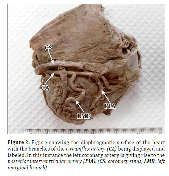
The single coronary artery in this instance arose from the left aortic cusp in the normal location of the left coronary artery and displayed the major characteristics of the left coronary artery (Figure 1). After it has arisen from the left cusp, the artery immediately divided into the left anterior descending artery or anterior interventricular branch and the circumflex artery in the expected pattern of branching in a “normal” left coronary artery. The left anterior interventricular branch continued in the anterior inter-ventricular sulcus on the sternocostal surface of the heart towards the apex of the heart, as is characteristic of this branch. At the apex, the terminal aspect of the left anterior descending branch continued onto the right margin as the right marginal artery. The circumflex branch of the left coronary artery continued onto the diaphragmatic surface of the heart giving off the larger left marginal branch and then terminated as the posterior interventricular artery in the posterior interventricular sulcus (Figure 2). In a normal coronary circulation in which both coronary arteries are present, the artery which gives rise to the posterior interventricular artery is said to have dominance in the heart because the diaphragmatic surface of the heart is largest and the posterior interventricular branch would supply this region. In this particular instance, the left coronary artery is dominant because of the absence of the right coronary artery, which again is a fairly rare occurrence, even in a “normal” coronary circulation where both coronary arteries are present, but is not unusual.
Figure 1. The sternocostal surface of the heart with the branches of the single coronary artery with the anterior interventricular artery (AIA) giving the right marginal branch. The right marginal branch (RMB) is indicated at the end of the point of the scissors. (Arrowhead: variant right coronary artery)
Figure 2. Figure showing the diaphragmatic surface of the heart with the branches of the circumflex artery (CA) being displayed and labeled. In this instance the left coronary artery is giving rise to the posterior interventricular artery (PIA). (CS: coronary sinus; LMB: left marginal branch)
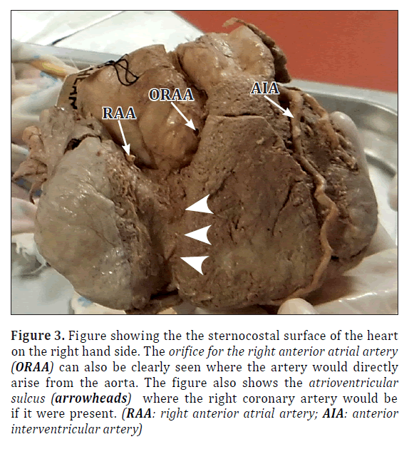
In a manner uncharacteristic of the single coronary artery circulation, the coronary orifice for the absent right coronary artery was patent [2]. Arising from this opening was the right anterior atrial artery which passed into the region between the superior vena cava and the aorta to supply the right atrium and the artery to the sinu-atrial node (Figure 3). It is important to note here that at first glance, the right anterior atrial artery was thought to be the right coronary artery by two highly experienced anatomists due to the relatively small size of the heart and the smaller diameter of the coronary vessels. However, upon further dissection, it was discovered that in fact that the initial diagnosis was mistaken and the vessel was actually the right anterior atrial artery.
Figure 3. Figure showing the the sternocostal surface of the heart on the right hand side. The orifice for the right anterior atrial artery (ORAA) can also be clearly seen where the artery would directly arise from the aorta. The figure also shows the atrioventricular sulcus (arrowheads) where the right coronary artery would be if it were present. (RAA: right anterior atrial artery; AIA: anterior interventricular artery)
Discussion
Information regarding the coronary circulation has grown with the increased frequency of cardiac surgeries, the use of non-invasive techniques to explore the coronary tree for pre-operative purposes or to investigate clinical symptoms, as well as the growing numbers of educational dissections of the heart. However, the information regarding the atrial arterial supply is relatively isolated towards the origins of the arteries to the sinu-atrial and atrioventricular nodes. An extensive search into the arterial supply to the atria has indicated that the artery to the sinu-atrial node arises from a number of origins as previously discussed, but to the best of our knowledge, the origin being presented in this study has not been mentioned in literature before. The variant which presented in this case study arose over the right cusp of the aorta where the right coronary artery would normally arise. During the initial inspection, it was thought, without further dissection, that the artery was in fact the right coronary artery. However, upon further examination and dissection, it was discovered that the mentioned variation was present instead. An extensive search of the literature regarding arterial supply to the atria reveals that a variation of this nature has not yet been documented. It is important to raise awareness of this unusual presentation to prepare the surgeon in the event that such a variation appears on the operating table.
References
- Trivellato M, Angelini P, Leachman RD. Variations in coronary artery anatomy: normal versus abnormal. Cardiovasc Dis. 1980; 7: 357–370.
- De Oliviera Silva-Junior G, Wilson da Silva Miranda S, Mandarim-de-Lacerda CA. Origin and development of the coronary arteries. Int J Morphol. 2009; 27: 891–898.
- Sadler TW. Medical Embryology. 11th Ed., London, Lippincott, Williams and Wilkins. 2009; 183–190.
- Yamanaka O, Hobbs RE. Coronary artery anomalies in 126,595 patients undergoing coronary arteriography. Cathet Cardiovasc Diagn. 1990; 21: 28–40.
- DiDo LJ, Lopes AC, Caetano AC, Prates JC. Variations of the origin of the artery of the sinoatrial node in normal human hearts. Surg Radiol Anat. 1995; 17: 19–26.
- Taimur SD, Khan SR, Haq MM, Mansur M, Haque H, Sultan AU. Congenital absence of right coronary artery without any other associated anomalies – a case report. University Heart Journal. 2010; 6: 45–47.
- Ogden JA, Goodyer AVN. Patterns of distribution of the single coronary artery. Yale J Biol Med. 1970; 43: 11-21.
- Eckart RE, Scoville SL, Campbell CL, Shry EA, Stajduhar KC, Potter RN, Pearse LA, Virmani R. Sudden death in young adults: a 25-year review of autopsies in military recruits. Ann Intern Med. 2004; 141: 829–834.
- James TN, Burch GE. The atrial coronary arteries in man. Circulation. 1958; 17: 90–98.
- Gavrielatos G, Nerantzis CE. Post mortem coronary angiographic visualization of a rare sinus node artery anatomical variant. Hellenic J Cardiol. 2011; 52: 84–85.
- Hutchinson MC. A study of the atrial arteries in man. J Anat. 1978; 125: 39–54.
- Onciu M, Tuta LA, Baz R, Leonte T. Specifics of the blood supply of the sinoatrial node. Rev Med Chir Soc Med Nat Iasi. 2006; 110: 667–673. (Romanian)
- Okmen AS, Okmen E. Sinoatrial node artery arising from posterolateral branch of right coronary artery: definition by screening consecutive 1500 coronary angiographies. Anadolu Karadiyol Derg. 2009; 9: 481–485.
- Nerantzis CE, Anninos H, Kouysaffis PN. Variation in the blood supply of the sinus node. Surg Radiol Anat. 2010; 32: 983–984.


