Dangerous anatomic variation of internal carotid artery – a rare case report
Reena Agrawal* and Sanjeeb Kumar Agrawal
Department of Anatomy, Meenakshi Medical College & Research Institute, Meenakshi University, Enathur, Kancheepuram, Aarthi Scans, Chennai, Tamil Nadu, India
- *Corresponding Author:
- Reena Agrawal, MS
Associate Professor, Department of Anatomy, Meenakshi Medical College & Research Institute, Meenakshi University, Enathur, Kancheepuram - 631552 Tamil Nadu, India
Tel: +91 989 4059749
E-mail: sanjeeb100@rediffmail.com
Date of Received: April 25th, 2011
Date of Accepted: August 10th, 2011
Published Online: October 7th, 2011
© Int J Anat Var (IJAV). 2011; 4: 174–176.
[ft_below_content] =>Keywords
internal carotid artery, tortuous, pharynx
Introduction
The internal carotid artery (ICA) has a straight cervical course up to the cranial base, and does not emit branches in this course. In 10 to 40% of the cases there are anatomic variations in this course, and the most common are curvatures, elbowing and notches [1]. Anatomical variants of the ICA have been reported with a large variability of pattern and degree. Among these variants, an unusual retropharyngeal course of ICA can mimic parapharyngeal neoplasm and pose a risk of vascular injury during pharyngeal intervention. Devastating complications can result from biopsy, surgical and anesthetic procedures. Medial deviation of the ICA is a rare cause of widening of the retropharyngeal space. In children, the diagnosis of these variations must always be predicted, especially in patients prone to adenotonsillectomy, in which incidental lesions during the surgical process may have catastrophic consequences [2,3]. In elder people, such variations may often be associated with arteriosclerosis and thrombosis that may affect the blood flow and cause encephalic ischemic processes [1].
Case Report
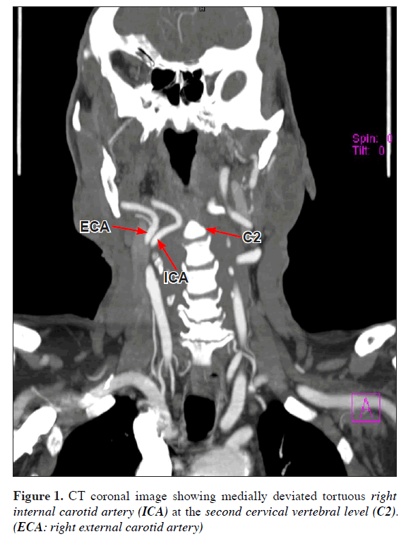
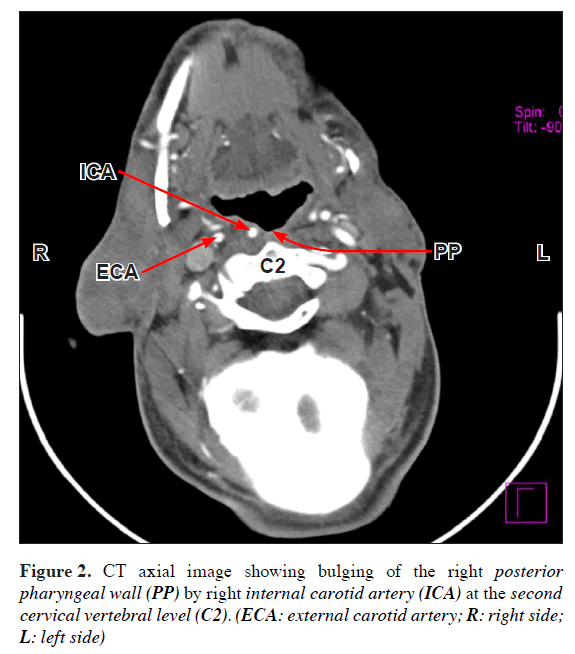
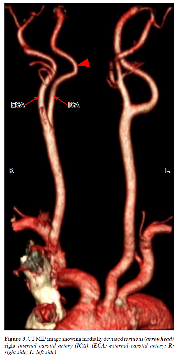
A 74-year-old male patient was referred for computed tomography (CT) to our radiology department due to dysphagia and foreign body sensation in the posterior pharynx for one month. The patient’s medical history was hypertension, without known coronary artery or cerebrovascular disease. By contrast-enhanced multi-slice CT scan (by Siemens SOMATOM Sensation 64), coronal (Figure 1), axial (Figure 2) and maximum intensity projection (MIP) (Figure 3) images were obtained. A retropharyngeal tortuous right internal carotid artery was found at the second cervical vertebral level (Figures 1, 2, 3). In this case, CT scan prior to intervention offered a concise diagnosis; therefore a clinical catastrophe can be avoided.
Discussion
The anatomic course of the ICA may be as follows: (a) a straight course to the base of skull; (b) an S- or C-shaped elongation with a medial, lateral, or ventrodorsal displacement; (c) a kinking of one or more of the segments; and (d) coiling of the artery that may appear as a double loop. The etiology of the various variations has been ascribed to embryologic, pathologic, and aging effects. As for the ICA cervical variant etiology, we believe many cases are congenital, but that, especially in elders, atherosclerotic processes and fibromuscular dysplasia may be implied [1]. Embryologically, the carotid artery originates from the third aortic arch and the dorsal aorta. Usually, the dorsal aortic root descends into the chest by the eighth week of development, thereby straightening the course of ICA [4]. However, it has been postulated that incomplete straightening of the carotid vessels enables the embryonic angulation to persist, resulting in congenitally tortuous or aberrant ICAs in the retropharyngeal space. Anatomic descriptions of tortuous ICA in the otolaryngology literature range from mild kinking to complete circular loop formation [4,5]. Congenitally tortuous courses may become more pronounced in the elders secondary to atherosclerosis and/or hypertension [4]. Medial dislocation of the ICA into retropharyngeal space at the level of the posterior pharynx typically presents on physical examination as a pulsating submucosal mass along the posterior pharyngeal wall. The mass is usually found in an asymptomatic patient during a routine head and neck examination [6]. Symptomatic patients may present with complaints of dysphagia, abnormal voice, or foreign body sensation in the posterior pharynx [6]. Occasionally, the variation is confused with a unilateral tonsillitis, peritonsillar abscess, or parapharyngeal neoplasm, and discovered only on closer inspection [7]. Displacement of the ICA into the retropharyngeal space carries a risk of major complications if such variant is not recognized before pharyngeal surgery. It is especially hazardous when the artery comes into contact with the tonsillar fossa or the posterior pharyngeal wall [6,7]. Such hidden asymptomatic variant may result in a life-threatening hemorrhage during tonsillectomy, uvulopalatopharyngoplasty, or incision and drainage of a peritonsillar abscess [6]. Retropharyngeal carotid artery could pose a significant additional risk of arterial puncture or arterial injection of local anesthetics when performing the transoral approach to block the glossopharyngeal nerve in the pharynx. Prominent forward and medial displacement of the pharyngeal tissues by an aberrant carotid artery (or other carotid variation causing a mass effect) may disrupt the anatomical balance of the pharyngeal orifice and can be a causative or potentiating factor in obstructive sleep apnea [8,9]. Imaging studies can be used to confirm the location of the ICA in order to reduce the risk of the hemorrhagic complications associated with retropharyngeal ICAs. Doppler ultrasonography, computed tomography angiography, and magnetic resonance angiography imaging can all be employed for assessment of the course, caliber, and contour of the vessels [6,10]. This condition requires no intervention. However, once it is diagnosed, the presence of retropharyngeal ICAs should be clearly documented in the patient’s medical chart to avoid complications should the patient require any type of intraoral or cervical surgical procedure in the future. Thus we can say that retropharyngeal ICA has a number of clinically relevant implications for the anesthesiologist as well as the surgeon.
References
- Paulsen F, Tillman B, Christofides C, Richter W, Koebke J. Curving and looping of the internal carotid artery in relation to the pharynx: frequency, embryology and clinical implications. J Anat. 2000; 197: 373–381.
- Wasserman JM, Sclafani SJ, Goldstein NA. Intraoperative evaluation of a pulsatile oropharyngeal mass during adenotonsillectomy. Int J Pediatr Otorhinolaryngol. 2006; 70: 371–375.
- Kay DJ, Mehta V, Goldsmith AJ. Perioperative adenotonsillectomy management in children: current practices. Laryngoscope. 2003, 113: 592–597.
- Shanley DJ. Bilateral aberrant cervical internal carotid arteries. Neuroradiology. 1992; 35: 55–56.
- Fix TJ, Daffner RH, Deeb ZL. Carotid transposition: Another cause of wide retropharyngeal soft tissues. AJR Am J Roentgenol. 1996; 167: 1305–1307.
- Galletti B, Bucolo S, Abbate G, Calabrese G, Romano G, Quattrocchi C, Freni F. Internal carotid artery transposition as risk factor in pharyngeal surgery. Laryngoscope. 2002; 112: 1845–1848.
- Tsunoda K, Takanosawa M, Matsuda K. Aberrant internal carotid artery in the mouth. Lancet. 1997; 350: 340.
- Tsuiki S, Isono S, Ishikawa T, Yamashiro Y, Tatsumi K, Nishino T. Anatomical balance of the upper airway and obstructive sleep apnea. Anesthesiology. 2008; 108: 1009–1015.
- Schmal F, Stoll W. Differential diagnosis and management of retropharyngeal space-occupying lesions. HNO. 2002; 50: 418–423. (German)
- Palacios E, Kirsch D, Rojas R. Anomalous course of the carotid arteries in the retropharyngeal space poses a surgical risk. Ear Nose Throat J. 2005; 84: 336–337.
Reena Agrawal* and Sanjeeb Kumar Agrawal
Department of Anatomy, Meenakshi Medical College & Research Institute, Meenakshi University, Enathur, Kancheepuram, Aarthi Scans, Chennai, Tamil Nadu, India
- *Corresponding Author:
- Reena Agrawal, MS
Associate Professor, Department of Anatomy, Meenakshi Medical College & Research Institute, Meenakshi University, Enathur, Kancheepuram - 631552 Tamil Nadu, India
Tel: +91 989 4059749
E-mail: sanjeeb100@rediffmail.com
Date of Received: April 25th, 2011
Date of Accepted: August 10th, 2011
Published Online: October 7th, 2011
© Int J Anat Var (IJAV). 2011; 4: 174–176.
Abstract
The aim is to report a case of retropharyngeal tortuous right internal carotid artery in a 74-year-old male patient. Anatomical variants of the carotid artery have been reported with a large variability of pattern and degree. Devastating complications can result from biopsy, surgical and anesthetic procedures in presence of a retropharyngeal internal carotid artery. A retropharyngeal mass is usually found in an asymptomatic patient during a routine head and neck examination. Symptomatic patients may present with complaints of dysphagia, abnormal voice, or foreign body sensation in the posterior pharynx.
-Keywords
internal carotid artery, tortuous, pharynx
Introduction
The internal carotid artery (ICA) has a straight cervical course up to the cranial base, and does not emit branches in this course. In 10 to 40% of the cases there are anatomic variations in this course, and the most common are curvatures, elbowing and notches [1]. Anatomical variants of the ICA have been reported with a large variability of pattern and degree. Among these variants, an unusual retropharyngeal course of ICA can mimic parapharyngeal neoplasm and pose a risk of vascular injury during pharyngeal intervention. Devastating complications can result from biopsy, surgical and anesthetic procedures. Medial deviation of the ICA is a rare cause of widening of the retropharyngeal space. In children, the diagnosis of these variations must always be predicted, especially in patients prone to adenotonsillectomy, in which incidental lesions during the surgical process may have catastrophic consequences [2,3]. In elder people, such variations may often be associated with arteriosclerosis and thrombosis that may affect the blood flow and cause encephalic ischemic processes [1].
Case Report
A 74-year-old male patient was referred for computed tomography (CT) to our radiology department due to dysphagia and foreign body sensation in the posterior pharynx for one month. The patient’s medical history was hypertension, without known coronary artery or cerebrovascular disease. By contrast-enhanced multi-slice CT scan (by Siemens SOMATOM Sensation 64), coronal (Figure 1), axial (Figure 2) and maximum intensity projection (MIP) (Figure 3) images were obtained. A retropharyngeal tortuous right internal carotid artery was found at the second cervical vertebral level (Figures 1, 2, 3). In this case, CT scan prior to intervention offered a concise diagnosis; therefore a clinical catastrophe can be avoided.
Discussion
The anatomic course of the ICA may be as follows: (a) a straight course to the base of skull; (b) an S- or C-shaped elongation with a medial, lateral, or ventrodorsal displacement; (c) a kinking of one or more of the segments; and (d) coiling of the artery that may appear as a double loop. The etiology of the various variations has been ascribed to embryologic, pathologic, and aging effects. As for the ICA cervical variant etiology, we believe many cases are congenital, but that, especially in elders, atherosclerotic processes and fibromuscular dysplasia may be implied [1]. Embryologically, the carotid artery originates from the third aortic arch and the dorsal aorta. Usually, the dorsal aortic root descends into the chest by the eighth week of development, thereby straightening the course of ICA [4]. However, it has been postulated that incomplete straightening of the carotid vessels enables the embryonic angulation to persist, resulting in congenitally tortuous or aberrant ICAs in the retropharyngeal space. Anatomic descriptions of tortuous ICA in the otolaryngology literature range from mild kinking to complete circular loop formation [4,5]. Congenitally tortuous courses may become more pronounced in the elders secondary to atherosclerosis and/or hypertension [4]. Medial dislocation of the ICA into retropharyngeal space at the level of the posterior pharynx typically presents on physical examination as a pulsating submucosal mass along the posterior pharyngeal wall. The mass is usually found in an asymptomatic patient during a routine head and neck examination [6]. Symptomatic patients may present with complaints of dysphagia, abnormal voice, or foreign body sensation in the posterior pharynx [6]. Occasionally, the variation is confused with a unilateral tonsillitis, peritonsillar abscess, or parapharyngeal neoplasm, and discovered only on closer inspection [7]. Displacement of the ICA into the retropharyngeal space carries a risk of major complications if such variant is not recognized before pharyngeal surgery. It is especially hazardous when the artery comes into contact with the tonsillar fossa or the posterior pharyngeal wall [6,7]. Such hidden asymptomatic variant may result in a life-threatening hemorrhage during tonsillectomy, uvulopalatopharyngoplasty, or incision and drainage of a peritonsillar abscess [6]. Retropharyngeal carotid artery could pose a significant additional risk of arterial puncture or arterial injection of local anesthetics when performing the transoral approach to block the glossopharyngeal nerve in the pharynx. Prominent forward and medial displacement of the pharyngeal tissues by an aberrant carotid artery (or other carotid variation causing a mass effect) may disrupt the anatomical balance of the pharyngeal orifice and can be a causative or potentiating factor in obstructive sleep apnea [8,9]. Imaging studies can be used to confirm the location of the ICA in order to reduce the risk of the hemorrhagic complications associated with retropharyngeal ICAs. Doppler ultrasonography, computed tomography angiography, and magnetic resonance angiography imaging can all be employed for assessment of the course, caliber, and contour of the vessels [6,10]. This condition requires no intervention. However, once it is diagnosed, the presence of retropharyngeal ICAs should be clearly documented in the patient’s medical chart to avoid complications should the patient require any type of intraoral or cervical surgical procedure in the future. Thus we can say that retropharyngeal ICA has a number of clinically relevant implications for the anesthesiologist as well as the surgeon.
References
- Paulsen F, Tillman B, Christofides C, Richter W, Koebke J. Curving and looping of the internal carotid artery in relation to the pharynx: frequency, embryology and clinical implications. J Anat. 2000; 197: 373–381.
- Wasserman JM, Sclafani SJ, Goldstein NA. Intraoperative evaluation of a pulsatile oropharyngeal mass during adenotonsillectomy. Int J Pediatr Otorhinolaryngol. 2006; 70: 371–375.
- Kay DJ, Mehta V, Goldsmith AJ. Perioperative adenotonsillectomy management in children: current practices. Laryngoscope. 2003, 113: 592–597.
- Shanley DJ. Bilateral aberrant cervical internal carotid arteries. Neuroradiology. 1992; 35: 55–56.
- Fix TJ, Daffner RH, Deeb ZL. Carotid transposition: Another cause of wide retropharyngeal soft tissues. AJR Am J Roentgenol. 1996; 167: 1305–1307.
- Galletti B, Bucolo S, Abbate G, Calabrese G, Romano G, Quattrocchi C, Freni F. Internal carotid artery transposition as risk factor in pharyngeal surgery. Laryngoscope. 2002; 112: 1845–1848.
- Tsunoda K, Takanosawa M, Matsuda K. Aberrant internal carotid artery in the mouth. Lancet. 1997; 350: 340.
- Tsuiki S, Isono S, Ishikawa T, Yamashiro Y, Tatsumi K, Nishino T. Anatomical balance of the upper airway and obstructive sleep apnea. Anesthesiology. 2008; 108: 1009–1015.
- Schmal F, Stoll W. Differential diagnosis and management of retropharyngeal space-occupying lesions. HNO. 2002; 50: 418–423. (German)
- Palacios E, Kirsch D, Rojas R. Anomalous course of the carotid arteries in the retropharyngeal space poses a surgical risk. Ear Nose Throat J. 2005; 84: 336–337.


