Double femoral veins and other variations in the lower limbs of a single cadaver
Chiranjit Samanta, Manotosh Banerjee, Satyajit Sangram, Sudeshna Majumdar*
Department of Anatomy, Nil Ratan Sircar Medical College, Kolkata-700014, West Bengal, India.
- *Corresponding Author:
- Sudeshna Majumdar, MS, DNB
Professor, Department of Anatomy, Nil Ratan Sircar Medical College, Kolkata-700014, West Bengal, India.
Tel: +91 (943) 3007363
E-mail: sudeshnamajumdar.2007@rediffmail.com
Date of Received: March 23rd, 2015
Date of Accepted: September 22nd, 2015
Published Online: August 16th, 2016
© Int J Anat Var (IJAV). 2016; 9: 25–28.
[ft_below_content] =>Keywords
double femoral veins, long saphenous vein, medial & lateral circumflex femoral arteries, sacrotuberous ligament, sciatic nerve
Introduction
The femoral vein accompanies the femoral artery, beginning at the adductor opening of the popliteal vein and ending posterior to the inguinal ligament as the external iliac vein. The vein is postero-lateral to the femoral atery in the distal adductor canal. More proximally, in the canal and in the distal femoral triangle, the vein lies posterior to the artery. Proximally at the base of the triangle, the vein runs medial to the artery [1].
Long saphenous vein (great saphenous vein) is the longest vein of the body, starts distally as a continuation of the medial marginal vein of the foot and ends in the femoral vein after passing through the saphenous opening of the deep fascia of the thigh with tributaries like the posteromedial and anterolateral veins of the thigh [1].
The lateral and medial circumflex femoral arteries arise from the profunda femoris artery (a branch of the femoral artery)in proximal thigh and take part in the formation of anastomotic ring around the femoral neck, cruciate and trochanteric anastomoses, etc.[1].
The sacrotuberous ligament is attached by its broad base to the posterior superior iliac spine, the lateral margins of the lower sacrum, upper coccyx. Its oblique fibers are attached to the medial margin of the ischial tuberosity. It then spreads along the ischial ramus as the falciform ligament, which blends with the fascial sheath of the internal pudendal vessels and the pudendal nerve. The lower part of the ligament continues with the tendon of biceps femoris and gluteus maximus to be attached to their posterior surfaces by the lowest fibers [1].
The sciatic nerve is 2cm wide at its origin and it is the thickest nerve of the body with the root value: L4-5, S1-3 (ventral rami). It leaves the pelvis via the greater sciatic foramen, below the piriformis and above the superior gemellus muscle, descends in the gluteal region and along the back of the thigh dividing into tibial (L4-5, S1-3) and common peroneal or common fibular (L4-5, S1-2) nerves at a varying level proximal to the knee [1]. Very often these two nerves arise separately from the sacral plexus. They may be separated in the greater sciatic foramen by the piriformis muscle and pass into the thigh as contiguous but separate structures [2].
The aim of this case report was to know about the variations of the lower limb as that knowledge might contribute in the fields of gross and surgical anatomy, anesthesiology, physical medicine, etc.
Case Report
While doing the routine dissection for MBBS students in November, 2014, multiple variations were found in the inferior extremities of a male cadaver in the Department of Anatomy, NRS Medical College, Kolkata. The subject was about sixty years old. Dissection was done properly in both the lower limbs, structures were observed carefully and relevant photographs were taken.
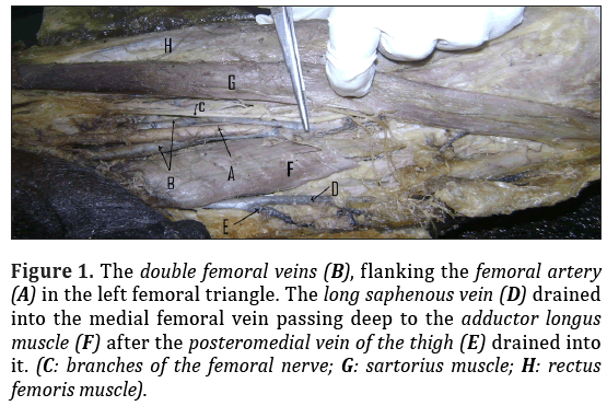
In the left lower limb, there were two femoral veins flanking the femoral artery in the lower part of the femoral triangle. But in the upper part of the triangle, the lateral vein passed deep to the femoral artery to come to its medial side and the two veins ran upwards side by side. Near the apex of the femoral triangle, the lateral femoral vein passed superficial to the femoral artery. Moreover, the long saphenous vein received the posteromedial vein of the thigh and ran deep to the adductor longus muscle to drain into the medial femoral vein in the same limb (Figure 1).
Figure 1: The double femoral veins (B), flanking the femoral artery (A) in the left femoral triangle. The long saphenous vein (D) drained into the medial femoral vein passing deep to the adductor longus muscle (F) after the posteromedial vein of the thigh (E) drained into it. (C: branches of the femoral nerve; G: sartorius muscle; H: rectus femoris muscle).
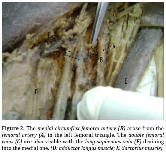
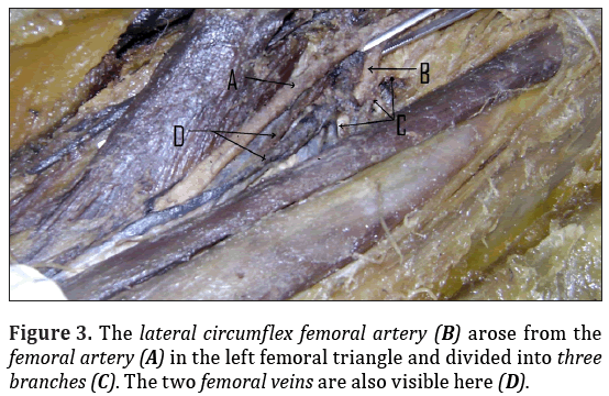
The left-sided medial and the lateral circumflex femoral arteries arose from the femoral artery directly instead of the profunda femoris artery. The medial circumflex passed medially deep to the double femoral veins and between the psoas major and pectineus muscles. The lateral circumflex passed between the divisions of the femoral nerve and posterior to the sartorius and rectus femoris muscles to divide into ascending, transverse and descending branches (Figures 2, 3).
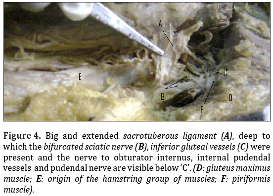
In the right lower limb no such variation was found. However, in the right gluteal region, the sacrotuberous ligament was found to be big enough to cover not only the pudendal nerve and internal pudendal vessels, but also the structures like sciatic nerve,inferior gluteal vessels and nerve, tricipital tendon of gemellus superior and inferior with obturator internus and was attached with the gluteus maximus, piriformis and hamstring muscles (Figure 4).
Figure 4: Big and extended sacrotuberous ligament (A), deep to which the bifurcated sciatic nerve (B), inferior gluteal vessels (C) were present and the nerve to obturator internus, internal pudendal vessels and pudendal nerve are visible below ‘C’. (D: gluteus maximus muscle; E: origin of the hamstring group of muscles; F: piriformis muscle).
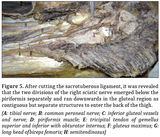
After dissecting this ligament, we found that the right sciatic nerve had been divided into tibial and common peroneal nerves beforehand and they emerged separately below the piriformis muscle to run in the gluteal region, between the greater trochanter and the ischial tuberosity. They were crossed by the long head of biceps femoris and descended in the back of thigh as contiguous but separate structures (Figure 5).
Figure 5: After cutting the sacrotuberous ligament, it was revealed
that the two divisions of the right sciatic nerve emerged below the
piriformis separately and ran downwards in the gluteal region as
contiguous but separate structures to enter the back of the thigh.
(A: tibial nerve; B: common peroneal nerve; C: inferior gluteal vessels
and nerve; D: piriformis muscle; E: tricipital tendon of gemellus
superior and inferior with obturator internus; F: gluteus maximus; G: long head ofbiceps femoris; H: semitendinosus)
In the left lower limb no such variation was found. The sciatic nerve divided into tibial and common peroneal components in the popliteal fossa.
Discussion
The femoral vein may be doubled in part, or throughout its length. It may divide into two and encircle the femoral artery [3]. Double femoral veins were found in 16 limbs (14.0%), among a total of 114 specimens of lower limbs, harvested from 60 adult cadavers by Yan and Rein (2012). They came to the conclusion that it is reliable and safe to use femoral vein as a vascular graft because of the existence of the great saphenous vein. So the present case has significance in vascular surgery [4].
The long saphenous vein is often harvested for grafts in peripheral and coronary arterial surgery [1]. In this case the long saphenous vein, passing deep to the adductor longus muscle, joined the medial femoral vein.
In the proximal thigh, the lateral circumflex femoral artery may arise from the femoral artery and the medial circumflex femoral artery often originates from the femoral artery [1]. Occasionally, both circumflexes arise independently from the femoral artery [5].
The tension developed in the gluteus maximus, biceps femoris, piriformis can influence the sacrotuberous ligament as they are attached with this ligament, so also the obturator internus and hamstring muscles and they have the potential to influence the force transmission through this ligament which has a role in physiotherapy [6]. In the present case the extended sacrotuberous ligament was attached with these muscles.
Smoll (2010) found that the prevalence of high division of sciatic nerve (SN) in cadavers was 16.9% and in surgical case series was 16.2% [7]. Bhattacharya et al. (2013) reported a case with high division of the right SN into common peroneal and tibial nerves in the gluteal region after emerging from the lower border of piriformis. Such variation may cause complications during intramuscular injections or surgery in gluteal region (surgical injury may occur during posterior hip operations) [8,9]. Bapuli et al. stated about a similar case, whereas, Pais et al. (2013) found two cases with high division of the sciatic nerve in the lower part of gluteal region. They concluded that this variation might result in sciatica, piriformis syndrome, coccygodynia, and failed or incomplete block of SN during popliteal block anesthesia. Furthermore, it has importance in neurology, sports medicine [9,10].
Conclusion
This case, with all these variations, will enhance our knowledge in gross anatomy and has important clinical implications in general and vascular surgery, anesthesiology, neurology, physical medicine and sports medicine.
Acknowledgement
We express our gratitude to Dr. Hironmoy Roy, Assistant Professor, Department of Anatomy, North Bengal Medical College, West Bengal, India, for his earnest help to complete the formalities of this case report. We are also grateful to all the members of the Department of Anatomy, Nil Ratan Sircar Medical College, Kolkata, West Bengal, India, regarding this work.
References
- Standring S, Mahadevan V, Collins P, Healy JC, Lee J, Niranjan NS, eds. Gray’s Anatomy, The Anatomical Basis of Clinical Practice. 40th Ed., Spain, Philadelphia, Churchill Livingstone Elsevier. 2008; reprinted in 2011; 1337–1338, 1350, 1366, 1379–1382.
- Bergman RA, Afifi AK, Miyauchi R. Illustrated Encyclopedia of Human Anatomic Variation: Opus III: Nervous System: Plexuses. Sacral Plexus. http://www.anatomyatlases.org/ AnatomicVariants/NervousSystem/Text/SacralPlexus.shtml (accessed March, 2015).
- Bergman RA, Afifi AK, Miyauchi R. Illustrated Encyclopedia of Human Anatomic Variation: Opus II: Cardiovascular System: Veins: Lower Limb: Femoral Vein. http:// www.anatomyatlases.org/AnatomicVariants/Cardiovascular/Text/Veins/Femoral.shtml (accessed March, 2015)
- Yan J, Ren W. Applied anatomical study on feasibility and safety of femoral vein as a vascular graft material. ZhongguoXiu Fu Chang_JianWaiKeZaZhi. 2012; 26: 102–105.
- Bergman RA, Afifi AK, Miyauchi R. Illustrated Encyclopedia of Human Anatomic Variation: Opus II: Cardiovascular System: Arteries: Lower Limb. Medial and Lateral Femoral Circumflex Arteries. http://www.anatomyatlases.org/AnatomicVariants/Cardiovascular/ Text/Arteries/FemoralCircumflexMedLat.shtml (accessed March, 2015).
- Woodley SJ, Kennedy E, Mercer SR. Anatomy in practice: the sacrotuberous ligament. New Zealand, J Physiotherapy.2005; 33: 91–94.
- Smoll NR. Variations of the piriformis and sciatic nerve with clinical consequence: a review. Clin Anat. 2010; 23: 8–17.
- Santanu B, Pitbaran C, Sudeshna M, Hasi D. Different neuromuscular variations in the gluteal region. Int J Anat Var (IJAV). 2013; 6: 136–139.
- Bapuli C, Biswas S, Banerjee M, Amam SA, Majumdar S, Goswami AK, Biswas M, Sinha B. Unilateral high division of the sciatic nerve with divided piriformis. Indian J Basic Appl Med Res (IJBAMR).2013; 3: 16–20.
- Pais D, Casal D, Bettencourt Pires MA, Furtado A, Bilhim T, Angélica-Almeida M, Goyri- O’Neill J. Sciatic nerve high division: two different anatomical variants. Acta Med Port. 2013; 26: 208–211.
Chiranjit Samanta, Manotosh Banerjee, Satyajit Sangram, Sudeshna Majumdar*
Department of Anatomy, Nil Ratan Sircar Medical College, Kolkata-700014, West Bengal, India.
- *Corresponding Author:
- Sudeshna Majumdar, MS, DNB
Professor, Department of Anatomy, Nil Ratan Sircar Medical College, Kolkata-700014, West Bengal, India.
Tel: +91 (943) 3007363
E-mail: sudeshnamajumdar.2007@rediffmail.com
Date of Received: March 23rd, 2015
Date of Accepted: September 22nd, 2015
Published Online: August 16th, 2016
© Int J Anat Var (IJAV). 2016; 9: 25–28.
Abstract
In a single male cadaver multiple variations were found in the lower limbs during routine dissection for undergraduate students in the Department of Anatomy, NRS Medical College, Kolkata, India, in November, 2014.
In the left lower limb of the cadaver, there were two femoral veins, flanking the femoral artery. The long saphenous vein passed deep to the adductor longus to drain into the medial femoral vein. Moreover, the medial and the lateral circumflex femoral arteries arose directly from the femoral artery.
In the right lower limb, the sacrotuberous ligament was big enough and was attached with the gluteus maximus, piriformis and hamstring group of muscles. There was also high division of the sciatic nerve into tibial and common peroneal components emerging separately in the gluteal region.
This case report will augment our knowledge in gross and surgical anatomy, physical medicine and nerve block (regional anaesthesia) in relation to inferior extremity.
-Keywords
double femoral veins, long saphenous vein, medial & lateral circumflex femoral arteries, sacrotuberous ligament, sciatic nerve
Introduction
The femoral vein accompanies the femoral artery, beginning at the adductor opening of the popliteal vein and ending posterior to the inguinal ligament as the external iliac vein. The vein is postero-lateral to the femoral atery in the distal adductor canal. More proximally, in the canal and in the distal femoral triangle, the vein lies posterior to the artery. Proximally at the base of the triangle, the vein runs medial to the artery [1].
Long saphenous vein (great saphenous vein) is the longest vein of the body, starts distally as a continuation of the medial marginal vein of the foot and ends in the femoral vein after passing through the saphenous opening of the deep fascia of the thigh with tributaries like the posteromedial and anterolateral veins of the thigh [1].
The lateral and medial circumflex femoral arteries arise from the profunda femoris artery (a branch of the femoral artery)in proximal thigh and take part in the formation of anastomotic ring around the femoral neck, cruciate and trochanteric anastomoses, etc.[1].
The sacrotuberous ligament is attached by its broad base to the posterior superior iliac spine, the lateral margins of the lower sacrum, upper coccyx. Its oblique fibers are attached to the medial margin of the ischial tuberosity. It then spreads along the ischial ramus as the falciform ligament, which blends with the fascial sheath of the internal pudendal vessels and the pudendal nerve. The lower part of the ligament continues with the tendon of biceps femoris and gluteus maximus to be attached to their posterior surfaces by the lowest fibers [1].
The sciatic nerve is 2cm wide at its origin and it is the thickest nerve of the body with the root value: L4-5, S1-3 (ventral rami). It leaves the pelvis via the greater sciatic foramen, below the piriformis and above the superior gemellus muscle, descends in the gluteal region and along the back of the thigh dividing into tibial (L4-5, S1-3) and common peroneal or common fibular (L4-5, S1-2) nerves at a varying level proximal to the knee [1]. Very often these two nerves arise separately from the sacral plexus. They may be separated in the greater sciatic foramen by the piriformis muscle and pass into the thigh as contiguous but separate structures [2].
The aim of this case report was to know about the variations of the lower limb as that knowledge might contribute in the fields of gross and surgical anatomy, anesthesiology, physical medicine, etc.
Case Report
While doing the routine dissection for MBBS students in November, 2014, multiple variations were found in the inferior extremities of a male cadaver in the Department of Anatomy, NRS Medical College, Kolkata. The subject was about sixty years old. Dissection was done properly in both the lower limbs, structures were observed carefully and relevant photographs were taken.
In the left lower limb, there were two femoral veins flanking the femoral artery in the lower part of the femoral triangle. But in the upper part of the triangle, the lateral vein passed deep to the femoral artery to come to its medial side and the two veins ran upwards side by side. Near the apex of the femoral triangle, the lateral femoral vein passed superficial to the femoral artery. Moreover, the long saphenous vein received the posteromedial vein of the thigh and ran deep to the adductor longus muscle to drain into the medial femoral vein in the same limb (Figure 1).
Figure 1: The double femoral veins (B), flanking the femoral artery (A) in the left femoral triangle. The long saphenous vein (D) drained into the medial femoral vein passing deep to the adductor longus muscle (F) after the posteromedial vein of the thigh (E) drained into it. (C: branches of the femoral nerve; G: sartorius muscle; H: rectus femoris muscle).
The left-sided medial and the lateral circumflex femoral arteries arose from the femoral artery directly instead of the profunda femoris artery. The medial circumflex passed medially deep to the double femoral veins and between the psoas major and pectineus muscles. The lateral circumflex passed between the divisions of the femoral nerve and posterior to the sartorius and rectus femoris muscles to divide into ascending, transverse and descending branches (Figures 2, 3).
In the right lower limb no such variation was found. However, in the right gluteal region, the sacrotuberous ligament was found to be big enough to cover not only the pudendal nerve and internal pudendal vessels, but also the structures like sciatic nerve,inferior gluteal vessels and nerve, tricipital tendon of gemellus superior and inferior with obturator internus and was attached with the gluteus maximus, piriformis and hamstring muscles (Figure 4).
Figure 4: Big and extended sacrotuberous ligament (A), deep to which the bifurcated sciatic nerve (B), inferior gluteal vessels (C) were present and the nerve to obturator internus, internal pudendal vessels and pudendal nerve are visible below ‘C’. (D: gluteus maximus muscle; E: origin of the hamstring group of muscles; F: piriformis muscle).
After dissecting this ligament, we found that the right sciatic nerve had been divided into tibial and common peroneal nerves beforehand and they emerged separately below the piriformis muscle to run in the gluteal region, between the greater trochanter and the ischial tuberosity. They were crossed by the long head of biceps femoris and descended in the back of thigh as contiguous but separate structures (Figure 5).
Figure 5: After cutting the sacrotuberous ligament, it was revealed
that the two divisions of the right sciatic nerve emerged below the
piriformis separately and ran downwards in the gluteal region as
contiguous but separate structures to enter the back of the thigh.
(A: tibial nerve; B: common peroneal nerve; C: inferior gluteal vessels
and nerve; D: piriformis muscle; E: tricipital tendon of gemellus
superior and inferior with obturator internus; F: gluteus maximus; G: long head ofbiceps femoris; H: semitendinosus)
In the left lower limb no such variation was found. The sciatic nerve divided into tibial and common peroneal components in the popliteal fossa.
Discussion
The femoral vein may be doubled in part, or throughout its length. It may divide into two and encircle the femoral artery [3]. Double femoral veins were found in 16 limbs (14.0%), among a total of 114 specimens of lower limbs, harvested from 60 adult cadavers by Yan and Rein (2012). They came to the conclusion that it is reliable and safe to use femoral vein as a vascular graft because of the existence of the great saphenous vein. So the present case has significance in vascular surgery [4].
The long saphenous vein is often harvested for grafts in peripheral and coronary arterial surgery [1]. In this case the long saphenous vein, passing deep to the adductor longus muscle, joined the medial femoral vein.
In the proximal thigh, the lateral circumflex femoral artery may arise from the femoral artery and the medial circumflex femoral artery often originates from the femoral artery [1]. Occasionally, both circumflexes arise independently from the femoral artery [5].
The tension developed in the gluteus maximus, biceps femoris, piriformis can influence the sacrotuberous ligament as they are attached with this ligament, so also the obturator internus and hamstring muscles and they have the potential to influence the force transmission through this ligament which has a role in physiotherapy [6]. In the present case the extended sacrotuberous ligament was attached with these muscles.
Smoll (2010) found that the prevalence of high division of sciatic nerve (SN) in cadavers was 16.9% and in surgical case series was 16.2% [7]. Bhattacharya et al. (2013) reported a case with high division of the right SN into common peroneal and tibial nerves in the gluteal region after emerging from the lower border of piriformis. Such variation may cause complications during intramuscular injections or surgery in gluteal region (surgical injury may occur during posterior hip operations) [8,9]. Bapuli et al. stated about a similar case, whereas, Pais et al. (2013) found two cases with high division of the sciatic nerve in the lower part of gluteal region. They concluded that this variation might result in sciatica, piriformis syndrome, coccygodynia, and failed or incomplete block of SN during popliteal block anesthesia. Furthermore, it has importance in neurology, sports medicine [9,10].
Conclusion
This case, with all these variations, will enhance our knowledge in gross anatomy and has important clinical implications in general and vascular surgery, anesthesiology, neurology, physical medicine and sports medicine.
Acknowledgement
We express our gratitude to Dr. Hironmoy Roy, Assistant Professor, Department of Anatomy, North Bengal Medical College, West Bengal, India, for his earnest help to complete the formalities of this case report. We are also grateful to all the members of the Department of Anatomy, Nil Ratan Sircar Medical College, Kolkata, West Bengal, India, regarding this work.
References
- Standring S, Mahadevan V, Collins P, Healy JC, Lee J, Niranjan NS, eds. Gray’s Anatomy, The Anatomical Basis of Clinical Practice. 40th Ed., Spain, Philadelphia, Churchill Livingstone Elsevier. 2008; reprinted in 2011; 1337–1338, 1350, 1366, 1379–1382.
- Bergman RA, Afifi AK, Miyauchi R. Illustrated Encyclopedia of Human Anatomic Variation: Opus III: Nervous System: Plexuses. Sacral Plexus. http://www.anatomyatlases.org/ AnatomicVariants/NervousSystem/Text/SacralPlexus.shtml (accessed March, 2015).
- Bergman RA, Afifi AK, Miyauchi R. Illustrated Encyclopedia of Human Anatomic Variation: Opus II: Cardiovascular System: Veins: Lower Limb: Femoral Vein. http:// www.anatomyatlases.org/AnatomicVariants/Cardiovascular/Text/Veins/Femoral.shtml (accessed March, 2015)
- Yan J, Ren W. Applied anatomical study on feasibility and safety of femoral vein as a vascular graft material. ZhongguoXiu Fu Chang_JianWaiKeZaZhi. 2012; 26: 102–105.
- Bergman RA, Afifi AK, Miyauchi R. Illustrated Encyclopedia of Human Anatomic Variation: Opus II: Cardiovascular System: Arteries: Lower Limb. Medial and Lateral Femoral Circumflex Arteries. http://www.anatomyatlases.org/AnatomicVariants/Cardiovascular/ Text/Arteries/FemoralCircumflexMedLat.shtml (accessed March, 2015).
- Woodley SJ, Kennedy E, Mercer SR. Anatomy in practice: the sacrotuberous ligament. New Zealand, J Physiotherapy.2005; 33: 91–94.
- Smoll NR. Variations of the piriformis and sciatic nerve with clinical consequence: a review. Clin Anat. 2010; 23: 8–17.
- Santanu B, Pitbaran C, Sudeshna M, Hasi D. Different neuromuscular variations in the gluteal region. Int J Anat Var (IJAV). 2013; 6: 136–139.
- Bapuli C, Biswas S, Banerjee M, Amam SA, Majumdar S, Goswami AK, Biswas M, Sinha B. Unilateral high division of the sciatic nerve with divided piriformis. Indian J Basic Appl Med Res (IJBAMR).2013; 3: 16–20.
- Pais D, Casal D, Bettencourt Pires MA, Furtado A, Bilhim T, Angélica-Almeida M, Goyri- O’Neill J. Sciatic nerve high division: two different anatomical variants. Acta Med Port. 2013; 26: 208–211.


