Dual palmaris longus variation in which muscles originated from antebrachial fascia
Received: 25-Oct-2018 Accepted Date: Jan 03, 2019; Published: 10-Jan-2019, DOI: 10.37532/1308-4038.19.12.005
Citation: Diramali A, Kosif R, Meyvaci SS, et al. Dual palmaris longus variation in which muscles originated from antebrachial fascia. Int J Anat Var. Mar 2019;12(1): 005-007.
This open-access article is distributed under the terms of the Creative Commons Attribution Non-Commercial License (CC BY-NC) (http://creativecommons.org/licenses/by-nc/4.0/), which permits reuse, distribution and reproduction of the article, provided that the original work is properly cited and the reuse is restricted to noncommercial purposes. For commercial reuse, contact reprints@pulsus.com
Abstract
Introduction: Palmaris longus originates from medial epicondyle and attaches to palmar aponeurosis. This muscle is largely used for tendon transfers by surgeons so understanding of its variations is necessary anatomists and clinicians.
Case Report: This report discusses a case of variation in origin of palmaris longus muscles in both extremities of a male cadaver seen in routine anatomy dissection in Department of Anatomy.
Discussion and Conclusion: Palmaris longus is one of the most variable muscles in the human body. The most common variation is agenesis and the total percentage of all other variations is about 9%. Previous studies specified that palmaris longus may begin from antebrachial fascia, biceps brachii, flexor carpi radialis, flexor carpi ulnaris or flexor digitorum superficialis. In our cadaver in both upper extremities palmaris longus started from antebrachial fascia. To prevent unwanted mistakes in surgical procedures and solve unsual clinical symptoms, a clinician must remember variations..
Keywords
Palmaris longus; Variation; Origin; Antebrachial fascia
Introduction
The history of the concept and contents of anatomical variations is essentially the same as that of the anatomy, or more accurately, the basis of the normal structure and composition of the human body. Moore already described the normal as “within the normal range of variation”. Increase in surgical possibilities made variations more important than before [1].
Palmaris longus (PL) is a variable muscle that is one of the major muscles of the human body. It is a short, thin and fusiform muscle that originates from the medial epicondyle of humerus, extends through flexor carpi radialis and flexor carpi ulnaris above flexor digitalis superficialis and its distal tendon attaches to the transverse carpal ligament with palmar aponeurosis [2-5]. Of all the muscles, 91% has this appearance except for agenesis and it can easily be shown with the opposition movement of little finger and thumb [3,6].
The function of the muscle is to support the wrist during flexion and to stretch palmar aponeurosis. It is innervated by median nerve and the bloody supply of the muscle comes from anterior recurrent ulnar artery. Fleckenstein et al. showed that seven of 11 individuals received support from PL in the wrist flexion against resistance [7]. Since PL is located just beneath the skin, it is commonly used for tendon transfers by hand surgeons, and for lip and chin restorations and eyelid reconstruction by plastic surgeons. Another clinical importance is that it acts as a median nerve protector and its variations may cause median nerve entrapment syndromes [1,2,4,7].
Case Report
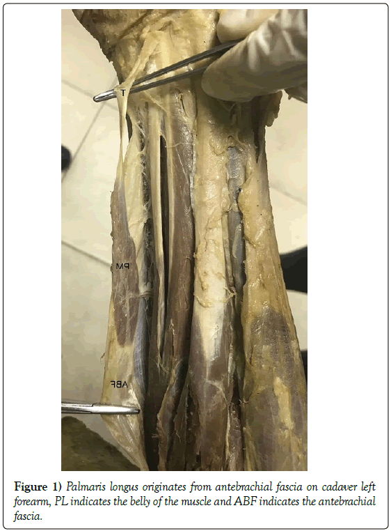
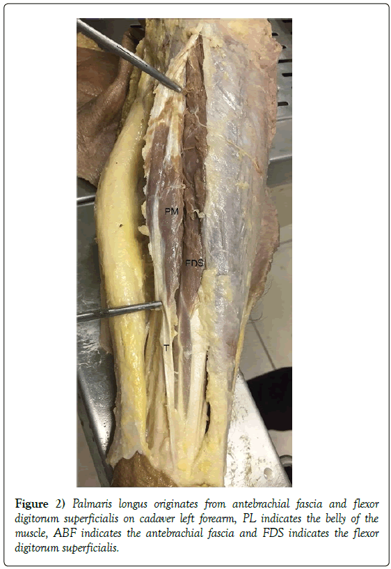
It is observed that the origins of the PLs in both extremities was different from their appearance in conventional text books during routine anatomy dissection of a 65-year-old male cadaver fixed in a 10% formalin solution. The dissection was deepened; the muscles were exposed and photographed. The left PL started from antebrachial fascia whereas right PL originated from both antebrachial fascia and flexor digitorum superficialis. The muscles ended in the transverse carpal ligament and palmar aponeurosis as thin tendons on both sides (Figures 1 and 2). Tendon and muscle measurements were performed with the help of electronic digital calliper. The muscle length, muscle width and tendon length measurements of the right side were 85,23 mm, 21,66 mm and 168,03 mm, respectively; on the left side was atrophic; the muscle length was 64,70 mm, width was 15,19 mm and the tendon length were 129,19 mm.
Discussion and Conclusion
After their study including 1600 cadaver extremities, Reimann et al. grouped the variations of PL by a number of varieties (e.g., total agenesis, location or ratio of muscle to tendon; different starting and ending points, duplication or triplication; the presence of accessory fibres). The most common variation was the agenesis of the muscle (12.8%) [5].
Another study was conducted with 562 patients of Poursinia Hospital and examined 1124 upper extremities of the patients; that study used Schaeffer, Pushpakumar, Thomson and Mishra II tests to detect PL and declared that the incidence of muscle agenesis was 13.2%, which was consistent with the report by Remann et al. [8].
Agenesis is usually bilateral. Its unilateral absence is more common in women and in left extremities. Flexor carpi ulnaris muscle undertakes the task of strengthening the palmar aponeurosis. The total percentage of all other variations is about 9% [2,5,9,10]. The frequencies of muscle duplication were reported as 0.8% and 3.1 % by Reimann et al. and Gruber et al., respectively [5].
Structural differences can be seen in three ways: Proximal, central and distal. The most common form is the proximal. Distal location is named as “inverted palmaris longus muscle” [5,9]. Ramesh et al. reported that only one of the 60 cadavers had a PL that was completely formed by muscular tissue except for the short tendinous structures at the beginning and ending points [11].
In a different classification made by Olewnik depended on standard dissection which realised on 80 upper limbs; the palmaris longus muscle was present in 92.5% of specimens. In addition, three types of palmaris longus muscle were identified associated with the morphology of its insertion (types I-III) and these groups were further subdivided into three subgroups (A-C) according to the ratio of the length of the muscle belly and its tendon. The most frequent was type I (78.8%), where the tendon attached to the palmar aponeurosis. The tendon-to-belly ratio was 1–1.5 (41.1%) in the subtype B of type I [12].
Although previous studies specified that PL may begin from bicipital aponeurosis, antebrachial fascia of the mid-forearm, intermuscular septum, coronoid process, biceps brachii, flexor carpi radialis, flexor carpi ulnaris or flexor digitorum superficialis, none of them was associated with pathology [5,9].
In our cadaver, right PL muscle originated from antebrachial fascia and the left one originated from both antebrachial fascia and the flexor digitorum superficialis. It was located centrally, and it almost reached the mid one third of the forearm. The ending point of the muscle is highly variable. It may end in antebrachial fascia, flexor carpi ulnaris tendon, flexor retinaculum, abductor pollicis brevis, hypothenar muscles and hamatum, pisiform or scaphoid bones [9,10].
Bozkurt et al. observed that distally located PL of a female cadaver starting from medial epicondyle of the right hand gave fibres to abductor digiti minimi as well as flexor retinaculum and palmar aponeurosis [13]. Gune et al. performed a similar, routine dissection of a male cadaver; they found that PMLs of the both extremities were distally located, and the tendon was separated into three parts ending in palmar aponeurosis, fascia covering thenar muscles and the abductor digiti minimi muscle [14]. The variations in the distal part of the muscle are important due to the proximity of the ulnar and median nerves [9,10].
Sunil et al. performed a dissection of an adult male cadaver and discovered that the tendon of PL was divided into two on the right side and into three on the left side. On both sides, they gave fibers to accessorius ad flexorem digiti minimi, an accessory muscle attaching to fifth metacarpal body. They thought that the ulnar artery had a rounded course in their cadaver due to the closeness of this accessory muscle [10].
Markeson et al. observed a subcutaneous mass in a male patient that was admitted due to the complaint of intermittent pain and paraesthesia in the innervation region of left median nerve for three months. That mass was located on the connection point of the mid and distal one third and it was moving with flexion and extension. After surgery, they decided that the mass was a variant of PL [6].
Pires et al. observed a remarkable variation of the reversed palmaris longus in a male cadaver with no known lead to death was fixated with a phenol solution and dissected for teaching purposes. The referred muscle was abnormally enlarged and was mainly consists of muscle tissue. Although it inserted itself into the flexor retinaculum, it is originated by a tendinous slip and became fully fleshy. It had regular origins and attachments points were as usual [15].
Kocabıyık et al. performed a study on 24 human foetuses’ upper limbs aged 17-40 weeks of gestation. The palmaris longus muscles were absent in both forearms in 7 and in one forearm in 5 foetuses. Moreover, most foetuses had a typical palmaris longus muscle and tendon shape. Reversed palmaris longus muscle was found in 2 forearms in two foetuses. Long muscular belly and short tendon of palmaris longus muscle was observed in one of 24 foetuses but in both forearms [16].
Similarly, Tiengo et al. had a female patient who had a tingling sensation on palmar side of the right hand and the first three fingers for five months. She had no nocturnal symptoms and negative Tinel sign. The excised mass was located epifascially to grow with the hand’s flexion movement. Histologic examination defined the mass as PL [6].
In our cadaver, palmaris longus muscles on both sides were ending in palmar aponeurosis with thin tendons. His medical history did not reveal any entrapment symptoms of median or ulnar nerves and his hand muscles were not atrophic. The clinical importance of a symptomatic PL is evident. However, an asymptomatic muscle is also interesting with regard to local radiological examinations and possible problems during endoscopic wrist surgery [9].
Excision of these unusual muscles is controversial and should be considered in each case. Wesser et al. did not see surgery as necessary since these variations could be functional units of the fingers. Similarly, Vichare did not remove these muscles unless they had clinical symptoms. Kostakoğlu et al. preferred surgery because of their potential for pain and compression syndromes [17]. In surgical procedures involving the upper extremity (e.g., tendon reconstructions, carpal tunnel or Guyon canal decompressions), all variations of the palmaris longus muscle should be considered.
REFERENCES
- Enye LA, Saalu LC, Osinubi AA. The Prevalence of Agenesis of Palmaris Longus Muscle amongst Students in Two Lagos-Based Medical Schools. Int J Morphol. 2010;28:849-54.
- Natsis K, Levva S, Totlis T, et al. Three-headed reversed palmaris longus muscle and its clinical significance. Ann Anat. 2007;189:97-101.
- Norbury JW, Morrison EJ. Palmaris Longus Tendinopathy Diagnosed with Ultrasound: A Case Report. PMR. 2018.
- Pekala PA, Henry BM, Pekala JR, et al. Congenital absence of the palmaris longus muscle: A meta-analysis comparing cadaveric and functional studies. J Plast Reconstr Aesthet Surg. 2017;70:1715-24.
- Reimann AF, Daseler EH, Anson BJ, et al. The palmaris longus muscle and tendon. A study of 1600 extremities. The Anatomical Record. 1944;89:495-505.
- Markeson D, Basu I, Kulkarni MK. The dual tendon palmaris longus variant causing dynamic median nerve compression in the forearm. J Plast Reconstr Aesthet Surg. 2012;65:220-2.
- Fowlie C, Fuller C, Pratten MK. Assessment of the presence/absence of the palmaris longus muscle in different sports, and elite and non-elite sport populations. Physiotherapy. 2012:98;138-142.
- Nasiri E, Pourghasem M, Moladoust H. The prevalence of absence of the palmaris longus muscle tendon in the north of Iran: A comparative study. Iran Red Crescent Med J. 2016;18:1-4.
- Fazan VPS. Reversed palmaris longus muscle and medıan nerve relationships. Case report and literature review. Braz J Morphol Sci. 2007;24:88-91.
- Sunil V, Rajanna S, Gitanjali, et al. Variation in the insertion of the palmaris longus tendon. Singapore Med J. 2015;56:7-9.
- Ramesh P, Aswin P, Pratibha G. A fleshy palmaris longus muscle. Anat J Africa. 2015;4:436-9.
- Olewnik L, Wysiadecki G, Polguj M, et al. Anatomical variations of the palmaris longus muscle including its relation to the median nerve- a proposal for a new classification. BMC Musculoskeletal Disorders. 2017;18:539.
- Bozkurt MC, Tagil SM, Ersoy M, et al. Muscle variations and abnormal branching and course of the ulnar nerve in the forearm and hand. Cli Anat. 2004;17:64-6.
- Gune, Pote AJ, Patil AD, et al. Bilateral reversed palmaris longus muscle with trifid insertion, a rare variation. Int J Res Med Sci. 2014;2:741-3.
- Pires L, Perisse JP, Araujo GCS, et al. Hypertrophic reversed palmaris longus muscle: a cadaveric finding. Folia Morphol. 2017;77:403-5.
- Kocabıyık N, Yıldız S, Develi S, et al. Morphometric analysis of the palmaris longus muscle: a fetal study. Anat. 2013;6:42-7.
- Caetano EB, Neto JJS, Ribas LAA, et al. Accessory muscle of the flexor digitorum superficialis and its clinical implications. Rev Bras Ortop. 2017;52:731-4.


