Duplicated vas deferens identified in post-mortem analysis
Received: 24-Dec-2020 Accepted Date: Feb 05, 2021; Published: 12-Feb-2021, DOI: 10.37532/1308-4038.14(2).163-164
This open-access article is distributed under the terms of the Creative Commons Attribution Non-Commercial License (CC BY-NC) (http://creativecommons.org/licenses/by-nc/4.0/), which permits reuse, distribution and reproduction of the article, provided that the original work is properly cited and the reuse is restricted to noncommercial purposes. For commercial reuse, contact reprints@pulsus.com
Abstract
The identification of a duplicated Vas Deferens is significant during common operations such as vasectomies, groin explorations and hernioplasties. The reports are rare and incidental. Consequences of failure to identify this anomaly may yield effects such as sperm granulomas and vasovenous fistulas. We present the first publication of a duplicated vas from an Australian cadaveric dissection.
Keywords
Duplicated Vas Deferens; Anatomical variation; Australia; General surgery; Urology
Introduction
The literature offers only a few published cases of Duplicated Vas Deferens. Although the incidence of the anomaly is rare, it may be under-reported or under-recognised [1]. The identification of this anomaly may avoid iatrogenic intraoperative injury. Duplicated Vas Deferens may be identified as a secondary pathology during inguinal surgery. The significance of intraoperative failure of its identification is significant to specialists such as urologists, general surgeons and any proceduralist who operates in this area.
Findings
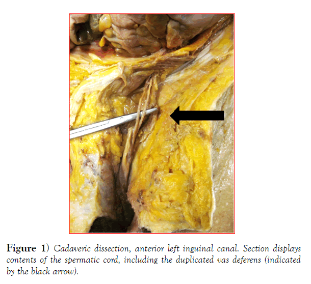
During a routine anatomical dissection, conducted at The University of Melbourne Anatomical Department, a duplicated Vas Deferens was identified. There were no associated renal anomalies noted during dissection. Both the testis and epididymis were normal. From a review of the cadaver’s premortal, limited clinical history, there were no documented symptoms that may have led to surgical discovery of this anomaly. This is in accordance with current knowledge of a largely asymptomatic pathology.
Discussion
This is the first published finding of a duplicated Vas Deferens within Australia. While this discovery was made post-mortem, it is significant because of the relative rarity of this abnormality, with an estimated prevalence of below 1 in 2000 males (0.05%) [1-3]. Anomalies of vas deferens can be categorised as absence, duplication, ectopia, hypoplasia and diverticulum. Some anomalies may be associated with other congenital abnormalities such as ipsilateral renal agenesis and cystic fibrosis, but no cases of abnormalities of the kidneys, ureters, or bladder have been demonstrated with duplication of the vas deferens [1,4].
The embryologic etiology of a duplicated vas deferens has not yet been clearly established. Around week seven of gestation, when the development of the male reproductive organs begins, the paramesonephric duct regresses and the spermatogenic cord appear in the fetal testis [5]. The mesonephric duct gives rise to the epididymal canal and ejaculatory duct while central portion of the mesonephric duct specifically gives rise to the vas deferens. Hence, duplication of the vas may result from duplication of the fetal mesonephric system or transversal division of the mesonephric duct during organogenesis [5,6]. Gill et al. argued that aberrant ductal structures in resected pediatric hernia sacs are embryologic remnants, persistent mesonephric tubules which failed to incorporate into the efferent tubules of the testes [7]. As these remnants deteriorate during puberty, the anomalies are rarely seen in adults. Most of these findings are incidental.
Previously reported cases of this rare anomaly were identified during procedures such as: orchidopexy; inguinal hernia repair; vasectomy; varicocoelectomy; and, radical prostatectomy [1,8,9]. In Australia, a total of 324 618 groin hernia repairs were performed on adults between 2000 and 2015 [10]. Groin repairs, particularly via laparoscopic methods, have been increasing on a yearly basis. Intraoperative identification during inguinal hernia repair is critical to avoid potential complications such as infertility, chronic testicular pain, and spermatic granulomas [11-13]. The rate of such complications may be higher if the duplication is unilateral and hence, harder to identify. Post-operative imaging may define any other extra testicular abnormalities [4].
In hypoplastic or absence anomalies, the failure of identification carries the same consequences. During vasectomy, failure to identify a duplicated vas deferens may lead to failure of the surgery and significant legal ramifications. Literature review has revealed that only 2 out of 29 such cases were found during a vasectomy. While it has not yet been reported if patient fertility is affected by duplication of the vas, it is plausible that fertility might be negatively impacted. Poly-vas deferentia may yield asthenospermia, or reduced spermatic motility [1], which has further been associated with a decline in fertility [14]. Given the current rarity of poly vasa deferentia, high rate of asymptomatic cases and, wide age range of affected patients, a study to ascertain this as a direct side effect of this pathology is impractical [1,11].
At time of writing, only 29 other cases were published since 1959 [15]. Our reported duplication fulfills the requirement of a type I poly-vasa deferential classification [4]. (Figure 1) displays a clear, and complete, bifurcation of the vas deferens within the anterior inguinal canal. Polymorphism and an ectopic ureter were both absent in this case thus ruling out the other proposed classifications. Furthermore, we observed left sided vas duplication; however duplication is equally probable on either side [4].
Vohra and Morgentaler suggest that calling an anomaly true duplication of the vas deferens should be restricted to cases in which a duplicate vas is identified within the spermatic cord and should be distinguished from the term double vas deferens, which is used in several case reports in the literature to describe an ectopic ureter draining into the seminal vesicle [5]. If the duplicated vas deferens is separated for only a short distance, then the duplication is said to be partial. To definitively classify the anomaly, Liang et al proposed the below classification (Table 1).
| Type 1 | Classical duplication of vas deferens, partial or complete. Most fall within this category. |
| Type 2 | Multiple vasa deferentia with polyorchidism |
| Type 3 | False poly vas representing the double vasa deferentia where an ectopic ureter drains into the ejaculatory system |
Table 1: Classification of poly vas deferens [4].
Conclusion
We present a case study that identifies a type I vas deferens duplication. Given that the overall reports of vas duplication are rare and incidental, likely under-recognised or under-reported, this finding is significant given the increasing popularity of groin surgery in nations like Australia. Awareness of this anomaly will prevent future iatrogentic complications and their ramifications of asthenospermia and overall fertility.
Acknowledgement
We thank the University of Melbourne Anatomy Department for granting permission for us to use their specimens. We would additionally like to thank all staff within this department for their assistance in specimen preparation.
REFERENCES
- Lee JN, Kim BS, Kim HT, et al. A Case of Duplicated Vas Deferens Found Incidentally during Varicocelectomy. World J Mens Health. 2013;31:268-71.
- Terawaki K, Satake R, Takano N, et al. A rare case of duplicated vas deferens and epididymis. J Pediatr Surg Case Rep. 2014.2:541-3.
- Akay F, Atug F, Turkeri L. Partial duplication of the vas deferens at the level of inguinal canal. Int J Urology. 2005. 12(8): p. 773-775.
- Liang MK, Subramanian A, Weedin J, et al. True duplication of the vas deferens: a case report and review of literature. Int Urol Nephrol. 2012;44:385-91.
- Vohra S, Morgentaler A. Congenital anomalies of the vas deferens, epididymis, and seminal vesicles. Urology. 1997;49:313-21.
- Karaman A, Karaman I, Yagız B, et al. Partial duplication of vas deferens: How important is it? J Indian Assoc Pediatr Surg. 2010;15:135-6.
- Gill B, Favale D, Kogan SJ, et al. Significance of accessory ductal structures in hernia sacs. J Urol. 1992;148:697-8.
- Khandelwal R, Punia S, Vashistha N, et al. Duplication of vas deferens-A rare anomaly with review of literature. Int J Surg Case Rep. 2011;2:241-2.
- Shariat SF, Naderi ASA, Miles B, et al. Anomalies of the wolffian duct derivatives encountered at radical prostatectomy. Rev Urol. 2005;7:75-80.
- Kevric J, Papa N, Toshniwal S, et al. Fifteen-year groin hernia trends in Australia: the era of minimally invasive surgeons. ANZ J Surg. 2018;88:E298-E302.
- Silich RC, McSherry CK. Spermatic granuloma an uncommon complication of the tension-free hernia repair. Surg Endosc. 1996;10:537-9.
- Chintamani, Khandelwal R, Tandon M, et al. Isolated unilateral duplication of vas deferens, a surgical enigma: a case report and review of the literature. 2009;2:167.
- Breitinger MC, Roszkowski EV, Bauermeister AJ, et al. Duplicate Vas Deferens Encountered during Inguinal Hernia Repair: A Case Report and Literature Review. Surg Case Rep. 2016;1-4.
- Barak S, Baker HWG. Clinical Management of Male Infertility, in Endotext, K.R. Feingold, et al., Editors. 2000: South Dartmouth (MA).
- Coetzee T. Double vas deferens: a case report. BJU. 1959;31:336-9.


