Duplication of Renal Arteries with Unusual Extra Renal Division Patterns and Positional Relationship to Hilar Structures
Received: 04-Apr-2022, Manuscript No. ijav-22-4698; Editor assigned: 06-Apr-2022, Pre QC No. ijav-22-4698(PQ); Reviewed: 25-Apr-2022 QC No. ijav-22-4698; Revised: 27-Apr-2022, Manuscript No. ijav-22-4698(R); Published: 30-Apr-2022, DOI: 10.37532/1308-4038.15(4).185
Citation: Tessema CB. Duplication of Renal Arteries With Unusual Extra Renal Division Patterns and Positional Relationship to Hilar Structures. Int J Anat Var. 2021;15(4):166-168.
This open-access article is distributed under the terms of the Creative Commons Attribution Non-Commercial License (CC BY-NC) (http://creativecommons.org/licenses/by-nc/4.0/), which permits reuse, distribution and reproduction of the article, provided that the original work is properly cited and the reuse is restricted to noncommercial purposes. For commercial reuse, contact reprints@pulsus.com
Abstract
During the dissection of an 80-year-old male cadaver, left unilateral duplication of renal arteries consisting of an accessory renal artery forming the superior polar artery and a main renal artery that divided into prominent extra renal divisions were detected. In another 77-year-old female cadaver, bilateral double renal arteries with different extra renal division patterns, including a superior polar division from one of the double renal arteries on each side, were noted. In both cases, the extra renal divisions had variable positional relationships to the other hilar structures of the kidneys.
The presence of such variations and their co-occurrence could be associated with potential risk of various medical conditions like secondary hypertension and could also complicate outcomes of clinical procedures. So, awareness of such variations would be crucial in the diagnosis and treatment of renal, urologic and other associated diseases, and in the reduction of the risk of complications.
Keywords
Accessory renal arteries; Double renal arteries; Extra renal division
Introduction
The kidneys receive about 20 – 25% of the cardiac output delivered to them by renal arteries, which are one of the paired lateral branches of the abdominal aorta. Each renal artery before reaching the hilum of the kidneys gives branches to the suprarenal gland, renal pelvis, ureter, perinephric tissue and renal capsule. In ideal condition, each renal artery terminates near the hilum of the kidneys by dividing into anterior and posterior branches running on the respective sides of the renal pelvis that further divide into renal segmental arteries [1]. Though it is often described that a single renal artery supplying each kidney is found in 70% of the cases, there are variable data indicating extensive variations of the renal arteries. As demonstrated by a dissection study of 30 kidneys of 15 preserved cadavers, about 86.6% of them showed a single artery supplying each kidney [2]. Whereas, a CT renal angiographic study of 248 cases revealed a lower percentage (43.35%) of single renal artery supplying each kidney [3]. Another recent CT renal angiographic investigation done on 200 patients also noted that renal arterial variation accounts for 37% of renal vascular variations, which is frequently seen in males than in females. The commonest of these variations being the presence of accessory renal artery [4]. According to many other studies, renal arterial variations included the unilateral or bilateral presence of multiple (2 - 6) renal arteries and their early (extra renal, pre-hilar) division (branching) patterns [4-7]. It is also demonstrated by a number of studies that the pattern of terminal division of renal arteries is widely variable. A CT renal angiographic research done on 204 patients, noted that the extra renal (pre-hilar) division pattern has a prevalence of 14.7% [8]. As all these studies indicate, there is a wide range of variability in the origin, number and division patterns of renal arteries. The frequency of these variations reflects the complexity of the renal organogenesis representing vestiges of ascent of the kidneys during the 6th – 9th week of gestation from the pelvis to the final lumbar destination. This ascent is accompanied by sequential sprouting of branches from the aorta while the preceding lower branches regress [9]. Chan PL et al. [10] in their case report of 21and 41-year-old female patients with hypertension and accessory renal arteries concluded that accessory renal arteries are common and may contribute (depending on the anatomy and hemodynamic characteristics) to resistant secondary hypertension through renin dependent hyperaldosteronism.
Materials and Methods
During the dissection study of the retroperitoneal space of 80-year-old male and 77-year-old female cadavers, arterial variations in the number, division patterns and relations to hilar structures of the kidneys were noted. The arteries were carefully dissected, cleaned and painted red and photographs were taken for illustrations. In the male cadaver, where there were two arteries, the larger artery was considered as the main renal artery and the smaller one was taken as accessory renal artery. The bilateral double renal arteries with comparable sizes in the female cadaver were considered as superior and inferior renal arteries, while the corresponding extra renal branches in both cadavers were designated as superior, middle and inferior for a purpose of description.
Case Report
Case 1
In the 80-year-old male cadaver, double left renal arteries (superior and inferior) with an unusual division pattern of the inferior renal artery and its relation to hilar structures were observed:
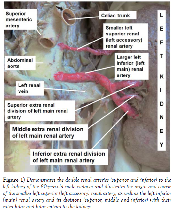
1. The smaller left superior renal artery (left accessory renal artery) arose directly from the left lateral part of the abdominal aorta at the level between the origins of superior mesenteric artery and the left main renal artery and entered the medial aspect of the superior pole of the left kidney as superior polar artery.
2. The larger left inferior renal artery (left main renal artery), at first, ran superior and posterior to the left renal vein. Then it crossed anterior to the renal vein and descended obliquely to bifurcate into superior and inferior extra renal divisions. A smaller middle extra renal division branched from the inferior extra renal division short after the bifurcation. These three extra renal divisions (superior, middle and inferior) of the left main renal artery then entered the left kidney through the hilum anterior to all other hilar structures (Figure 1).
Figure 1) Demonstrates the double renal arteries (superior and inferior) to the left kidney of the 80-year-old male cadaver and illustrates the origin and course of the smaller left superior (left accessory) renal artery, as well as the left inferior (main) renal artery and its divisions (superior, middle and inferior) with their extra hilar and hilar entries to the kidneys.
Case 2
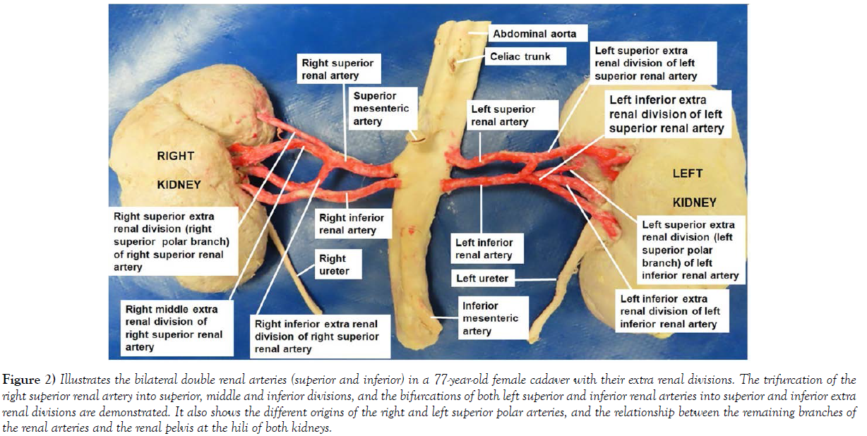
In the 77-year-old female cadaver, bilateral duplication of the renal arteries was detected, where each kidney was supplied by a pair of renal arteries (right and left superior and inferior renal arteries); both of which branched from the abdominal aorta inferior to the level of the origin of the superior mesenteric artery. These bilateral double renal arteries ramified into extra renal divisions before reaching the hilum of the kidneys. The right superior renal artery divided into three extra renal divisions, one of which was extra hilar and entered the medial aspect of the superior pole of the right kidney as right superior polar branch, while the middle and inferior divisions were hilar branches that entered the kidney through the hilum posterior to the renal pelvis. The right inferior renal artery continued undivided and entered the lower corner of the hilum of the right kidney anterior to the renal pelvis. In contrary to those on the right side, the left superior and inferior renal arteries divided into two extra renal divisions each. Even though, both divisions of the left superior renal artery were hilar, which entered the left kidney anterior to the renal pelvis, the inferior division descended caudally anterior to the inferior renal artery, turned laterally at about a right angle and entered the left kidney at the lower corner of the hilum anterior to renal pelvis. The inferior extra renal division of the left inferior renal artery was hilar and entered the kidney through hilum posterior to the renal pelvis, while its superior extra renal division was extra hilar and entered the medial aspect of the superior pole of the left kidney as a left superior polar artery (Figure 2).
Figure 2) Illustrates the bilateral double renal arteries (superior and inferior) in a 77-year-old female cadaver with their extra renal divisions. The trifurcation of the right superior renal artery into superior, middle and inferior divisions, and the bifurcations of both left superior and inferior renal arteries into superior and inferior extra renal divisions are demonstrated. It also shows the different origins of the right and left superior polar arteries, and the relationship between the remaining branches of the renal arteries and the renal pelvis at the hili of both kidneys.
Discussion
Though, a single renal artery supplying each kidney is frequently described to be found in 70% of the cases, current literature review shows inconsistency, which varies from 86.6% to as low as 43.35%. There are also similar inconsistencies in the various study results related to the existence of variant renal arteries that range between 13.30% and 56.65%. An earlier CT angiographic study done on 284 patient noted multiple renal arteries in 19.35% of the cases and out of which the one with a larger caliber is considered as the dominant renal artery while the rest are taken as accessory. It is obvious that the current finding in the 80-year-old male cadaver also goes in line with this description where the inferior remarkably larger one is a dominant (main) renal artery, while the superior and smaller one is an accessory renal artery. However, in the case of the bilateral pairs of renal arteries in the 77-year-old female cadaver, they seem to have comparable size. Therefore, the author simply considered them as superior and inferior renal arteries rather than main and accessory renal arteries. The renal arteries also have variability in their branching patterns to the kidneys. Many previous studies, have documented that variable extra renal branching patterns are frequent encounters, which were also observed in both cadavers in the current report, which confirms the earlier documentations. However, in addition to their duplications and variable extra renal branching patterns, the two cases in this report are unique in that the origin of the superior polar artery is completely different in the left kidney of the male cadaver and in the two kidneys of the female cadaver. In the male cadaver it is a continuation of the accessory renal artery, whereas, in the female cadaver it arose from the right superior renal artery on the right side and from the inferior renal artery on the left side. Moreover, the oblique descent of the left main renal artery anterior to the left renal vein in the male cadaver and the inferior branch of the left superior renal artery anterior to the inferior renal artery in the female cadaver to enter the respective left kidneys anterior to the hilar structures including the renal pelvis, are also peculiar and could increase their liability to iatrogenic injury during surgery. It is also remarkable that, in the female cadaver, the hilar right inferior renal artery entered the kidney anterior to the renal pelvis but the inferior division of left inferior renal artery entered the left kidney posterior to the renal pelvis at the hilum.
The co-existence of such variations in the number, branching and distribution patterns, and relationships in different cadavers and even on the opposite sides of the same cadaver, like in this report, could be challenging to professionals concerned with the diagnosis and treatment of patients with renal, urologic or other related diseases. It could also be a cause of minor or major health compromises in otherwise living healthy kidney donors. Therefore, the author believes that considering the inclusion of such variations of clinical importance during preclinical anatomy teaching can equip students with the indispensable knowledge that fosters awareness during clinical practice.
Conclusion
The presence of such renal arterial variations is associated with potential risk of various medical conditions like secondary hypertension and the cooccurrence of multiple variations like this could have a detrimental effect on outcomes of different procedures and increases the risk of complications. Therefore, awareness of such variations would help in the differential diagnosis of causes of hypertension, renal and urologic diseases, and in the planning and deployment of appropriate strategies to reduce the risk of complications during procedures like interventional radiology, harvesting healthy donor kidneys, renal transplantation, endovascular repair of abdominal aortic aneurysm and other nephron-urologic surgeries. Hence, it is worth of consideration in the pre-clinical and clinical teachings.
Acknowledgement
I am thankful to the donors and their families for their invaluable donation and consent for education, research and publication. My thanks also go to the department of biomedical sciences for the encouragement and uninterrupted support. I am also grateful to Denelle Kees, Chelsey Swanson and John Opland for their immense assistance during the dissection of this cadaver in the gross anatomy lab.
Conflicts Of Interest
None.
REFERENCES
- Standring S. Gray’s anatomy the anatomical basis of clinical practice, 40th Ed., Churchill Livingstone, Elsevier. 2008; 1231-1233.
- Aristotle S, Sundarapandian, Felicia C. Anatomical variations in the blood supply of the kidneys. J Clin Diagn Res. 2013; 7(8): 1555 -1557.
- Majos M, Stefanczyk L, Szemraj-Rogucka Z et al. Does the type of renal artery anatomic variant determine the diameter of main vessel supplying a kidney? A study based on CT data with a particular focus on the presence of multiple renal arteries. Surg Radiol Anat. 2018; 40: 381-388.
- Famurewa OC, Asaleye CM, Ibitoye BO et al. Variation of renal vascular anatomy in a Nigerian population: a computerized tomography study. Niger J Clin Pract 2018; 21(7):840-846.
- Budhiraja V, Rastogi R, Jain V. Anatomical variations of renal artery and its clinical correlations: a cadaveric study from central India. J Morphol Sci 2013; 30(4): 228-233.
- Daescu E, Zahoi DE, Alexa A et al. Morphological variability of renal artery branching pattern: a brief review and anatomic study. Rom J Morphol Embryol. 2012; 53 (2): 287-291
- Recto C, Pilia AM, Campi R et al. Renal artery variation: a 20,782 kidney review. IJAE. 2019; 124 (2):153-163.
- Sungura R, Mathenge I, Onyambu C. The radiological scrutiny of the relation between renal vascular dimensions and anatomical variation of renal arteries: The must to know before renal transplantation. J Kidney. 2017; 3: 158.
- Kem DC, Lyons DF, Wenzl J et al. Renin-dependent hypertension Caused by non-focal stenotic aberrant artery- proof of a new syndrome. Hypertension. 2005; 46: 380-385.
- Chan PL, Tan FHS. Renin dependent hypertension caused by accessory renal arteries. Clinical hypertension. 2018; 24: 15.
Indexed at, Google Scholar, Crossref
Indexed at, Google Scholar, Crossref
Indexed at, Google Scholar, Crossref
Indexed at, Google Scholar, Crossref
Indexed at, Google Scholar, Crossref
Indexed at, Google Scholar, Crossref


