Duplication of the inferior vena cava – report of a rare congenital variation
Arindom Banerjee*, Siddarth Maharana, I. Anil Kumar and P. Jhansi
Department of Anatomy, Konaseema Institute of Medical Sciences and Research Foundation, Amalapuram, Andhra-Pradesh, India
- *Corresponding Author:
- Dr. Arindom Banerjee
Associate Professor, Department of Anatomy, Konaseema Institute of Medical Sciences and Research Foundation, Amalapuram, NH – 214, East-Godavari District, Andhra-Pradesh, 533201, India
Tel: +91 (996) 6275513
E-mail: arindomdoc@yahoo.co.in
Date of Received: January 2nd, 2012
Date of Accepted: September 22nd, 2012
Published Online: December 31st, 2012
© Int J Anat Var (IJAV). 2012; 5: 141–143.
[ft_below_content] =>Keywords
double inferior vena cava, congenital variation, embryonic cardinal venous system, retroperitoneal surgery, thrombo embolic diseases
Introduction
The inferior vena cava (IVC) develops during the 6th to 10th week of embryogenesis from many sources like the posterior cardinal vein, the subcardinal and supracardinal veins and various anastomoses between these veins. Any deviation in this complex process of embryogenesis may lead to variations in the final development of the IVC. Duplication of the IVC in one such relatively rare condition which majority of cases is clinically silent and diagnosed incidentally by imaging (including computed tomography [CT] and magnetic resonance [MR] imaging) done for other reasons. It is a relatively uncommon condition with a reported incidence of 0.2–3%[1].The percentage of intraoperative findings varies in different series between 0.2-0.6%of cases. Variations in the IVC are hence indicative of defective angiogenesis and are of immense surgical importance especially retroperitoneal surgeries and in cases of thromboembolism.
Case Report
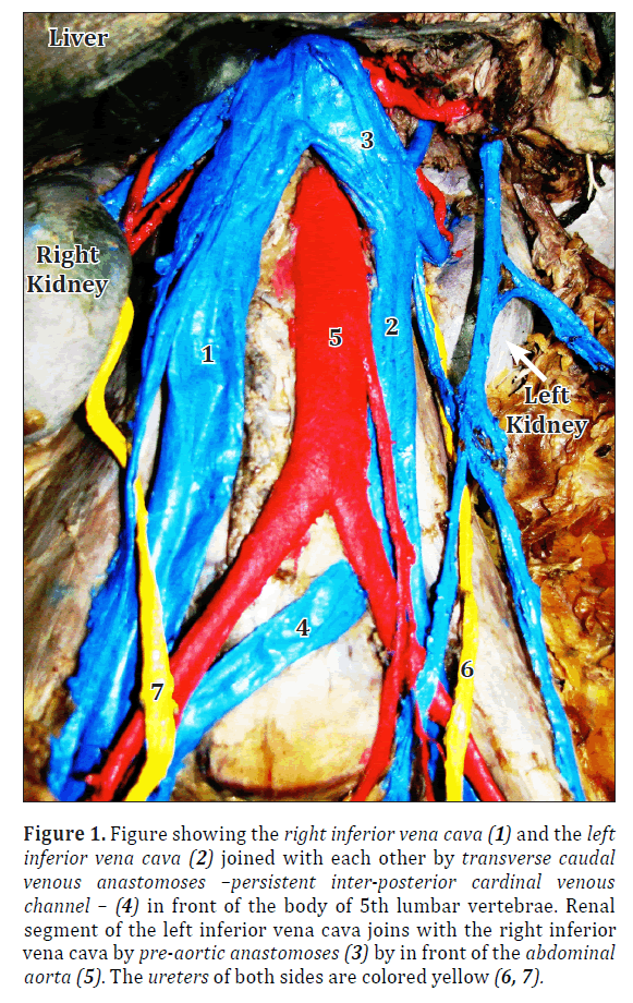
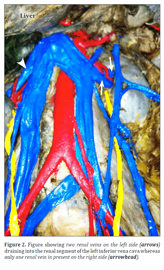
The present case was observed during abdomen dissection in an adult male cadaver for undergraduates in the Department of Anatomy, Konaseema Institute of Medical Sciences and Research foundation, Amalapuram. In this case apart from the IVC present normally on the right side, another similar vein was also seen running parallel to it on the left side. This left sided IVC was formed by the junction of left internal and external iliac vein and continued upwards laying on left side of lumbar segment of vertebral column up to the liver where it joined with the right IVC and ascended cranially as a common trunk behind the liver to open normally into the right atrium. The common iliac veins before continuing as IVC on either side were interconnected by transverse venous anastomoses which was situated dorsal to the common iliac arteries and ventral to the body of the fifth lumbar vertebrae. Moreover, the two IVC’s were again united ventral to the abdominal aorta at the level of the entry of the left renal vein (Figure 1). On the right side, one renal vein drained into the right sided IVC whereas on the left side two renal veins were founded draining into the left IVC. The right testicular vein joined the right vena-caval trunk at the normal level but the left one was found to join the left IVC just below the point of drainage of the left renal vein (Figure 2).
No defect was being found in intersegmental lumbar veins.
Figure 1. Figure showing the right inferior vena cava (1) and the left inferior vena cava (2) joined with each other by transverse caudal venous anastomoses –persistent inter-posterior cardinal venous channel – (4) in front of the body of 5th lumbar vertebrae. Renal segment of the left inferior vena cava joins with the right inferior vena cava by pre-aortic anastomoses (3) by in front of the abdominal aorta (5). The ureters of both sides are colored yellow (6, 7).
Discussion
Embryogenesis of IVC is complex process of successive appearance of three pairs of primitive veins (posterior cardinal, subcardinal and supracardinal veins) interconnecting, anastomozing and followed by regression. This process begins at the sixth week of gestation and is completed by the tenth week [1]. Development of the IVC can broadly be divided from cranio-caudally into the common hepatic segment, the hepatic, renal and the post renal segments. In the present case the common hepatic segment and the hepatic segment of IVC are normal in development.Below the liver two separate IVC’s are present on either side of the abdominal aorta possessing the usual tributaries (renal veins, suprarenal and gonadal veins). On the right side the development of the renal and post-renal part of the IVC is almost normal (i.e.,right posterior subcardinal vein and right supra-subcardinal anastomoses). The right common iliac vein was derived from the caudal part of right posterior cardinal vein caudal to the inter-posterior cardinal venous anastomoses. The renal segment on the left side was derived from the inter-subcardinal anastomoses (pre-aortic anastomoses), which normally constitutes the part of the left renal vein crossing anterior to the abdominal aorta [2]. Post renal segment is developmentally almost similar to that of the right side. The IVC’s of the two side are interconnected by a venous channel which is the persistent inter posterior cardinal venous channel.
Double IVC is a rare variation and only seen in 0.2–3% cases as reported by earlier researchers. In this present case the sources of development of renal segments of IVC’s of both the sides differ. The renal segment of left IVC was derived from the pre-aortic anastomosis, which usually gives rise to the development of left renal vein. The cardinal venous system on the left side usually disappears in the course of development, but persisted in this case to form the left IVC which represents the left post-renal segment like that of the right side. The embryological posterior cardinal venous anastomoses persisted and joined the two IVC.
Renal veins are formed by anastomoses of the subcardinal veins and supracardinal veins and according to previous researches it is found that supernumerary renal veins if present are more common on the right side [3]. But in this case two renal veins are seen on the left and single one is seen on the right side. A similar case was reported by Rischbieth, in which he found double origin of left renal vein which eventually united together and drained in the left IVC. In his study also there was only one renal vein present on the right side [4].
The majority of cases of double IVC are diagnosed incidentally by imaging for other reasons, but these variations can have significant clinical implications. Radiologically, the presence of double IVC can be mistaken as a pathological lesion such as lymphadenopathy[5,6] or left pyelo-ureteric dilatation[7].The presence of double IVC may also complicate retroperitoneal surgery. The double IVC can be inadvertently injured or ligated during retroperitoneal surgery[8].
Moreover, there are several case reports of thromboembolic events occurring in patients with double IVC. There appears to be an increased incidence of thrombosis formation in double IVC, but the exact cause is unknown[9].
Therefore, it is very important to have a comprehensive knowledge about the variations in the anatomy of IVC so that they can be diagnosed pre-operatively to assist safe surgical interventions.
Acknowledgement
The authors would like to thank Dr. P.C. Maharana, Professor and Head of the Department of Anatomy, KIMS & RF, Amalapuram, for his valuable advice and guidance in giving the final shape to this manuscript.
References
- Bass JE, Redwine MD, Kramer LA, Huynh PT, Harris JH Jr. Spectrum of congenital anomalies of the inferior vena cava: cross-sectional imaging findings. Radiographics. 2000; 20:639–652.
- Williams PL, Bannister H, Berry MM, Collins P, Dyson M, Dussek JE, Fergusson MWJ, eds. Gray’s Anatomy. 38th Ed., London, Churchill Livingstone.1995; 324–325.
- Biswas S, Chattopadhyay JC, Panicker H, Anbalagan J, GhoshSK. Variations in renal and testicular veins–a case report. J Anat Soc India. 2006; 55:69–71.
- Rischbieth H. Anomaly of the inferior vena cava: duplication of the post-renal segment. J Anat Physiol. 1914;48: 287–292.
- KlimbergI, Wajsman Z. Duplicated inferior vena cava simulating retroperitoneal lymphadenopathy in a patient with embryonal cell carcinoma of the testicle. J Urol. 1986; 136: 678–679.
- Faer MJ, Lynch RD, Evans HO, Chin FK. Inferior vena cava duplication: demonstration by computed tomography. Radiology. 1979; 130:707–709.
- Cohen SI, Hochsztein P, Cambio J, Sussett J. Duplicated inferior vena cava misinterpreted by computerized tomography as metastatic retroperitoneal testicular tumour. J Urol. 1982; 128:389–391.
- Shingleton WB, Hutton M, Resnick MI. Duplication of inferior vena cava: its importance in retroperitoneal surgery. Urology. 1994; 43:113–115.
- Kouroukis C, Leclerc JR. Pulmonary embolism with duplicated inferior vena cava. Chest. 1996; 109: 1111–1113.
Arindom Banerjee*, Siddarth Maharana, I. Anil Kumar and P. Jhansi
Department of Anatomy, Konaseema Institute of Medical Sciences and Research Foundation, Amalapuram, Andhra-Pradesh, India
- *Corresponding Author:
- Dr. Arindom Banerjee
Associate Professor, Department of Anatomy, Konaseema Institute of Medical Sciences and Research Foundation, Amalapuram, NH – 214, East-Godavari District, Andhra-Pradesh, 533201, India
Tel: +91 (996) 6275513
E-mail: arindomdoc@yahoo.co.in
Date of Received: January 2nd, 2012
Date of Accepted: September 22nd, 2012
Published Online: December 31st, 2012
© Int J Anat Var (IJAV). 2012; 5: 141–143.
Abstract
Double inferior vena cava is a congenital variation resulting from the persistence of the embryonic cardinal venous system. Duplication of the inferior vena cava is a rare occurrence where the left side cardinal venous system persisted as the left inferior vena cava.
Just above the union of the external and internal iliac veins on each side, the two symmetrical trunks were connected by a transverse trunk which is the embryological remnant of the inter posterior cardinal venous anastomoses and was situated dorsal to the common iliac arteries and ventral to the body of the fifth lumbar vertebra. These venous variations though generally silent may have significant clinical implications, especially during retroperitoneal surgery and in the treatment of thromboembolic diseases.
Keywords
double inferior vena cava, congenital variation, embryonic cardinal venous system, retroperitoneal surgery, thrombo embolic diseases
Introduction
The inferior vena cava (IVC) develops during the 6th to 10th week of embryogenesis from many sources like the posterior cardinal vein, the subcardinal and supracardinal veins and various anastomoses between these veins. Any deviation in this complex process of embryogenesis may lead to variations in the final development of the IVC. Duplication of the IVC in one such relatively rare condition which majority of cases is clinically silent and diagnosed incidentally by imaging (including computed tomography [CT] and magnetic resonance [MR] imaging) done for other reasons. It is a relatively uncommon condition with a reported incidence of 0.2–3%[1].The percentage of intraoperative findings varies in different series between 0.2-0.6%of cases. Variations in the IVC are hence indicative of defective angiogenesis and are of immense surgical importance especially retroperitoneal surgeries and in cases of thromboembolism.
Case Report
The present case was observed during abdomen dissection in an adult male cadaver for undergraduates in the Department of Anatomy, Konaseema Institute of Medical Sciences and Research foundation, Amalapuram. In this case apart from the IVC present normally on the right side, another similar vein was also seen running parallel to it on the left side. This left sided IVC was formed by the junction of left internal and external iliac vein and continued upwards laying on left side of lumbar segment of vertebral column up to the liver where it joined with the right IVC and ascended cranially as a common trunk behind the liver to open normally into the right atrium. The common iliac veins before continuing as IVC on either side were interconnected by transverse venous anastomoses which was situated dorsal to the common iliac arteries and ventral to the body of the fifth lumbar vertebrae. Moreover, the two IVC’s were again united ventral to the abdominal aorta at the level of the entry of the left renal vein (Figure 1). On the right side, one renal vein drained into the right sided IVC whereas on the left side two renal veins were founded draining into the left IVC. The right testicular vein joined the right vena-caval trunk at the normal level but the left one was found to join the left IVC just below the point of drainage of the left renal vein (Figure 2).
No defect was being found in intersegmental lumbar veins.
Figure 1. Figure showing the right inferior vena cava (1) and the left inferior vena cava (2) joined with each other by transverse caudal venous anastomoses –persistent inter-posterior cardinal venous channel – (4) in front of the body of 5th lumbar vertebrae. Renal segment of the left inferior vena cava joins with the right inferior vena cava by pre-aortic anastomoses (3) by in front of the abdominal aorta (5). The ureters of both sides are colored yellow (6, 7).
Discussion
Embryogenesis of IVC is complex process of successive appearance of three pairs of primitive veins (posterior cardinal, subcardinal and supracardinal veins) interconnecting, anastomozing and followed by regression. This process begins at the sixth week of gestation and is completed by the tenth week [1]. Development of the IVC can broadly be divided from cranio-caudally into the common hepatic segment, the hepatic, renal and the post renal segments. In the present case the common hepatic segment and the hepatic segment of IVC are normal in development.Below the liver two separate IVC’s are present on either side of the abdominal aorta possessing the usual tributaries (renal veins, suprarenal and gonadal veins). On the right side the development of the renal and post-renal part of the IVC is almost normal (i.e.,right posterior subcardinal vein and right supra-subcardinal anastomoses). The right common iliac vein was derived from the caudal part of right posterior cardinal vein caudal to the inter-posterior cardinal venous anastomoses. The renal segment on the left side was derived from the inter-subcardinal anastomoses (pre-aortic anastomoses), which normally constitutes the part of the left renal vein crossing anterior to the abdominal aorta [2]. Post renal segment is developmentally almost similar to that of the right side. The IVC’s of the two side are interconnected by a venous channel which is the persistent inter posterior cardinal venous channel.
Double IVC is a rare variation and only seen in 0.2–3% cases as reported by earlier researchers. In this present case the sources of development of renal segments of IVC’s of both the sides differ. The renal segment of left IVC was derived from the pre-aortic anastomosis, which usually gives rise to the development of left renal vein. The cardinal venous system on the left side usually disappears in the course of development, but persisted in this case to form the left IVC which represents the left post-renal segment like that of the right side. The embryological posterior cardinal venous anastomoses persisted and joined the two IVC.
Renal veins are formed by anastomoses of the subcardinal veins and supracardinal veins and according to previous researches it is found that supernumerary renal veins if present are more common on the right side [3]. But in this case two renal veins are seen on the left and single one is seen on the right side. A similar case was reported by Rischbieth, in which he found double origin of left renal vein which eventually united together and drained in the left IVC. In his study also there was only one renal vein present on the right side [4].
The majority of cases of double IVC are diagnosed incidentally by imaging for other reasons, but these variations can have significant clinical implications. Radiologically, the presence of double IVC can be mistaken as a pathological lesion such as lymphadenopathy[5,6] or left pyelo-ureteric dilatation[7].The presence of double IVC may also complicate retroperitoneal surgery. The double IVC can be inadvertently injured or ligated during retroperitoneal surgery[8].
Moreover, there are several case reports of thromboembolic events occurring in patients with double IVC. There appears to be an increased incidence of thrombosis formation in double IVC, but the exact cause is unknown[9].
Therefore, it is very important to have a comprehensive knowledge about the variations in the anatomy of IVC so that they can be diagnosed pre-operatively to assist safe surgical interventions.
Acknowledgement
The authors would like to thank Dr. P.C. Maharana, Professor and Head of the Department of Anatomy, KIMS & RF, Amalapuram, for his valuable advice and guidance in giving the final shape to this manuscript.
References
- Bass JE, Redwine MD, Kramer LA, Huynh PT, Harris JH Jr. Spectrum of congenital anomalies of the inferior vena cava: cross-sectional imaging findings. Radiographics. 2000; 20:639–652.
- Williams PL, Bannister H, Berry MM, Collins P, Dyson M, Dussek JE, Fergusson MWJ, eds. Gray’s Anatomy. 38th Ed., London, Churchill Livingstone.1995; 324–325.
- Biswas S, Chattopadhyay JC, Panicker H, Anbalagan J, GhoshSK. Variations in renal and testicular veins–a case report. J Anat Soc India. 2006; 55:69–71.
- Rischbieth H. Anomaly of the inferior vena cava: duplication of the post-renal segment. J Anat Physiol. 1914;48: 287–292.
- KlimbergI, Wajsman Z. Duplicated inferior vena cava simulating retroperitoneal lymphadenopathy in a patient with embryonal cell carcinoma of the testicle. J Urol. 1986; 136: 678–679.
- Faer MJ, Lynch RD, Evans HO, Chin FK. Inferior vena cava duplication: demonstration by computed tomography. Radiology. 1979; 130:707–709.
- Cohen SI, Hochsztein P, Cambio J, Sussett J. Duplicated inferior vena cava misinterpreted by computerized tomography as metastatic retroperitoneal testicular tumour. J Urol. 1982; 128:389–391.
- Shingleton WB, Hutton M, Resnick MI. Duplication of inferior vena cava: its importance in retroperitoneal surgery. Urology. 1994; 43:113–115.
- Kouroukis C, Leclerc JR. Pulmonary embolism with duplicated inferior vena cava. Chest. 1996; 109: 1111–1113.


