Embryological basis of bilateral double parotid ducts: a rare anatomical variation
Rajesh B. Astik* and Urvi H. Dave
Department of Anatomy, GSL Medical College, Rajahmundry, District – East Godavari, Andhra Pradesh, India
- *Corresponding Author:
- Dr. Rajesh B. Astik
Associate Professor, Department of Anatomy, GSL Medical College NH-5, Rajahmundry District – East Godavari Andhra Pradesh, 533296, India
Tel: +91 883 2484999
E-mail: astikrajesh@yahoo.co.in
Date of Received: January 7th, 2011
Date of Accepted: August 6th, 2011
Published Online: August 10th, 2011
© Int J Anat Var (IJAV). 2011; 4: 141–143.
[ft_below_content] =>Keywords
facial nerve, masseter muscle, parotid gland, sialography, Stensen’s duct
Introduction
The paired parotid glands are the largest salivary glands. In 20% of cases, a small, usually detached, part called the accessory parotid gland lies between the zygomatic arch above and the parotid duct below. Parotid duct begins within the anterior part of the parotid gland by the confluence of two or three ducts which ascend or descend respectively at right angles to the main duct. The duct appears at the anterior border of the upper part of the gland and passes horizontally across masseter, approximately midway between the angle of the mouth and the zygomatic arch. It crosses masseter, turns medially at its anterior border at almost a right angle to traverse the buccal fat pad and buccinator. While crossing masseter, the parotid duct lies between the upper and lower buccal branches of the facial nerve, and may receive the accessory parotid duct. The duct then runs obliquely forwards between buccinator and the oral mucosa to open on a small papilla opposite the crown of upper second molar tooth [1].
The parotid gland develops as the result of epithelial–mesenchymal interactions between the ectodermal epithelial lining of the oral cavity and the subjacent neural crest derived mesenchyme. It can be recognized at stage 15 (8 mm) as an elongated furrow running dorsally from the angle of the mouth between the mandibular and maxillary prominences. The groove, which is converted into a tube, persists as the parotid duct. The secretory part of the parotid gland is formed by epithelial cells sprouting from the terminal ends of the branching parotid duct [1]. Early division of the parotid duct and epithelial sprouts from both ducts might lead to development of two parotid ducts and their ramifications within the parotid gland.
The knowledge of such a rare variation is essential for surgeons who are planning for surgery in this region to avoid injury to facial nerve and accidental damage to main parotid duct.
Case Report
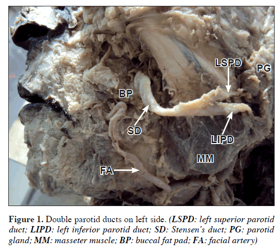
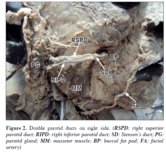
During routine dissection in the Department of Anatomy, we found double parotid ducts bilaterally in a 50-year-old male cadaver of Asian origin. Both parotid ducts were carefully traced from their origin in the parotid gland to fusion with each other at the anterior border of the masseter muscle to form Stensen’s duct on both sides, and measured with digital caliper. Measurements of diameters of both parotid ducts were taken at posterior border of masseter muscle on both sides with digital caliper. Diameter of both Stensen’s duct was measured 0.5 cm from anterior border of masseter muscle.
The lengths of superior and inferior parotid ducts on the left side were 28 and 34 mm, respectively (Figure 1). On the right side, the lengths of superior and inferior ducts were 29 and 36 mm, respectively (Figure 2). Diameters of superior and inferior ducts on the left side were 2.4 mm and 2.6 mm, respectively; and on the right side 2.5 mm and 2.2 mm, respectively. Diameter of Stensen’s duct on the left side was 2.9 mm and on the right side 2.8 mm.
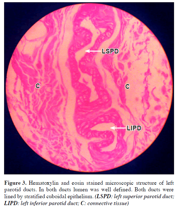
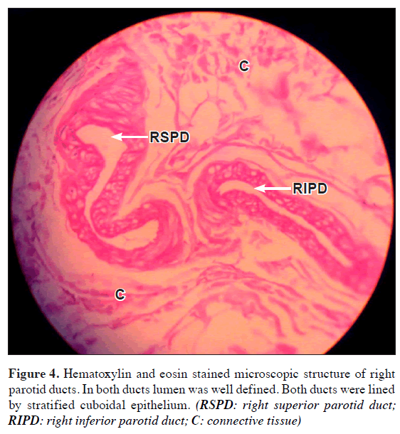
Microscopic examination of both main parotid ducts of both sides was performed using hematoxylin and eosin staining. Both parotid ducts of the left side (Figure 3) and of the right side (Figure 4) showed well-defined lumen and were lined by stratified cuboidal epithelium.
Discussion
Normally the Stensen’s duct exits from anterior border of the parotid gland and opens as a sole parotid duct in the oral cavity. It is lined by stratified cuboidal or stratified columnar epithelium [1].
Aktan et al. reported double parotid ducts on the right side of face in a cadaver. They considered these ducts as extension of collecting ducts, which should be fused within parotid gland normally. Both ducts were fused with each other before reaching the buccinator muscle [2]. Similar variation was found by Fernandes et al. on the right side, but origins of both ducts have not been traced in parotid gland [3]. Bailey reported incidence of double parotid duct in 7% of population [4]. But presence of double parotid ducts on both sides is not reported hitherto.
The average dimensions of the parotid duct are 50 mm long and 3 mm wide [1]. We found diameters of superior and inferior ducts on the left side 2.4 mm and 2.6 mm, respectively; and on the right side 2.5 mm and 2.2 mm, respectively. Diameter of Stensen’s duct on the left side was 2.9 mm and on the right side 2.8 mm. The knowledge of true dimensions of excretory ducts is important in duct endoscopy and lithotripsy [5].
The knowledge of presence of double parotid ducts is essential in diagnosis of congenital fistula from accessory parotid gland by CT sialography and CT fistulography [6], and avoiding iatrogenic injury to buccal branch of facial nerve in parotid gland surgery, parotid duct surgery and some facial cosmetic surgery [7].
The presence of double parotid ducts can be explained on the basis of developmental morphology of the parotid gland. Avery et al. described development of the parotid gland in six stages: induction of bud formation from the oral epithelium by the underlying mesenchyme; formation and growth of epithelial cord; initiation of branching in terminal parts of the cord; lobule formation through repetitive branching of the epithelial cord; canalization of the cords to form ducts; and cytodifferentiation. The growth, cytodifferentiation and morphogenesis of the parotid gland depend on both intrinsic and extrinsic factors. The programmed pattern of cell-specific gene expression is the genetic script established early in development, while extrinsic factors include cell-cell and cell-matrix interactions and growth factors. An intact basal lamina and the presence of mesenchyme are required for normal branching [8]. The synthesis and deposition of both types I and III collagen appear to be required for parotid gland branching morphogenesis [9]. Type IV collagen and, to a limited extent, laminin have been shown to play a role in the regulation of the differentiation of parotid gland secretory cells and to some extent ductal cells. Collagen synthesis stabilizes and maintains the branch points. Specific growth factors presented by extracellular molecular proteins appear to regulate events such as branching and lobule elongation [8].
The cell-matrix interactions and growth factors are increasingly important to the understanding of how the developmental signals are transmitted or mediated by extracellular matrix molecules of the basement membrane and associated mesenchyme regulate specific gene expression and cellular function leading to morphogenesis and cytodifferentiation of parotid gland and development of such variant.
References
- Standring S, ed. Gray’s Anatomy. The Anatomical Basis of Clinical Practice. 39th Ed., Philadelphia, Elsevier. 2005: 515–517, 604, 613.
- Aktan ZA, Bilge O, Pinar YA, Ikiz AO. Duplication of the parotid duct: a previously unreported anomaly. Surg Radiol Anat. 2001; 23: 353–354.
- Fernandes ACS, Lima GR, Rossi AM, Aguiar CM. Parotid gland with double duct: an anatomic variation description. Int J Morphol. 2009; 27: 129–132.
- Bailey L. Short Practice of Surgery. 15th Ed., London, Lewis. 1971: 533.
- Zenk J, Hosemann WG, Iro H. Diameters of the main excretory ducts of the adult human submandibular and parotid gland: a histologic study. Oral Surg Oral Med Oral Pathol Oral Radiol Endod. 1998; 85: 576–580.
- Moon WK, Han MH, Kim IO, Sung MW, Chang KH, Choo SW, Han MC. Congenital fistula from ectopic accessory parotid gland: diagnosis with CT sialography and CT fistulography. AJNR Am J Neuroradiol. 1995; 16: 997–999.
- Pogrel MA, Schmidt B, Ammar A. The relationship of the buccal branch of the facial nerve to the parotid duct. J Oral Maxillofac Surg. 1996; 54: 71–73.
- Avery JK, Steele PF, Avery N. Oral Development and Histology. 3rd Ed., New York, Thieme. 2002: 293–296.
- Spooner BS, Faubion JM. Collagen involvement in branching morphogenesis of embryonic lung and salivary gland. Dev Biol. 1980; 77: 84–102.
Rajesh B. Astik* and Urvi H. Dave
Department of Anatomy, GSL Medical College, Rajahmundry, District – East Godavari, Andhra Pradesh, India
- *Corresponding Author:
- Dr. Rajesh B. Astik
Associate Professor, Department of Anatomy, GSL Medical College NH-5, Rajahmundry District – East Godavari Andhra Pradesh, 533296, India
Tel: +91 883 2484999
E-mail: astikrajesh@yahoo.co.in
Date of Received: January 7th, 2011
Date of Accepted: August 6th, 2011
Published Online: August 10th, 2011
© Int J Anat Var (IJAV). 2011; 4: 141–143.
Abstract
The secretion of the parotid gland reaches the oral cavity through single parotid duct (Stensen’s duct). During routine dissection in the Department of Anatomy, we found double parotid ducts bilaterally in a 50-year-old male cadaver of Asian origin. On both sides the two ducts fused at the anterior border of the masseter muscle to form one duct, which opens in the oral cavity opposite the crown of upper second molar tooth. Microscopic study of parotid ducts on both sides and measurement of diameters of superior and inferior ducts on both sides revealed both ducts were main parotid ducts.
Variation in branching morphogenesis of the parotid duct should be sought on the basis of its development. The operating surgeon should be familiar with such variation during surgery on parotid gland to avoid injury to buccal branch of the facial nerve or to the main parotid duct.
Keywords
facial nerve, masseter muscle, parotid gland, sialography, Stensen’s duct
Introduction
The paired parotid glands are the largest salivary glands. In 20% of cases, a small, usually detached, part called the accessory parotid gland lies between the zygomatic arch above and the parotid duct below. Parotid duct begins within the anterior part of the parotid gland by the confluence of two or three ducts which ascend or descend respectively at right angles to the main duct. The duct appears at the anterior border of the upper part of the gland and passes horizontally across masseter, approximately midway between the angle of the mouth and the zygomatic arch. It crosses masseter, turns medially at its anterior border at almost a right angle to traverse the buccal fat pad and buccinator. While crossing masseter, the parotid duct lies between the upper and lower buccal branches of the facial nerve, and may receive the accessory parotid duct. The duct then runs obliquely forwards between buccinator and the oral mucosa to open on a small papilla opposite the crown of upper second molar tooth [1].
The parotid gland develops as the result of epithelial–mesenchymal interactions between the ectodermal epithelial lining of the oral cavity and the subjacent neural crest derived mesenchyme. It can be recognized at stage 15 (8 mm) as an elongated furrow running dorsally from the angle of the mouth between the mandibular and maxillary prominences. The groove, which is converted into a tube, persists as the parotid duct. The secretory part of the parotid gland is formed by epithelial cells sprouting from the terminal ends of the branching parotid duct [1]. Early division of the parotid duct and epithelial sprouts from both ducts might lead to development of two parotid ducts and their ramifications within the parotid gland.
The knowledge of such a rare variation is essential for surgeons who are planning for surgery in this region to avoid injury to facial nerve and accidental damage to main parotid duct.
Case Report
During routine dissection in the Department of Anatomy, we found double parotid ducts bilaterally in a 50-year-old male cadaver of Asian origin. Both parotid ducts were carefully traced from their origin in the parotid gland to fusion with each other at the anterior border of the masseter muscle to form Stensen’s duct on both sides, and measured with digital caliper. Measurements of diameters of both parotid ducts were taken at posterior border of masseter muscle on both sides with digital caliper. Diameter of both Stensen’s duct was measured 0.5 cm from anterior border of masseter muscle.
The lengths of superior and inferior parotid ducts on the left side were 28 and 34 mm, respectively (Figure 1). On the right side, the lengths of superior and inferior ducts were 29 and 36 mm, respectively (Figure 2). Diameters of superior and inferior ducts on the left side were 2.4 mm and 2.6 mm, respectively; and on the right side 2.5 mm and 2.2 mm, respectively. Diameter of Stensen’s duct on the left side was 2.9 mm and on the right side 2.8 mm.
Microscopic examination of both main parotid ducts of both sides was performed using hematoxylin and eosin staining. Both parotid ducts of the left side (Figure 3) and of the right side (Figure 4) showed well-defined lumen and were lined by stratified cuboidal epithelium.
Discussion
Normally the Stensen’s duct exits from anterior border of the parotid gland and opens as a sole parotid duct in the oral cavity. It is lined by stratified cuboidal or stratified columnar epithelium [1].
Aktan et al. reported double parotid ducts on the right side of face in a cadaver. They considered these ducts as extension of collecting ducts, which should be fused within parotid gland normally. Both ducts were fused with each other before reaching the buccinator muscle [2]. Similar variation was found by Fernandes et al. on the right side, but origins of both ducts have not been traced in parotid gland [3]. Bailey reported incidence of double parotid duct in 7% of population [4]. But presence of double parotid ducts on both sides is not reported hitherto.
The average dimensions of the parotid duct are 50 mm long and 3 mm wide [1]. We found diameters of superior and inferior ducts on the left side 2.4 mm and 2.6 mm, respectively; and on the right side 2.5 mm and 2.2 mm, respectively. Diameter of Stensen’s duct on the left side was 2.9 mm and on the right side 2.8 mm. The knowledge of true dimensions of excretory ducts is important in duct endoscopy and lithotripsy [5].
The knowledge of presence of double parotid ducts is essential in diagnosis of congenital fistula from accessory parotid gland by CT sialography and CT fistulography [6], and avoiding iatrogenic injury to buccal branch of facial nerve in parotid gland surgery, parotid duct surgery and some facial cosmetic surgery [7].
The presence of double parotid ducts can be explained on the basis of developmental morphology of the parotid gland. Avery et al. described development of the parotid gland in six stages: induction of bud formation from the oral epithelium by the underlying mesenchyme; formation and growth of epithelial cord; initiation of branching in terminal parts of the cord; lobule formation through repetitive branching of the epithelial cord; canalization of the cords to form ducts; and cytodifferentiation. The growth, cytodifferentiation and morphogenesis of the parotid gland depend on both intrinsic and extrinsic factors. The programmed pattern of cell-specific gene expression is the genetic script established early in development, while extrinsic factors include cell-cell and cell-matrix interactions and growth factors. An intact basal lamina and the presence of mesenchyme are required for normal branching [8]. The synthesis and deposition of both types I and III collagen appear to be required for parotid gland branching morphogenesis [9]. Type IV collagen and, to a limited extent, laminin have been shown to play a role in the regulation of the differentiation of parotid gland secretory cells and to some extent ductal cells. Collagen synthesis stabilizes and maintains the branch points. Specific growth factors presented by extracellular molecular proteins appear to regulate events such as branching and lobule elongation [8].
The cell-matrix interactions and growth factors are increasingly important to the understanding of how the developmental signals are transmitted or mediated by extracellular matrix molecules of the basement membrane and associated mesenchyme regulate specific gene expression and cellular function leading to morphogenesis and cytodifferentiation of parotid gland and development of such variant.
References
- Standring S, ed. Gray’s Anatomy. The Anatomical Basis of Clinical Practice. 39th Ed., Philadelphia, Elsevier. 2005: 515–517, 604, 613.
- Aktan ZA, Bilge O, Pinar YA, Ikiz AO. Duplication of the parotid duct: a previously unreported anomaly. Surg Radiol Anat. 2001; 23: 353–354.
- Fernandes ACS, Lima GR, Rossi AM, Aguiar CM. Parotid gland with double duct: an anatomic variation description. Int J Morphol. 2009; 27: 129–132.
- Bailey L. Short Practice of Surgery. 15th Ed., London, Lewis. 1971: 533.
- Zenk J, Hosemann WG, Iro H. Diameters of the main excretory ducts of the adult human submandibular and parotid gland: a histologic study. Oral Surg Oral Med Oral Pathol Oral Radiol Endod. 1998; 85: 576–580.
- Moon WK, Han MH, Kim IO, Sung MW, Chang KH, Choo SW, Han MC. Congenital fistula from ectopic accessory parotid gland: diagnosis with CT sialography and CT fistulography. AJNR Am J Neuroradiol. 1995; 16: 997–999.
- Pogrel MA, Schmidt B, Ammar A. The relationship of the buccal branch of the facial nerve to the parotid duct. J Oral Maxillofac Surg. 1996; 54: 71–73.
- Avery JK, Steele PF, Avery N. Oral Development and Histology. 3rd Ed., New York, Thieme. 2002: 293–296.
- Spooner BS, Faubion JM. Collagen involvement in branching morphogenesis of embryonic lung and salivary gland. Dev Biol. 1980; 77: 84–102.


