Empty sella syndrome with herniation of gyrus rectus
2 Federal University of Ceara, Brazil
3 Sugery Department, Federal University of Ceara, Brazil
Received: 18-Apr-2018 Accepted Date: Apr 19, 2018; Published: 26-Apr-2018
Citation: Teixeira AAR. Empty sella syndrome with herniation of gyrus rectus. J Neurol Clin Neurosci. 2018;2(2):04.
This open-access article is distributed under the terms of the Creative Commons Attribution Non-Commercial License (CC BY-NC) (http://creativecommons.org/licenses/by-nc/4.0/), which permits reuse, distribution and reproduction of the article, provided that the original work is properly cited and the reuse is restricted to noncommercial purposes. For commercial reuse, contact reprints@pulsus.com
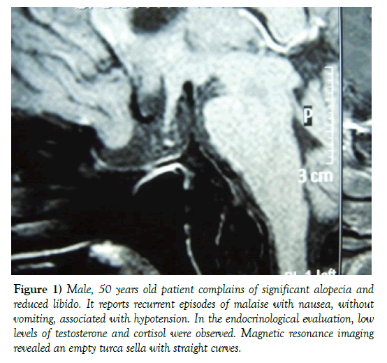
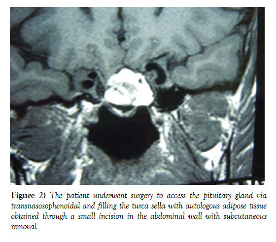
The straight curves were raised to their original position and the sella floor was redone with a bony fragment of the sphenoid face. The patient presented improvement of arterial hypotension, remaining in testosterone replacement (Figures 1 and 2) [1-3].
Figure 1) Male, 50 years old patient complains of significant alopecia and reduced libido. It reports recurrent episodes of malaise with nausea, without vomiting, associated with hypotension. In the endocrinological evaluation, low levels of testosterone and cortisol were observed. Magnetic resonance imaging revealed an empty turca sella with straight curves.
Figure 2) The patient underwent surgery to access the pituitary gland via transnasosophenoidal and filling the turca sella with autologous adipose tissue obtained through a small incision in the abdominal wall with subcutaneous removal
REFERENCES
- Auer MK, Stieg MR, Crispin A, et al. Primary Empty Sella Syndrome and the Prevalence of Hormonal Dysregulation: A Systematic Review. Deutsches Ärzteblatt International. 2018;115(7):99-105.
- Goedgezelschap A, Dejaeger E. Een 74-jarige patiënte met een recidiverend shockbeeld veroorzaakt door een onderliggend hypopituïtarisme. Tijdschr Gerontol Geriatr. 2015;46(6):320-6.
- Paroder V, Miller T, Cohen MM, et al. Absent Sella Turcica: A Case Report and a Review of the Literature. Fetal Pediatr Pathol. 2013;32(5):375-83.


