Epitransverse process: a rare outgrowth from atlas vertebra
Parul Kaushal*
Department of Anatomy, All India Institute of Medical Sciences, New Delhi, India
- *Corresponding Author:
- Parul Kaushal, PhD Student
Room number 1037, Department of Anatomy, All India Institute of Medical Sciences, New Delhi, India
Tel: +91 11 9958323452
E-mail: parulkaushal7@gmail.com
Date of Received: November 27th, 2009
Date of Accepted: July 10th, 2010
Published Online: August 9th, 2010
© Int J Anat Var (IJAV). 2010; 3: 108–109.
[ft_below_content] =>Keywords
atlas, epitransverse process, vertebra, cranio-vertebral, transverse process
Introduction
Acute injuries of the upper cervical spine as a cause of severe disability and death following trauma has at all times been an interesting phase of anatomical study [1]. The cranio-vertebral region has been considered an unstable, ontogenetically restless zone and thus susceptible to many variants, anomalies and malformations [2]. A number of authors have called attention to changes in the upper cervical spine as a common cause for headaches, neck pains and movements at the atlanto-occipital joint. The present case presents a rare outgrowth from the transverse process of atlas vertebra and discusses it’s the clinical significance to the surgeons and radiologists.
Case Report
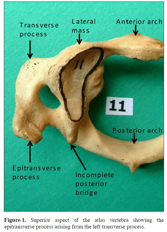
During routine osteology discussion class for the undergraduate students in the Department of Anatomy, Government Medical College, Amritsar, an unusual process arising from the left transverse process of one of the thirty atlas vertebrae was observed (Figure 1). The direction of the process was upwards and laterally, towards the jugular process of the occipital bone.
Discussion
During the development, the occipital bone is formed from the union of four to five somites, which normally fuse together to encircle the foramen magnum. The last occipital somite or pro-atlas somite, may fail to fully incorporate into the occiput, resulting in occipital or pro-atlas vertebrae. Manifestations of occipital vertebrae may include third condyle, paramastoid process, epitransverse process and various occipital ossicles. The process observed in the present case corresponds to the epitransverse process. Such a process was observed in one of the 59 adult male and 48 female adult vertebral columns studied by Tulsi at South Australian museum and Department of Anatomy, University of Adelaide, and it made contact with a facet on the jugular process [3]. Taitz reported a prominent pillar of bone extending from the jugular process of the occipital bone called paracondylar process to meet the epitransverse process in one of the 214 adult skeletons studied [4]. These studies point towards the formation of an additional pseudoarthrosis, thereby forming a three joint mechanism which may precipitate a moderate lateral dislocation of atlas. Presence of this process may even act as a shim, and lead to lateral tilt of the head manifesting as skeletal torticollis [5]. The movements at the atlanto-occipital joint are also disturbed by the presence of such variant process in the cranio-vertebral region [3,4]. Autopsy of 9 cases (63%) with traumatic basal subarachnoid hemorrhage by Gross, revealed developmental malformations including epitransverse process, posterior ponticle and foramen arcuale, thereby correlating the rupture of vertebral artery to the presence of developmental disorders in the cervico-occipital region [6]. Morphology of the upper cervical region is important to assess vertebral bony and vascular anomalies and the presence of variant bony structures may lead to difficulty in surgical procedures. Transverse process of atlas is one of a number of useful landmarks in head and neck surgery and its specific role as a beacon for internal jugular vein and cranial nerves IX, X, XI, XII has been emphasized [7]. Owing to its close proximity to these vital structures, the epitransverse process assumes clinical significance in case of radical neck dissection surgeries, increased intracranial pressure and compressive neuropathic symptoms. This type of bony process may be overlooked on both anteroposterior and lateral radiographs because of superimposed anatomy. The knowledge of this variant process can be of significance for accurate diagnosis of symptoms and radiographs in the cervico-occipital region.
Acknowledgements
I thank the faculty of the Department of Anatomy, GMC, Amritsar for allowing me to study the vertebrae.
References
- Bohlman HH. Acute fractures and dislocations of the cervical spine. An analysis of three hundred hospitalized patients and review of the literature. J Bone Joint Surg Am. 1979; 61: 1119–1142.
- Schmorl G, Junghanns H. The Human Spine in Health and Disease. 2nd American Edition. New York, Grune and Stratton. 1971; 504.
- Tulsi RS. Some specific anatomical features of the atlas and axis: dens, epitransverse process and articular facets. Aust N Z J Surg. 1978; 48: 570–574.
- Taitz C. Bony observations of some morphological variations and anomalies of the craniovertebral region. Clin Anat. 2000; 13: 354–360.
- Bland JH. Disorders of the Cervical Spine. Toronto, W.B. Saunders Company. 1987; 298–312.
- Gross A. Traumatic basal subarachnoid hemorrhages: autopsy material analysis. Forensic Sci Int. 1990; 45: 53–61.
- Funas DW. Transverse process of the atlas: guide to high ligation of the internal jugular vein in radical neck dissections. Ann Surg. 1967; 165: 473–476.
Parul Kaushal*
Department of Anatomy, All India Institute of Medical Sciences, New Delhi, India
- *Corresponding Author:
- Parul Kaushal, PhD Student
Room number 1037, Department of Anatomy, All India Institute of Medical Sciences, New Delhi, India
Tel: +91 11 9958323452
E-mail: parulkaushal7@gmail.com
Date of Received: November 27th, 2009
Date of Accepted: July 10th, 2010
Published Online: August 9th, 2010
© Int J Anat Var (IJAV). 2010; 3: 108–109.
Abstract
Acute injuries of the upper cervical spine as a cause of severe disability and death following trauma has at all times been an interesting phase of anatomical study. The present case study describes a rare outgrowth from the left transverse process of the atlas vertebra. This process referred to as epitransverse process can be of high importance to many specialties and especially to surgeons performing radical neck dissections, radiologists for accurate diagnosis of bony malformations and manipulative therapists, as it may markedly influence the posture, stability and mobility at the atlanto-occipital joint.
-Keywords
atlas, epitransverse process, vertebra, cranio-vertebral, transverse process
Introduction
Acute injuries of the upper cervical spine as a cause of severe disability and death following trauma has at all times been an interesting phase of anatomical study [1]. The cranio-vertebral region has been considered an unstable, ontogenetically restless zone and thus susceptible to many variants, anomalies and malformations [2]. A number of authors have called attention to changes in the upper cervical spine as a common cause for headaches, neck pains and movements at the atlanto-occipital joint. The present case presents a rare outgrowth from the transverse process of atlas vertebra and discusses it’s the clinical significance to the surgeons and radiologists.
Case Report
During routine osteology discussion class for the undergraduate students in the Department of Anatomy, Government Medical College, Amritsar, an unusual process arising from the left transverse process of one of the thirty atlas vertebrae was observed (Figure 1). The direction of the process was upwards and laterally, towards the jugular process of the occipital bone.
Discussion
During the development, the occipital bone is formed from the union of four to five somites, which normally fuse together to encircle the foramen magnum. The last occipital somite or pro-atlas somite, may fail to fully incorporate into the occiput, resulting in occipital or pro-atlas vertebrae. Manifestations of occipital vertebrae may include third condyle, paramastoid process, epitransverse process and various occipital ossicles. The process observed in the present case corresponds to the epitransverse process. Such a process was observed in one of the 59 adult male and 48 female adult vertebral columns studied by Tulsi at South Australian museum and Department of Anatomy, University of Adelaide, and it made contact with a facet on the jugular process [3]. Taitz reported a prominent pillar of bone extending from the jugular process of the occipital bone called paracondylar process to meet the epitransverse process in one of the 214 adult skeletons studied [4]. These studies point towards the formation of an additional pseudoarthrosis, thereby forming a three joint mechanism which may precipitate a moderate lateral dislocation of atlas. Presence of this process may even act as a shim, and lead to lateral tilt of the head manifesting as skeletal torticollis [5]. The movements at the atlanto-occipital joint are also disturbed by the presence of such variant process in the cranio-vertebral region [3,4]. Autopsy of 9 cases (63%) with traumatic basal subarachnoid hemorrhage by Gross, revealed developmental malformations including epitransverse process, posterior ponticle and foramen arcuale, thereby correlating the rupture of vertebral artery to the presence of developmental disorders in the cervico-occipital region [6]. Morphology of the upper cervical region is important to assess vertebral bony and vascular anomalies and the presence of variant bony structures may lead to difficulty in surgical procedures. Transverse process of atlas is one of a number of useful landmarks in head and neck surgery and its specific role as a beacon for internal jugular vein and cranial nerves IX, X, XI, XII has been emphasized [7]. Owing to its close proximity to these vital structures, the epitransverse process assumes clinical significance in case of radical neck dissection surgeries, increased intracranial pressure and compressive neuropathic symptoms. This type of bony process may be overlooked on both anteroposterior and lateral radiographs because of superimposed anatomy. The knowledge of this variant process can be of significance for accurate diagnosis of symptoms and radiographs in the cervico-occipital region.
Acknowledgements
I thank the faculty of the Department of Anatomy, GMC, Amritsar for allowing me to study the vertebrae.
References
- Bohlman HH. Acute fractures and dislocations of the cervical spine. An analysis of three hundred hospitalized patients and review of the literature. J Bone Joint Surg Am. 1979; 61: 1119–1142.
- Schmorl G, Junghanns H. The Human Spine in Health and Disease. 2nd American Edition. New York, Grune and Stratton. 1971; 504.
- Tulsi RS. Some specific anatomical features of the atlas and axis: dens, epitransverse process and articular facets. Aust N Z J Surg. 1978; 48: 570–574.
- Taitz C. Bony observations of some morphological variations and anomalies of the craniovertebral region. Clin Anat. 2000; 13: 354–360.
- Bland JH. Disorders of the Cervical Spine. Toronto, W.B. Saunders Company. 1987; 298–312.
- Gross A. Traumatic basal subarachnoid hemorrhages: autopsy material analysis. Forensic Sci Int. 1990; 45: 53–61.
- Funas DW. Transverse process of the atlas: guide to high ligation of the internal jugular vein in radical neck dissections. Ann Surg. 1967; 165: 473–476.


