Extensive systemic and cutaneous metastasis – Extremely rare presentation of breast carcinoma
Received: 24-Mar-2018 Accepted Date: Apr 09, 2018; Published: 12-Apr-2018
Citation: Bansal LK, Sharma D, Aggarwal L. Extensive systemic and cutaneous metastasis– Extremely rare presentation of breast carcinoma. Surg Case Rep. 2018;2(1):12-13.
This open-access article is distributed under the terms of the Creative Commons Attribution Non-Commercial License (CC BY-NC) (http://creativecommons.org/licenses/by-nc/4.0/), which permits reuse, distribution and reproduction of the article, provided that the original work is properly cited and the reuse is restricted to noncommercial purposes. For commercial reuse, contact reprints@pulsus.com
Abstract
Cutaneous metastases from systemic malignancy are relatively uncommon. Breast carcinoma is one of the most common cancer in women which metastasize to skin. Skin metastasis found in 0.7% to 10.4% of all patient with systemic malignancy. Here we report a 36-year-old lady with advanced primary ductal carcinoma of right breast ( T4N1M1 ) stage 4 disease who presented us with 1 year history of right breast lump, progress from small 2 cm nodule in right breast, remain static for another 9 months and then suddenly progress to recent size of 15 × 15 cm with development of multiple skin swelling in abdomen, back, upper arm, and right thigh region simultaneously. These skin nodules are clinically mimicking lipoma. There is multiple mobile lump in left breast also mimicking fibroadenoma which are increasing in size and number in last 3 months. These swelling in skin and opposite breast are turn out to be metastatic nodules after histopathology report. After radiological examination metastatic nodules found in liver and spleen too. Mammography shows BIRADS 5 in bilateral breast. Patient was put on neoadjuvant chemotherapy. First cycle of paclitaxel based chemotherapy was started after histological confirmation.
Keywords
Systemic metastatis; Cutaneous metastasis; Breast carcinoma; Systemic malignancy; Skin metastasis; Chemotherapy; Alopecia neoplastica
Skin metastasis found in 0.7% to 10.4% of all patient with systemic malignancy (1). The breast, skin, stomach, lungs, uterus, large intestine, and kidneys are the most frequent organs to produce cutaneous metastases. Cancers that have the highest propensity to metastasize to the skin include melanoma (45% of cutaneous metastasis cases) and cancers of the breast (30%), nasal sinuses (20%), larynx (16%), and oral cavity (12%) (2). Cutaneous metastasis is defined as a neoplastic lesion affecting the dermis or the subcutaneous tissue that originates from another primary tumor (3). 0.7-9% of patients with cancer develop skin metastasis, which is considered a rare dermatological event (4). Metastatic tumors are usually round, discrete tumor lobules in the dermis, with a Grenz zone, and are usually unassociated with the epidermis (5). Carcinoma may produce initial inflammatory response mimicking cellulitis. This pattern is referred as inflammatory breast carcinoma. Occasionally, patients with metastatic breast cancer have a firm, scar like area in the skin. When this occurs on the scalp, hair may be lost, and the clinical appearance may mimic alopecia areata, except that the skin exhibits marked induration on palpation. This condition, known as alopecia neoplastica (6).
Case Report
A 36-year-old lactating woman with negative family history of neoplastic disease presented us with 1 year history of right breast lump, progress from small 2 cm nodule in upper inner quadrant of right breast, remain static for another 9 months and then suddenly progress to recent size of 15 cm × 15 cm in last 5 months occupying upper outer, inner and central region of right breast.
She also noticed multiple 2 to 5 cm swelling in opposite breast also around 20-25 in number. These swellings are freely mobile mimicking multiple fibroadenoma in left breast. She experience rapidly increasing size of left breast also in last 3 months.
On examination right breast lump is found to be fixed with nipple and areola complex. It was free from underlying muscle and chest wall. There are multiple dilated vein in chest and bilateral breast region.
After careful breast and axillary examination single mobile 2 cm ×2 cm central group of axillary lymph node was found. There are no supraclavicular or opposite axillary lymph node metastasis (Figure 1).
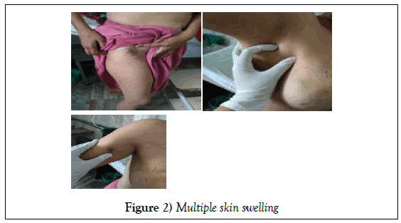
She also noticed multiple skin swelling in abdomen, back, upper arm, and right thigh region develop simultaneously size ranging from 1 to 4 cm increasing in number and size in last 3 months. These skin swelling are painless, freely mobile mimicking lipoma (Figure 2).
FNAC was obtained from both of the breast, axillary lymph node and skin swelling from abdomen, thigh and arm region. FNAC from both breast shows presence of ductal cells showing moderate to marked pleomorphism with anisocytonucleosis suggestive of bilateral invasive ductal carcinoma of breast. FNAC from axillary lymph node and skin swelling from abdomen, thigh and arm region showing malignant epithelial cell as seen in smears from breast swelling suggestive of metastatic subcutaneous deposits.
Tru cut biopsy also performed from the right breast lump. It is found to be triple negative invasive ductal carcinoma, NOS modified Nottingham grade- 3.
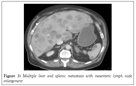
Bilateral mammography was done which shows BIRADS-5 in both breast. Skeletal survey done which include chest, spine, pelvis x-ray. These x-ray shows no bony metastasis. USG whole abdomen was done which shows multiple hypoechoic lesion in liver and spleen suggestive of multiple liver and splenic metastasis. USG findings are confirmed by CECT abdomen which shows multiple liver and splenic metastasis with mesenteric lymph node enlargement (Figure 3).
Discussion
Carcinoma breast metastasizes mainly to the lungs, bones, CNS and liver (7). In a retrospective study of carcinoma breast with metastatic disease, it is reported that cutaneous metastasis from breast carcinoma accounted for 5% of cases (8). But in any case, report it is not found bilateral carcinoma breast with extensive cutaneous and systemic metastasis.
Conclusion
Therefore, in my opinion it is probably very rare case of carcinoma breast with such atypical course and findings of the disease.
Conflict of Interest
The authors declare that there is no conflict of interest that could be perceived as prejudicing the impartiality of this review.
Patient Consent
The author ensure that the work described has been carried out in accordance with The Code of Ethics of the World Medical Association (Declaration of Helsinki) for experiments involving humans.
Informed consent was obtained for experimentation with human subjects. The privacy rights of human subjects always be observed.
I declare that consent has been obtained from patient or subject after full explanation of the purpose and nature of the procedures used.
I also declare that approval is not required in our study as patient is not harmed during the procedure.
REFERENCES
- Caldas FAA, Curtis JAG, Baldelin TAR, et al. Recidivas cutâneas de tumores mamários: formas de apresentação e diagnóstico diferencial. Rev Imagem. 2006;28:197–201.
- Casimiro LM, Corell JJV. Metástasis cutáneas de neoplasias internas. Med Cutan Iber Lat Am. 2009;37:117–129.
- Cassarino DS, Cabral ES, Kartha RV, et al. Primary dermal melanoma: distinct immunohistochemical findings and clinical outcome compared with nodular and metastatic melanoma. Arch Dermatol. 2008;144(1):49-56.
- De Giorgi V, Grazzini M, Alfaioli B, et al. Cutaneous manifestations of breast carcinoma. Dermatol Ther. 2010;23(6):581-9.
- Kordek R, Jassem J, Krzakowski M, et al. Onkologia. Podręcznikdla studentów i lekarzy. Via Medica, Gdańsk 2007.
- Lookingbill DP, Spangler N, Helm KF. Cutaneous metastases in patients with metastatic carcinoma: a retrospective study of 4020 patients. J Am Acad Dermatol. 1993;29:228-36.
- Requena L, Sangueza M, Sangueza OP, et al. Pigmented mammary Paget disease and pigmented epidermotropic metastases from breast carcinoma. Am J Dermatopathol. 2002;24(3):189-98.
- Vichapat V, Garmo H, Holmberg L, et al. Patterns of metastasis in women with metachronous contralateral breast cancer. Br J Cancer. 2012;107(2):221-3


