Fibroadenoma urinary bladder: An extremely rare case with review of literature
Received: 28-Apr-2018 Accepted Date: May 23, 2018; Published: 30-May-2018
Citation: Agarwal PK, Rimi P. Fibroadenoma urinary bladder: An extremely rare case with review of literature J Histol Histopathol Res 2018;2(1): 15-16.
This open-access article is distributed under the terms of the Creative Commons Attribution Non-Commercial License (CC BY-NC) (http://creativecommons.org/licenses/by-nc/4.0/), which permits reuse, distribution and reproduction of the article, provided that the original work is properly cited and the reuse is restricted to noncommercial purposes. For commercial reuse, contact reprints@pulsus.com
Abstract
BACKGROUND: Fibroadenoma, a commonly known tumour in the breast, is extremely rare in urinary bladder, probably the first case to the best of authors’ knowledge. Earlier adenofibroma was reported in the urinary bladder and other organs.
METHODS: Transurethral biopsy was performed in a clinically suspected malignant tumour of urinary bladder. Multiple tiny soft tissue pieces were fixed in 10% neutral formal saline for histopathological examination and submitted to the pathology department. Paraffin sections were prepared and stained with H: E stain for microscopic examination.
RESULTS: Histopathological diagnosis of fibroadenoma urinary bladder was made. Updated review of literature was searched to find out the incidence of this neoplasm. No other case of this neoplasm in urinary bladder could be seen.
CONCLUSION: Fibroadenoma is a common benign tumour of breast in young ladies. To the utter surprise of the author (PKA), classical histomorphological features of fibroadenoma breast were observed in this bladder tumour. This tempted the authors to report this case for the general information of all the specialists in the field, to keep in mind while reporting on tumours of urinary bladder. After the operation the patient had uneventful recovery and discharged from the hospital in sound health.
Keywords
Fibroadenoma of rare sites; Adenofibroma; Benign tumours of urinary bladder
Fibroadenoma and adenofibroma are biphasic neoplasms classified into the mixed epithelial and mesenchymal tumour group, both components being benign. The breast is the most common site for fibroadenoma in young ladies. However this tumour was reported outside the breast long back in 1891, arising from ovarian fimbria in four cases [1]. Recently, the author has come across fibroadenoma arising in urinary bladder, probably the first case at this site and that too in a male patient, which has not yet been reported in literature till date. Earlier in 1953, a solitary case of adenofibroma urinary bladder [2] was reported. More recently, a few sporadic cases of adenofibroma have been reported mainly located in female genital tract e.g. uterus and endocervix [3], fallopian tube [4-9] and ovary [10]. Only one reference is available of occurrence of adenofibroma in three cases of lungs, of which only one patient was male and other two were female patients, aged 40 and 55 years each [11]. Extreme rarity of the tumour in the urinary bladder in a male patient prompted the authors to document this case.
Case Presentation
A 52 year-old man, presented to the urology department of a tertiary care hospital in Northern India, with the chief complaints of hematuria since 1 week. On cystoscopy, urologist observed a polypoidal growth on the lateral wall of the bladder measuring approximately 3x2 cm in size, which had a narrow stalk. A clinical diagnosis of carcinoma urinary bladder was made, which is a common neoplasm at this site. No other abnormalities were noted. Routine haematological and biochemical investigations of blood and urine were within normal range. Transurethral resection of the growth was done; superficial as well as deep muscle was sampled to rule out any evidence of invasion. Both the specimens were submitted for histopathological examination. Post operatively antibiotics were started. He did well following surgery and no fresh complaints were noted. He was discharged from the hospital without any postoperative hassles.
Histopathological Examination
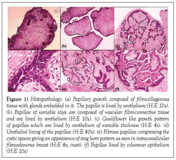
The histological Grossly, there were multiple greyish white soft tissue pieces from the superficial tumour, which collectively measured 2x1x1 cm in size. From deeper muscle two tiny bits of tissue were received separately to see if there was invasion of the tumour in the wall. Every bit of the tissue was processed for paraffin sectioning for microscopic examination. Sections from both the biopsies were stained with H&E stain. Serial sections of superficial biopsy pieces revealed papilliferous growths composed of proliferated avascular fibrocollagenous tissue and lined by urothelial cells. Scattered gland like structures, were embedded in the connective tissue stroma (Figure 1a). At places these papillae appeared leaf like (Figure 1b). The surface papillae were of variable sizes and presented cauliflower-like appearances under lower magnification (Figure 1c), each of these papillae were lined by urothelial epithelium of variable thickness (Figure 1d). Lobules of proliferated fibrocollagenous papillae compressed the cystic spaces which were lined by flattened epithelium and gave it an appearance of branching slit like spaces, very much like a stag horn pattern (Figure 1e; inset) as seen in fibroadenoma breast. A cluster of proliferated glandular structures were also seen, lined by columnar epithelium (Figure 1f). There was sparse lymphoplasmacytic cell infiltration in the stroma. However, no evidence of malignancy was noted in the serial sections examined. A histopathological diagnosis of fibroadenoma urinary bladder was made.
Figure 1: Histopathology: (a) Papillary growth composed of fibrocollagenous tissue with glands embedded in it. The papilla is lined by urothelium (H:E 10x). (b) Papillae of variable sizes are composed of vascular fibro-connective tissue and are lined by urothelium (H:E 10x). (c) Cauliflower like growth pattern of papillae which are lined by urothelium of variable thickness (H:E 4x). (d) Urothelial lining of the papillae (H:E 40x). (e) Fibrous papillae compressing the cystic spaces giving an appearance of stag horn pattern as seen in intracanalicular fibroadenoma breast (H:E 4x; inset). (f) Papillae lined by columnar epithelium (H:E 10x)
Sections from the deep muscle revealed normal muscle histology. There was no invasion by the tumour tissue.
Discussion
The diagnosis Fibroadenoma and adenofibroma although belonging to the same group of benign mixed mesenchymal and epithelial neoplasms have slight histomorphological differences as well as the clinical behaviour. Fibroadenoma breast may grow and develop into phylloides tumour, whereas adenofibromas arising in other organs have to be differentiated from low grade adenosarcoma as it is regarded a benign counterpart of mixed mullerian tumour, especially in the female genital tract. Thus it emphasises the need for differentiating fibroadenoma from adenofibroma [3].
Fibroadenoma is a common neoplasm of the breast in young women and is hormone dependent. Earlier in 1891, four cases of fibroadenoma were reported in the ovarian fimbria i.e., in an organ, other than the breast. These cases were clinically diagnosed as accessory ovary [1]. Occurrence of fibroadenoma in urinary bladder in a male patient is extremely rare and no such case has been reported in literature to the best of our knowledge. However, earlier Levi and Solomon reported a case of adenofibroma urinary bladder in a 46 year old man, who presented with burning during micturition and hematuria [2].
Diagnosis of adenofibroma has been histologically reserved for polyps composed of glands set in fibromatous mitotically inactive stroma [12]. These tumours are more commonly reported in female genital tract and rarely in other organ e.g. in lung, where also it revealed female dominance [one in male and two in females] [11] and a solitary case in male urinary bladder [2]. Origin of these tumours is a continued source of debate; some authors believe that some of the tumours currently classified as adenofibromas [on the basis of their low mitotic count and lack of significant nuclear atypia] are well differentiated adenosarcomas and recommend classifying adenofibroma into adenosarcoma of low malignant potential.
The present case under review presented as papillomatous growth in the urinary bladder, which was clinically diagnosed as transitional cell carcinoma. Thorough histopathological examination revealed very typical histomorphological features akin to intracanicular fibroadenoma breast due to the presence of branching slit like spaces [stag horn] lined by urothelium. During author [PKA]’s more than four decades of experience, as histopathologist, this is first case of fibroadenoma of male urinary bladder, which prompted the authors to document it, because of its extreme rarity.
Conclusion
Fibroadenoma, a biphasic neoplasm classified into the mixed epithelial and mesenchymal tumour group, both components being benign is extremely rare outside the breast, which is the most common site in young ladies. However fibroadenoma was reported outside the breast long back in 1891, arising from ovarian fimbria in four cases. Clinically these cases were suspected having accessory ovary, but on histopahological examination were diagnosed as fibroadenoma akin to breast tumour. Recently, the author [PKA] has come across this tumour, in male urinary bladder for the first time after a long experience as histopathologist. No reference of this tumour could be seen in the literature till date to the best of knowledge of authors.
Competing Interests
None
Authors Contributions
First author is Senior Consultant in Pathology and has diagnosed this case. Second author is Resident in Pathology and has helped in collecting and compiling the data.
Acknowledgements
The authors are grateful to Mr S. K. Shukla, of Pathology Department for his technical assistance.
REFERENCES
- Glendining B. Fibro-adenoma of the ovarian fimbria, and the question of the accessory ovary. Berl hlin Wochenschr 1891:1069-71.
- Levi M, Solomon C. Adenofibroma of urinary bladder: A case report. J Urol 1953;70(6):898-99.
- Agarwal PK, Husain N, Chandrawati P. Adenofibroma of uterus and endocervix. Histopathology 1991;18:79-80.
- Kanbour AI, Burgess F, Salazar H. Intramural adenofibroma of the fallopian tube. Light and electron microscopy. Cancer 1973;31:1433-39.
- Silverman AY, Artinian B, Sabin M. Serous cystadenofiboma of the fallopian tube: A case report. Am J Obstet Gynecol 1978;130:593-95.
- De La Fuente AA. Benign mixed mullerian tumor - adenofibroma of the fallopian tube. Histopathology 1982;6:661-66.
- Valerdiz Casasola S, Pardo Mindan J. Cystadenofibroma of fallopian tube. Appl Pathol 1989;7:256–59.
- Mondal SK. Adenofibroma and ectopic pregnancy of left fallopian tube: A rare coexistence. J Obstet Gynaecol Res 2010;36:690-92.
- Fukushima A, Shoji T, Tanaka S, et al. A case of fallopian tube adenofibroma: difficulties associated with differentiation from ectopic pregnancy. Clin Med Insights Case Rep 2014;7:135-7.
- Shukla S, Srivastava D, Acharya S. Serous adenofibroma of ovary: An eccentric presentation. J Cancer Res Ther 2015;11(4):1030.
- Kumar R, Desai S, Pai T, et al. Pulmonary adenofibroma: clinicopathological study of 3 cases of a rare benign lung lesion and review of the literature. Ann Diagn Pathol 2014;18(4):238-43.
- Robert A, Soslow MD, Teri A. Uterine pathology. (1st edn), Cambridge, UK. 2012;1:2.


