Foramen on the sacral ala and the lumbar transverse process: reviewing not a very common observation
Niladri Kumar Mahato*
Ohio Musculoskeletal and Neurological Institute (OMNI), Department of Biological Sciences, Ohio University, Athens, Ohio, USA
- *Corresponding Author:
- Niladri Kumar Mahato
Ohio Musculoskeletal and Neurological Institute (OMNI), Department of Biological Sciences, Ohio University, Athens, Ohio, 45701, USA
Tel: +1 (812) 603-3307
E-mail: mahatonk@yahoo.co.in
Date of Received: January 22nd, 2013
Date of Accepted: June 24th, 2013
Published Online: May 28th, 2014
© Int J Anat Var (IJAV). 2014; 7: 19–20.
[ft_below_content] =>Keywords
accessory, costo-transverse, facet joints, mamillary, retro-transverse
Introduction
A recently published study in this journal reported an example of a vertically oriented foramen at the sacral ala as a ‘new’ finding [1]. The current study/review is based on a brief survey of previously published literature on the occurrence of such foramina in the neural arches of the lumbosacral vertebrae, with discussion of the accepted clinical and embryologic significance of these foramina. Several perpendicularly oriented foramina have been reported running lateral or postero-lateral to the axial plane of the spinal canal at the lumbo-sacral region [2]. These formina reported by De Beers et al. in 1984, were from CT scans obtained from individuals suffering from low back pain. This included a foramen at the bases of the transverse and superior articular process of the L5, and vertical foramina passing through the medial part of the transverse process of L5, at the base of the sacral ala. Investigators have linked formation of these foramina due to osteoarthritic changes at the base of the facet joints [3–4], due to ossification of the mamillo-accessory ligament [5], or incomplete union of the costal and transverse ‘elements’ forming the transverse processes, during embryogenesis [6–7].
Case Report
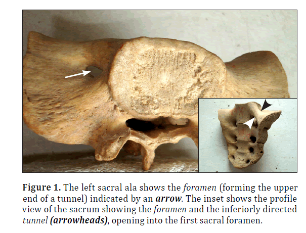
This study reports one such foramen occurring on the left sacral ala. This sacral sample was detected from the osteology collection at the Department of Forensic Medicine, Gandhi Medical College, Bhopal during a study conducted to investigate the morphology of sacra associated with lumbo-sacral transitional variations. This particular foramen (Figure 1) led to a bony tunnel that was about 17 mm in length. The cranial opening of the foramen was approximately 6 mm and 4 mm at its maximum and minimum diameters, respectively. The foramen was placed like a transversely oval gap on the left sacral ala about 4 mm lateral to the margin of the first sacral vertebral body. Situated just anterior to the mid-alar transverse plane, this bony tunnel ran inferiorly (slightly in an antero-medial direction) and opened caudally into the first anterior sacral foramen (Figure 1, inset) on the left side –and hence connected with the sacral canal, at the same level.
Discussion
One of the commonly accepted embryological evidence suggests that such foramina are formed by the ossification of a fibrous band called the mammillo-accessory ligament that extends between and connects the mammillary and accessory processes, at the base of the facet joints [3–4]. This ligament bridges a groove between the two processes forming a tunnel about 6 mm long [8]. This tunnel transmits vessels to the dorsal paraspinal muscles [9] and the medial division of the dorsal ramus from intervertebral space immediately above the foramen [10]. In case of ligamentous ossification, the foramen has been reported to be easily visible on plain radiographs [11] and during anatomic dissection appears like a “canal in the bone” containing most commonly the medial division [8]. These foramina have often been called “retro-transverse foramina” by Manners-Smith [9]. The dorsal rami medial branches supply motor innervations to the paraspinal musculature and carry sensory fibers from the facet joints. Thus entrapment of these nerves in the foramina discussed may lead to entrapment neuropathies [4]. Conceivably, congenital ossification have been the purported cause of formation of such foramina since both the mammillary and accessory processes are thought to derive embryologically from the so-called transverse element [12].
On the other hand, foramina at the transverse elements of the lumbo-sacral vertebrae may be positioned relatively anteriorly [2] or centered slightly antero-laterally on the transverse elements [6,9]. These groups of foramina, according to their position, have been suggested the name ‘costo-transverse foramina’ to separate them from the ‘retro-transverse foramina’ that are formed between the mammillary and accessory processes. Typically, the ‘costotransverse foramina’ might not transmit the medial division from the dorsal ramus. The development of lumbar transverse process is embryologically homologous to the ribs. Bulk of the anterior part of the transverse process arises from the ‘costal elements’, while a smaller medial and posterior part including the accessory process is documented to arise from the ‘transverse elements’. Structurally, lumbo-sacral transverse elements are homologous to the transverse process of the thoracic vertebrae [12]. Therefore, lumbar costo-transverse foramina may be likened to the cervical foramina transversaria [9]. Szawlowski [7] has postulated that remnants of anastomotic vessels running between the coastal and transverse elements in the lumbosacral region in the embryo may persist as structures within these foramina. Very early literature presents a foramen very similar as being reported in the study in discussion [7]. This foramen described by Szawlowski extended from a plane between the superior articular process and the sacral ala superiorly and then extended inferiorly to communicate with the first sacral foramen [2]. On account of plausible embryological explanations, remnants of embryonic lumbo-sacral longitudinal anastomotic vessels could be the most likely structures to pass through the foramen rather than the sympathetic trunk, the lumbo-sacral trunk or the iliac vessels.
To conclude, foramina at the sacral ala are not very uncommon findings. Detection of these aberrancies in the sacral ala may be associated with similar variations at the adjacent lumbar vertebrae. The contents of the foramina are more likely to be the remnants of embryonic lumbo-sacral anastomotic vessels. Pedicle or alar instrumentations in these individuals warrants careful per-operative imaging of these foramina.
References
- Singh R. A new foramen on posterior aspect of ala of first sacral vertebra. Int J Anat Var (IJAV). 2012; 5: 29–31.
- Beers GJ, Carter AP, McNary WF. Vertical foramina in the lumbosacral region: CT appearance. AJR Am J Roentgenol. 1984; 143: 1027–1029.
- Bogduk N, Long DM. The anatomy of the so-called “articular nerves” and their relationship to facet denervation in the treatment of low back pain. J Neurosurg. 1979; 51: 172–177.
- Bogduk N. The lumbar mamillo-accessory ligament: its anatomical and neurosurgical significance. Spine (Phila Pa 1976). 1981; 6: 162–167.
- Maigne JY, Maigne R, Guerin-Surville H. The lumbar mamillo-accessory foramen: a study of 203 lumbosacral spines. Surg Radiol Anat. 1991; 13: 29–32.
- Dwight T. A transverse foramen in the last lumbar vertebra. Anat Anz. 1902; 20: 571–572.
- Szawlowski J. Ueber einige seltene Variationen an der Wirbelsaeule beim Menschen. Anat Anz. 1901; 20: 305–320.
- Bradley KC. The anatomy of backache. Aust NZ J Surg. 1974; 44: 227–232.
- Manners-Smith T. The variability of the last lumbar vertebra. J Anat Physiol. 1909; 43: 146–160.
- Bogduk N, Wilson AS, Tynan W. The human lumbar dorsal rami. J Anat. 1982; 134: 383–397.
- Koehler A, Zimmer EA. Borderlands of the normal and early pathologic skeletal radiology. 3rd American Ed., Wilk SP, ed., New York, Grune & Stratton. 1968; 645–761.
- Gardner E, Gray DJ, O’Rahilly A. Anatomy: a regional study of human structure. 4th Ed., Philadelphia, Saunders. 1975; 60–555.
- Badgley CE. The articular facets in relation to low-back pain and sciatic radiation. J Bone Joint Surg. 1941; 23: 481–496.
Niladri Kumar Mahato*
Ohio Musculoskeletal and Neurological Institute (OMNI), Department of Biological Sciences, Ohio University, Athens, Ohio, USA
- *Corresponding Author:
- Niladri Kumar Mahato
Ohio Musculoskeletal and Neurological Institute (OMNI), Department of Biological Sciences, Ohio University, Athens, Ohio, 45701, USA
Tel: +1 (812) 603-3307
E-mail: mahatonk@yahoo.co.in
Date of Received: January 22nd, 2013
Date of Accepted: June 24th, 2013
Published Online: May 28th, 2014
© Int J Anat Var (IJAV). 2014; 7: 19–20.
Abstract
Detection of foramina at the sacral ala is not very uncommon and has been reported earlier in the literature. It has also been suggested that occurrence of such a foramen may be associated with similar findings at adjacent lumbar transverse processes. This report documents detection of a similar occurrence and reviews commonly accepted embryological explanations of this phenomenon. Pedicle or alar instrumentations in these individuals warrants careful per-operative imaging of these foramina due to the likelihood of these formina containing remnants of longitudinally anastomosing vascular structures, in most instances.
-Keywords
accessory, costo-transverse, facet joints, mamillary, retro-transverse
Introduction
A recently published study in this journal reported an example of a vertically oriented foramen at the sacral ala as a ‘new’ finding [1]. The current study/review is based on a brief survey of previously published literature on the occurrence of such foramina in the neural arches of the lumbosacral vertebrae, with discussion of the accepted clinical and embryologic significance of these foramina. Several perpendicularly oriented foramina have been reported running lateral or postero-lateral to the axial plane of the spinal canal at the lumbo-sacral region [2]. These formina reported by De Beers et al. in 1984, were from CT scans obtained from individuals suffering from low back pain. This included a foramen at the bases of the transverse and superior articular process of the L5, and vertical foramina passing through the medial part of the transverse process of L5, at the base of the sacral ala. Investigators have linked formation of these foramina due to osteoarthritic changes at the base of the facet joints [3–4], due to ossification of the mamillo-accessory ligament [5], or incomplete union of the costal and transverse ‘elements’ forming the transverse processes, during embryogenesis [6–7].
Case Report
This study reports one such foramen occurring on the left sacral ala. This sacral sample was detected from the osteology collection at the Department of Forensic Medicine, Gandhi Medical College, Bhopal during a study conducted to investigate the morphology of sacra associated with lumbo-sacral transitional variations. This particular foramen (Figure 1) led to a bony tunnel that was about 17 mm in length. The cranial opening of the foramen was approximately 6 mm and 4 mm at its maximum and minimum diameters, respectively. The foramen was placed like a transversely oval gap on the left sacral ala about 4 mm lateral to the margin of the first sacral vertebral body. Situated just anterior to the mid-alar transverse plane, this bony tunnel ran inferiorly (slightly in an antero-medial direction) and opened caudally into the first anterior sacral foramen (Figure 1, inset) on the left side –and hence connected with the sacral canal, at the same level.
Discussion
One of the commonly accepted embryological evidence suggests that such foramina are formed by the ossification of a fibrous band called the mammillo-accessory ligament that extends between and connects the mammillary and accessory processes, at the base of the facet joints [3–4]. This ligament bridges a groove between the two processes forming a tunnel about 6 mm long [8]. This tunnel transmits vessels to the dorsal paraspinal muscles [9] and the medial division of the dorsal ramus from intervertebral space immediately above the foramen [10]. In case of ligamentous ossification, the foramen has been reported to be easily visible on plain radiographs [11] and during anatomic dissection appears like a “canal in the bone” containing most commonly the medial division [8]. These foramina have often been called “retro-transverse foramina” by Manners-Smith [9]. The dorsal rami medial branches supply motor innervations to the paraspinal musculature and carry sensory fibers from the facet joints. Thus entrapment of these nerves in the foramina discussed may lead to entrapment neuropathies [4]. Conceivably, congenital ossification have been the purported cause of formation of such foramina since both the mammillary and accessory processes are thought to derive embryologically from the so-called transverse element [12].
On the other hand, foramina at the transverse elements of the lumbo-sacral vertebrae may be positioned relatively anteriorly [2] or centered slightly antero-laterally on the transverse elements [6,9]. These groups of foramina, according to their position, have been suggested the name ‘costo-transverse foramina’ to separate them from the ‘retro-transverse foramina’ that are formed between the mammillary and accessory processes. Typically, the ‘costotransverse foramina’ might not transmit the medial division from the dorsal ramus. The development of lumbar transverse process is embryologically homologous to the ribs. Bulk of the anterior part of the transverse process arises from the ‘costal elements’, while a smaller medial and posterior part including the accessory process is documented to arise from the ‘transverse elements’. Structurally, lumbo-sacral transverse elements are homologous to the transverse process of the thoracic vertebrae [12]. Therefore, lumbar costo-transverse foramina may be likened to the cervical foramina transversaria [9]. Szawlowski [7] has postulated that remnants of anastomotic vessels running between the coastal and transverse elements in the lumbosacral region in the embryo may persist as structures within these foramina. Very early literature presents a foramen very similar as being reported in the study in discussion [7]. This foramen described by Szawlowski extended from a plane between the superior articular process and the sacral ala superiorly and then extended inferiorly to communicate with the first sacral foramen [2]. On account of plausible embryological explanations, remnants of embryonic lumbo-sacral longitudinal anastomotic vessels could be the most likely structures to pass through the foramen rather than the sympathetic trunk, the lumbo-sacral trunk or the iliac vessels.
To conclude, foramina at the sacral ala are not very uncommon findings. Detection of these aberrancies in the sacral ala may be associated with similar variations at the adjacent lumbar vertebrae. The contents of the foramina are more likely to be the remnants of embryonic lumbo-sacral anastomotic vessels. Pedicle or alar instrumentations in these individuals warrants careful per-operative imaging of these foramina.
References
- Singh R. A new foramen on posterior aspect of ala of first sacral vertebra. Int J Anat Var (IJAV). 2012; 5: 29–31.
- Beers GJ, Carter AP, McNary WF. Vertical foramina in the lumbosacral region: CT appearance. AJR Am J Roentgenol. 1984; 143: 1027–1029.
- Bogduk N, Long DM. The anatomy of the so-called “articular nerves” and their relationship to facet denervation in the treatment of low back pain. J Neurosurg. 1979; 51: 172–177.
- Bogduk N. The lumbar mamillo-accessory ligament: its anatomical and neurosurgical significance. Spine (Phila Pa 1976). 1981; 6: 162–167.
- Maigne JY, Maigne R, Guerin-Surville H. The lumbar mamillo-accessory foramen: a study of 203 lumbosacral spines. Surg Radiol Anat. 1991; 13: 29–32.
- Dwight T. A transverse foramen in the last lumbar vertebra. Anat Anz. 1902; 20: 571–572.
- Szawlowski J. Ueber einige seltene Variationen an der Wirbelsaeule beim Menschen. Anat Anz. 1901; 20: 305–320.
- Bradley KC. The anatomy of backache. Aust NZ J Surg. 1974; 44: 227–232.
- Manners-Smith T. The variability of the last lumbar vertebra. J Anat Physiol. 1909; 43: 146–160.
- Bogduk N, Wilson AS, Tynan W. The human lumbar dorsal rami. J Anat. 1982; 134: 383–397.
- Koehler A, Zimmer EA. Borderlands of the normal and early pathologic skeletal radiology. 3rd American Ed., Wilk SP, ed., New York, Grune & Stratton. 1968; 645–761.
- Gardner E, Gray DJ, O’Rahilly A. Anatomy: a regional study of human structure. 4th Ed., Philadelphia, Saunders. 1975; 60–555.
- Badgley CE. The articular facets in relation to low-back pain and sciatic radiation. J Bone Joint Surg. 1941; 23: 481–496.


