Four headed triceps brachii muscle
Prabhjot Cheema1* and Rajan Singla2
1DHSJ Institute of Dental Sciences and Hospital, Panjab University, Chandigarh, India
2Government Medical College, Amritsar, India
- *Corresponding Author:
- Prabhjot Cheema
DHSJ Institute of Dental Sciences and Hospital, Panjab University, Chandigarh, India
Tel: +91 977 9073689
E-mail: manindergill2809@yahoo.com
Date of Received: July 28th, 2010
Date of Accepted: January 26th, 2011
Published Online: March 5th, 2011
© IJAV. 2011; 4: 43–44.
[ft_below_content] =>Keywords
fourth head, radial nerve, triceps brachii, variation
Introduction
Although variations of triceps brachii muscle are relatively uncommon, they have been occasionally reported by various authors [1]. The long head may split with one tendon attached to the shoulder capsule; the common tendon of insertion may be attached to the anconeous; a slip from the long head may attach to the coracoid process; slips may be found attaching to the triceps from the subscapularis; there may be fusion between the lateral head and the extensor carpi ulnaris; a duplicated lateral head may be present and a slip from the tendon of the latissimus dorsi may be attached to triceps brachii [2–4]. Sometimes the long head of triceps gets a muscular slip from the tendon of latissimus dorsi and is known as ‘latissimocondyloideus’ [5].
Macalister described following variations of triceps brachii: i) a fourth head arising from the medial part of humerus below its head by a long slender tendon and by an aponeurotic extension from the capsule of the shoulder, which blended with the medial head, ii) the splitting of long head with one part attached to capsule and other to tricipital spine or axillary border and iii) slips arising from the tendon of latissimus dorsi or teres major [3].
We report a case of fourth head of triceps brachii and its embryological and clinical significance.
Case Report
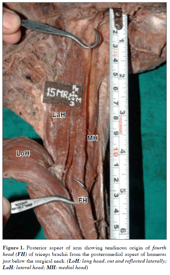
During routine cadaveric dissection in the Department of Anatomy, Government Medical College, Amritsar, India, we came across a four headed triceps brachii muscle on right side in a 40-year-old male cadaver. It originated as a tendinous slip from the postero-medial aspect of the humerus just below the surgical neck (Figure 1). This tendon was seen close to the shaft of humerus and directly posterior to the radial nerve and deep brachial artery in the radial sulcus. The tendon then passed distally and gave rise to a muscle belly, which after a short distance joined the long head of triceps brachii. The length of tendon and that of muscle belly was 5 cm and 8.5 cm, respectively. It was innervated by a branch from the radial nerve.
Discussion
The triceps brachii muscle is normally composed of three heads. The long head originates from the infraglenoid tubercle, the lateral head from the humerus superior to the radial groove and lateral intermuscular septum and the medial head from the humerus inferior to the radial groove and medial intermuscular septum. This entire muscle inserts onto the olecranon process and contributes to the extension of forearm. It is supplied by a branch from the radial nerve and deep brachial artery [6].
A fourth head of triceps may arise from various points on the humerus, scapula, shoulder joint capsule or the coracoid process [4]. In the present case, the proximal attachment of the fourth head of the triceps brachii was almost similar to as described by Fabrizio and Clemente [1]. The distal attachment of the muscle to the long head of triceps has been mentioned earlier by Macalister, which he described as the “splitting of the long head of triceps brachii” [3]. Clinicians diagnosing or treating patients with weakness or pain of posterior arm should consider variations of this compartment that may result in neurovascular compression [7].
Embryologically, during fifth week of development, mesoderm invades the upper limb bud to further condense into ventral and dorsal muscle masses [8]. The triceps muscle is derived from dorsal muscle mass of upper limb bud and it could be during this period that accessory muscles may have formed.
Clinically, close proximity of the tendon of fourth head to radial nerve and deep brachial artery may be a contributing factor in compression of the same, especially during strenuous exertion or muscular contraction leading to neurovascular compromise.
References
- Fabrizio PA, Clemente FR. Variation in the triceps brachii muscle: a fourth muscular head. Clin Anat. 1997; 10: 259–263.
- Wood J. On human muscular variations in their relation to comparative anatomy. J Anat Physiol. 1867; 1: 44–59.
- Nayak SR, Krishnamurthy A, Kumar M, Prabhu LV, Saralaya V, Thomas MM. Four headed biceps and triceps brachii muscles with neurovascular variation. Anat Sci Int. 2008; 83: 107–111.
- Anson BJ. Morris’ Human Anatomy. 12th Ed., New York, McGraw-Hill. 1966; 482–484.
- Bergman RA, Afifi AK, Miyauchi R. Anatomy Atlases: Illustrated Encyclopedia of Human Anatomic Variation. http://www.anatomyatlases.org/AnatomicVariants/MuscularSystem/Text/T/47Triceps.shtml (accessed May 2009)
- Williams PL, Warwick R, Dyson M, Bannister LH. Gray’s Anatomy. 37th Ed., New York, Churchill Livingstone. 1989; 615–616.
- Tubbs RS, Salter EG, Oakes WJ. Triceps brachii muscle demonstrating a fourth head. Clin. Anat. 2006; 19: 657–660.
- Schoenwolf GC, Bleyl SB, Brauer PR, Francis-West PH. Larsen’s Human Embryology. 4th Ed., Philadelphia, Churchill Livingstone. 2009; 241–242.
Prabhjot Cheema1* and Rajan Singla2
1DHSJ Institute of Dental Sciences and Hospital, Panjab University, Chandigarh, India
2Government Medical College, Amritsar, India
- *Corresponding Author:
- Prabhjot Cheema
DHSJ Institute of Dental Sciences and Hospital, Panjab University, Chandigarh, India
Tel: +91 977 9073689
E-mail: manindergill2809@yahoo.com
Date of Received: July 28th, 2010
Date of Accepted: January 26th, 2011
Published Online: March 5th, 2011
© IJAV. 2011; 4: 43–44.
Abstract
Variations of the triceps brachii muscle are infrequent. We report a case of four headed triceps brachii muscle which originated from the posteromedial aspect of humerus just below the surgical neck. The fibers of this extra head blended with the muscle fibers of long head of triceps. The embryological and clinical significance of the variation is discussed.
-Keywords
fourth head, radial nerve, triceps brachii, variation
Introduction
Although variations of triceps brachii muscle are relatively uncommon, they have been occasionally reported by various authors [1]. The long head may split with one tendon attached to the shoulder capsule; the common tendon of insertion may be attached to the anconeous; a slip from the long head may attach to the coracoid process; slips may be found attaching to the triceps from the subscapularis; there may be fusion between the lateral head and the extensor carpi ulnaris; a duplicated lateral head may be present and a slip from the tendon of the latissimus dorsi may be attached to triceps brachii [2–4]. Sometimes the long head of triceps gets a muscular slip from the tendon of latissimus dorsi and is known as ‘latissimocondyloideus’ [5].
Macalister described following variations of triceps brachii: i) a fourth head arising from the medial part of humerus below its head by a long slender tendon and by an aponeurotic extension from the capsule of the shoulder, which blended with the medial head, ii) the splitting of long head with one part attached to capsule and other to tricipital spine or axillary border and iii) slips arising from the tendon of latissimus dorsi or teres major [3].
We report a case of fourth head of triceps brachii and its embryological and clinical significance.
Case Report
During routine cadaveric dissection in the Department of Anatomy, Government Medical College, Amritsar, India, we came across a four headed triceps brachii muscle on right side in a 40-year-old male cadaver. It originated as a tendinous slip from the postero-medial aspect of the humerus just below the surgical neck (Figure 1). This tendon was seen close to the shaft of humerus and directly posterior to the radial nerve and deep brachial artery in the radial sulcus. The tendon then passed distally and gave rise to a muscle belly, which after a short distance joined the long head of triceps brachii. The length of tendon and that of muscle belly was 5 cm and 8.5 cm, respectively. It was innervated by a branch from the radial nerve.
Discussion
The triceps brachii muscle is normally composed of three heads. The long head originates from the infraglenoid tubercle, the lateral head from the humerus superior to the radial groove and lateral intermuscular septum and the medial head from the humerus inferior to the radial groove and medial intermuscular septum. This entire muscle inserts onto the olecranon process and contributes to the extension of forearm. It is supplied by a branch from the radial nerve and deep brachial artery [6].
A fourth head of triceps may arise from various points on the humerus, scapula, shoulder joint capsule or the coracoid process [4]. In the present case, the proximal attachment of the fourth head of the triceps brachii was almost similar to as described by Fabrizio and Clemente [1]. The distal attachment of the muscle to the long head of triceps has been mentioned earlier by Macalister, which he described as the “splitting of the long head of triceps brachii” [3]. Clinicians diagnosing or treating patients with weakness or pain of posterior arm should consider variations of this compartment that may result in neurovascular compression [7].
Embryologically, during fifth week of development, mesoderm invades the upper limb bud to further condense into ventral and dorsal muscle masses [8]. The triceps muscle is derived from dorsal muscle mass of upper limb bud and it could be during this period that accessory muscles may have formed.
Clinically, close proximity of the tendon of fourth head to radial nerve and deep brachial artery may be a contributing factor in compression of the same, especially during strenuous exertion or muscular contraction leading to neurovascular compromise.
References
- Fabrizio PA, Clemente FR. Variation in the triceps brachii muscle: a fourth muscular head. Clin Anat. 1997; 10: 259–263.
- Wood J. On human muscular variations in their relation to comparative anatomy. J Anat Physiol. 1867; 1: 44–59.
- Nayak SR, Krishnamurthy A, Kumar M, Prabhu LV, Saralaya V, Thomas MM. Four headed biceps and triceps brachii muscles with neurovascular variation. Anat Sci Int. 2008; 83: 107–111.
- Anson BJ. Morris’ Human Anatomy. 12th Ed., New York, McGraw-Hill. 1966; 482–484.
- Bergman RA, Afifi AK, Miyauchi R. Anatomy Atlases: Illustrated Encyclopedia of Human Anatomic Variation. http://www.anatomyatlases.org/AnatomicVariants/MuscularSystem/Text/T/47Triceps.shtml (accessed May 2009)
- Williams PL, Warwick R, Dyson M, Bannister LH. Gray’s Anatomy. 37th Ed., New York, Churchill Livingstone. 1989; 615–616.
- Tubbs RS, Salter EG, Oakes WJ. Triceps brachii muscle demonstrating a fourth head. Clin. Anat. 2006; 19: 657–660.
- Schoenwolf GC, Bleyl SB, Brauer PR, Francis-West PH. Larsen’s Human Embryology. 4th Ed., Philadelphia, Churchill Livingstone. 2009; 241–242.


