Four roots of median nerve and its surgical and clinical significance
Surekha Wamanrao Meshram1*, Kiran Janardhan Khobragade2, Sudhir Vishnupant Pandit1 and Jyoti Suryabhan Jadhav1
1Departmernt of Anatomy, Shri Vasantrao Naik Government Medical College, Yavatmal, Maharashtra, India.
2Hinduja Multispeciality Hospital, Mumbai, Maharashtra, India.
- *Corresponding Author:
- Dr. Surekha Wamanrao Meshram
Department of Anatomy, Shri Vasantrao Naik Government Medical College, Yavatmal, Maharashtra, India
Tel: +91 738 5030010
E-mail: drsurekhameshram@gmail.com
Date of Received: October 4th, 2011
Date of Accepted: August 22nd, 2012
Published Online: December 15th, 2012
© Int J Anat Var (IJAV). 2012; 5: 110–112.
[ft_below_content] =>Keywords
roots, median nerve, brachial plexus, variation
Introduction
Brachial plexus is a complex nerve bunch situated partly in neck and partly in axilla. It is formed by ventral divisions of anterior primary rami of C5–8 and T1 spinal nerves. It has roots, trunks and cords. Median nerve is formed by joining of lateral root (C5, C6, C7) and medial root (C8 and T1) to form composite median nerve having root value C5, C6, C7, C8 and T1. Lateral root of median nerve crosses the axillary artery from lateral to medial side and joins with medial root to form the main trunk of median nerve. Many variations are noted by many authors in the branching pattern of brachial plexus but variations in the formation of roots, trunks and cords are rare. In this study we present a rare case of four roots of formation of median nerve.
Case Report
During routine undergraduate dissection of a 65-year-old male cadaver in the Department of Anatomy, Shri Vasantrao Naik Government Medical College, Yavatmal, Maharashtra, it was found that in the right axilla formation of median nerve was variant.
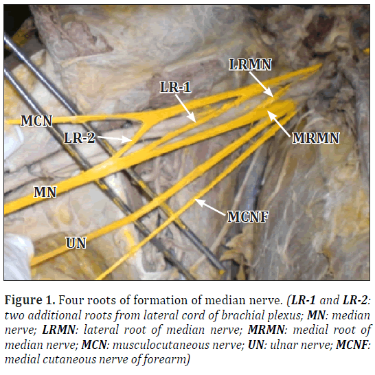
The right median nerve was formed by four roots, three were coming from lateral cord and one from medial cord of brachial plexus. The uppermost or highest root was noted to be at the level of first part of axillary artery where it gave origin to superior thoracic artery. The second root was found to be 2 cm below the first one and the third root was found 3cm below the second root. Highest root of median nerve crossed the axillary artery from lateral to medial side and joined with medial root immediately on medial side of artery. The remaining two roots were found to be passing obliquely in front of second and third part of axillary artery and joining individually with the medial root of median nerve and forming the main trunk of median nerve in front of third part of axillary artery (Figures 1,2). Dissection of both the upper limbs (axilla, arm, cubital fossa, forearm and palm) was done thoroughly to find out the relations and distribution of the variant right median nerve and the position of the left median nerve but the further course and distribution of the variant median nerve as well as left median nerve was found normal. The course and distribution of arterial pattern in arm and forearm was also normal. It was also found that all the three roots coming from lateral cord of brachial plexus were compressing the axillary artery.
Figure 1: Four roots of formation of median nerve. (LR-1 and LR-2: two additional roots from lateral cord of brachial plexus; MN: median nerve; LRMN: lateral root of median nerve; MRMN: medial root of median nerve; MCN: musculocutaneous nerve; UN: ulnar nerve; MCNF: medial cutaneous nerve of forearm)
Discussion
Median-musculocutaneous nerve communication has been studied by many authors but variations in the formation of median nerve were noted by only some earlier workers. Satyanarayana and Guhafound that the median nerve was formed by three roots coming from the lateral cord and one root coming from medial cord of brachial plexus [1]. Pandey and Shukla have found in 4.7% cases that the roots of median nerve joined on medial side of axillary artery, and in 2.3% cases the roots did not join but continued separately [2]. Eglseder and Goldman investigated that the median nerve was formed of two lateral roots in 14% of their specimens [3]. Chauhan and Roy reported formation of median nerve by two lateral and one medial root [4]. Same observation was reported by Saeed and Rufai [5]. Anrkooli et al., in their case report they observed formation of median nerve by fusion of three roots in both the arms of the same cadaver. There were two lateral roots and one medial root [6]. Sargon et al. found that the median nerve was formed by three branches, two from lateral cord and one from medial cord [7]. According to Hollinshead, anomalies of nerves are accompanied by abnormalities of vessels [8]. The variations of brachial plexus were associated with those of subclavian, axillary and brachial arteries but in the present study, we found no such associated vascular variation.
So many studies are done but in rare cases such type of variation of the formation of median nerve by three roots from lateral cord of brachial plexus which are compressing the axillary artery was not found.
Such variation can be explained in the light of embryologic development. At the seventh week of development limb musculature first observed as condensation of mesenchyme near the base of the limb buds. After elongation of the limb buds, these muscle splits into flexor and extensor compartments. The position of upper limb buds are opposite the lower five cervical and upper two thoracic segments. As soon as the buds form, ventral primary rami from these spinal nerves penetrate into the mesenchyme. At first, each ventral ramus divides into dorsal and ventral branches, but soon these branches unite to form named peripheral nerves that supply extensor and flexor group of muscles respectively. Early after the above rearrangement of nerves, they enter the limb bud and establish an intimate contact with the differentiating mesodermal condensation and this early contact between the nerve and muscle cell is a prerequisite for their complete functional differentiation. Thus we can say that guidance of developing axons is regulated by expression of chemo-attractants and chemo-repulsants in coordination with the mesenchymal cells during the development of muscles. As the embryonic somites migrate to form the extremities, they bring their own nerve supply, so that each dermatome and myotome retains its original segmental innervations.
There are two principal theories concerning the directional growth of nerve fibers the neurotropism or chemotropism hypothesis of Ramon y Cajal [9] and the principle of contact-guidance of Weiss [10]. According to these theories both cell-cell and cell-matrix interactions may responsible to find the final path of neurons. It is also found that under as well as over expression of certain transcription factors i.e. neural cell adhesion molecule (N-CAD), L1 & cadherins may responsible for development of variations in the brachial plexus.
Clinicians, surgeons and anesthesiologist should be aware of such variations while performing surgical procedure in this region to prevent postoperative complications. Injury to such a variant nerve may lead to severe manifestations including sensory, motor, vasomotor and trophic changes. It is also important for neurosurgeons in tumors of nerve sheath like schwannomas and neurofibromas. It is also important for the proper orthopedic treatment of diseases of cervical spines. Such variations are also clinically very important in post-traumatic evaluations and exploratory interventions of the arm for peripheral nerve repair.
References
- Satyanarayana N, Guha R. Formation of median nerve by four roots. J Coll Med Sci. 2008; 5: 105–107.
- Pandey SK, Shukla VK. Anatomical variations of the cords of brachial plexus and the median nerve. Clin Anat. 2007; 20: 150–156.
- Eglseder WA Jr, Goldman M. Anatomic variations of musculocutaneous nerve in the arm. Am J Orthop (Belle Mead NJ). 1997; 26: 777–780.
- Chauhan R, Roy TS. Communication between the median and musculocutaneous nerve - a case report. J Anat Soc India. 2002: 51: 72–75.
- Saeed M, Rufai AA. Median nerve and musculocutaneous nerves: variant formation and distribution. Clin Anat. 2003; 16: 453–457.
- Jafari AI, Mahmoudian AR, Karimfar MH. A rare bilateral variation in the formation of median nerve. J Iran Anat Sci. 2007; 4: 383–387.
- Sargon MF, Uslu SS, Celik HH, Aksit D. A variation of the median nerve at the level of brachial plexus. Bull Assoc Anat (Nancy). 1995; 79: 25–26.
- Hollinshead WH. Anatomy for Surgeons. Philadelphia, Harper & Row. 1982; 220–223.
- Cajal RS. Acción neurotrópica de los epitelios (algunos detalles sobre el mecanismo genético de las ramificaciones nerviosas intraepiteliales, sensitivas y sensoriales). Trab Lab Inv Bio. 1919; 17: 181–228.
- Weiss P. Nerve patterns: the mechanics of nerve growth. Growth. 1941; Suppl 5: 163–203.
Surekha Wamanrao Meshram1*, Kiran Janardhan Khobragade2, Sudhir Vishnupant Pandit1 and Jyoti Suryabhan Jadhav1
1Departmernt of Anatomy, Shri Vasantrao Naik Government Medical College, Yavatmal, Maharashtra, India.
2Hinduja Multispeciality Hospital, Mumbai, Maharashtra, India.
- *Corresponding Author:
- Dr. Surekha Wamanrao Meshram
Department of Anatomy, Shri Vasantrao Naik Government Medical College, Yavatmal, Maharashtra, India
Tel: +91 738 5030010
E-mail: drsurekhameshram@gmail.com
Date of Received: October 4th, 2011
Date of Accepted: August 22nd, 2012
Published Online: December 15th, 2012
© Int J Anat Var (IJAV). 2012; 5: 110–112.
Abstract
Variations in the formation of brachial plexus have been reported by many authors since decade. In this case study, it was found that in the right axilla median nerve along with its normal medial root and lateral root had two additional roots from lateral cord of brachial plexus. These two variant roots crossed the axillary artery from lateral to medial side and joined the main trunk of median nerve. In the left arm the formation of median nerve was as usual .The distribution of the variant median nerve was normal in arm, forearm and palm. The arterial pattern in the arm (axillary and brachial arteries) was also normal. Variations in median nerve has been reported by many investigators but such type of variation in which four roots of median nerve compressing the axillary artery is very rare. Knowledge of such type of variations is essential for surgeons dealing with surgery in axilla and upper arm.
-Keywords
roots, median nerve, brachial plexus, variation
Introduction
Brachial plexus is a complex nerve bunch situated partly in neck and partly in axilla. It is formed by ventral divisions of anterior primary rami of C5–8 and T1 spinal nerves. It has roots, trunks and cords. Median nerve is formed by joining of lateral root (C5, C6, C7) and medial root (C8 and T1) to form composite median nerve having root value C5, C6, C7, C8 and T1. Lateral root of median nerve crosses the axillary artery from lateral to medial side and joins with medial root to form the main trunk of median nerve. Many variations are noted by many authors in the branching pattern of brachial plexus but variations in the formation of roots, trunks and cords are rare. In this study we present a rare case of four roots of formation of median nerve.
Case Report
During routine undergraduate dissection of a 65-year-old male cadaver in the Department of Anatomy, Shri Vasantrao Naik Government Medical College, Yavatmal, Maharashtra, it was found that in the right axilla formation of median nerve was variant.
The right median nerve was formed by four roots, three were coming from lateral cord and one from medial cord of brachial plexus. The uppermost or highest root was noted to be at the level of first part of axillary artery where it gave origin to superior thoracic artery. The second root was found to be 2 cm below the first one and the third root was found 3cm below the second root. Highest root of median nerve crossed the axillary artery from lateral to medial side and joined with medial root immediately on medial side of artery. The remaining two roots were found to be passing obliquely in front of second and third part of axillary artery and joining individually with the medial root of median nerve and forming the main trunk of median nerve in front of third part of axillary artery (Figures 1,2). Dissection of both the upper limbs (axilla, arm, cubital fossa, forearm and palm) was done thoroughly to find out the relations and distribution of the variant right median nerve and the position of the left median nerve but the further course and distribution of the variant median nerve as well as left median nerve was found normal. The course and distribution of arterial pattern in arm and forearm was also normal. It was also found that all the three roots coming from lateral cord of brachial plexus were compressing the axillary artery.
Figure 1: Four roots of formation of median nerve. (LR-1 and LR-2: two additional roots from lateral cord of brachial plexus; MN: median nerve; LRMN: lateral root of median nerve; MRMN: medial root of median nerve; MCN: musculocutaneous nerve; UN: ulnar nerve; MCNF: medial cutaneous nerve of forearm)
Discussion
Median-musculocutaneous nerve communication has been studied by many authors but variations in the formation of median nerve were noted by only some earlier workers. Satyanarayana and Guhafound that the median nerve was formed by three roots coming from the lateral cord and one root coming from medial cord of brachial plexus [1]. Pandey and Shukla have found in 4.7% cases that the roots of median nerve joined on medial side of axillary artery, and in 2.3% cases the roots did not join but continued separately [2]. Eglseder and Goldman investigated that the median nerve was formed of two lateral roots in 14% of their specimens [3]. Chauhan and Roy reported formation of median nerve by two lateral and one medial root [4]. Same observation was reported by Saeed and Rufai [5]. Anrkooli et al., in their case report they observed formation of median nerve by fusion of three roots in both the arms of the same cadaver. There were two lateral roots and one medial root [6]. Sargon et al. found that the median nerve was formed by three branches, two from lateral cord and one from medial cord [7]. According to Hollinshead, anomalies of nerves are accompanied by abnormalities of vessels [8]. The variations of brachial plexus were associated with those of subclavian, axillary and brachial arteries but in the present study, we found no such associated vascular variation.
So many studies are done but in rare cases such type of variation of the formation of median nerve by three roots from lateral cord of brachial plexus which are compressing the axillary artery was not found.
Such variation can be explained in the light of embryologic development. At the seventh week of development limb musculature first observed as condensation of mesenchyme near the base of the limb buds. After elongation of the limb buds, these muscle splits into flexor and extensor compartments. The position of upper limb buds are opposite the lower five cervical and upper two thoracic segments. As soon as the buds form, ventral primary rami from these spinal nerves penetrate into the mesenchyme. At first, each ventral ramus divides into dorsal and ventral branches, but soon these branches unite to form named peripheral nerves that supply extensor and flexor group of muscles respectively. Early after the above rearrangement of nerves, they enter the limb bud and establish an intimate contact with the differentiating mesodermal condensation and this early contact between the nerve and muscle cell is a prerequisite for their complete functional differentiation. Thus we can say that guidance of developing axons is regulated by expression of chemo-attractants and chemo-repulsants in coordination with the mesenchymal cells during the development of muscles. As the embryonic somites migrate to form the extremities, they bring their own nerve supply, so that each dermatome and myotome retains its original segmental innervations.
There are two principal theories concerning the directional growth of nerve fibers the neurotropism or chemotropism hypothesis of Ramon y Cajal [9] and the principle of contact-guidance of Weiss [10]. According to these theories both cell-cell and cell-matrix interactions may responsible to find the final path of neurons. It is also found that under as well as over expression of certain transcription factors i.e. neural cell adhesion molecule (N-CAD), L1 & cadherins may responsible for development of variations in the brachial plexus.
Clinicians, surgeons and anesthesiologist should be aware of such variations while performing surgical procedure in this region to prevent postoperative complications. Injury to such a variant nerve may lead to severe manifestations including sensory, motor, vasomotor and trophic changes. It is also important for neurosurgeons in tumors of nerve sheath like schwannomas and neurofibromas. It is also important for the proper orthopedic treatment of diseases of cervical spines. Such variations are also clinically very important in post-traumatic evaluations and exploratory interventions of the arm for peripheral nerve repair.
References
- Satyanarayana N, Guha R. Formation of median nerve by four roots. J Coll Med Sci. 2008; 5: 105–107.
- Pandey SK, Shukla VK. Anatomical variations of the cords of brachial plexus and the median nerve. Clin Anat. 2007; 20: 150–156.
- Eglseder WA Jr, Goldman M. Anatomic variations of musculocutaneous nerve in the arm. Am J Orthop (Belle Mead NJ). 1997; 26: 777–780.
- Chauhan R, Roy TS. Communication between the median and musculocutaneous nerve - a case report. J Anat Soc India. 2002: 51: 72–75.
- Saeed M, Rufai AA. Median nerve and musculocutaneous nerves: variant formation and distribution. Clin Anat. 2003; 16: 453–457.
- Jafari AI, Mahmoudian AR, Karimfar MH. A rare bilateral variation in the formation of median nerve. J Iran Anat Sci. 2007; 4: 383–387.
- Sargon MF, Uslu SS, Celik HH, Aksit D. A variation of the median nerve at the level of brachial plexus. Bull Assoc Anat (Nancy). 1995; 79: 25–26.
- Hollinshead WH. Anatomy for Surgeons. Philadelphia, Harper & Row. 1982; 220–223.
- Cajal RS. Acción neurotrópica de los epitelios (algunos detalles sobre el mecanismo genético de las ramificaciones nerviosas intraepiteliales, sensitivas y sensoriales). Trab Lab Inv Bio. 1919; 17: 181–228.
- Weiss P. Nerve patterns: the mechanics of nerve growth. Growth. 1941; Suppl 5: 163–203.


