Fusion of the short lateral rotators of the thigh and posterior fibers of the gluteus medius with a variant course of the common fibular nerve
Received: 06-Sep-2019 Accepted Date: Dec 23, 2019; Published: 30-Dec-2019, DOI: 10.37532/1308-4038.20.13.71
Citation: Tessema CB. Fusion of the short lateral rotators of the thigh and posterior fibers of the gluteus medius with a variant course of the common fibular nerve. Int J Anat Var. 2020;13(1): 71-72.
This open-access article is distributed under the terms of the Creative Commons Attribution Non-Commercial License (CC BY-NC) (http://creativecommons.org/licenses/by-nc/4.0/), which permits reuse, distribution and reproduction of the article, provided that the original work is properly cited and the reuse is restricted to noncommercial purposes. For commercial reuse, contact reprints@pulsus.com
Abstract
During the dissection of an 80-year-old male cadaver, fused tendons of four lateral rotators of the thigh and the posterior portion of gluteus medius muscle that attached to the greater trochanter of femur through a common tendon were detected. The common fibular nerve coursed between the posterior fibers of gluteus medius and piriformis muscle and merged with the tibial nerve in the subgluteal space. Variations of the lateral rotators of the thigh and the course of the sciatic nerve divisions is widely reported but no report similar to this case has been found. The addition of the posterior fibers of the gluteus medius to the lateral rotators on the common tendon would probably reinforce the ability of these muscles in laterally rotating the thigh. Such variation of attachment with variant course of the common fibular nerve could also be associated with different pain syndromes in the gluteal region.
Keywords
External rotators; Gluteus medius; Common tendon; Variant course
Introduction
The short lateral rotator muscles of the thigh that belong to the deep group of gluteal muscles, include the piriformis, superior gemellus, obturator internus, inferior gemellus, and quadratus femoris. All of these muscles, with the exception of quadratus femoris, attach directly or indirectly to the greater trochanter. Sometimes, the posterior edge of the gluteus medius may blend with the piriformis muscle [1]. Frequently, the superior and inferior gemelli fuse with the corresponding sides of obturator internus tendon before it inserts to the greater trochanter forming a conjoint tendon [1,2]. This gemelli-obturator internus complex was called as triceps coxae muscle [2]. The tendon of piriformis muscle can be partially blended with the common tendon of obturator internus-gemelli complex [3].
The separate tibial and common fibular parts of the sciatic nerve are held together by connective tissue and frequently pass through the greater sciatic foramen inferior to the piriformis muscle [2]. Occasionally they remain separate in which case the tibial nerve passes inferior to the piriformis muscle, while the common fibular nerve pierces through or passes superior to the piriformis muscle [2,4-6]. The short lateral rotator muscles and the surrounding tissue are closely related to the sciatic nerve in the subgluteal space and could involve in non-discogenic extra-pelvic sciatic and other nerve entrapments causing deep gluteal syndrome [7,8]. The sciatic nerve takes variable course in relation to the piriformis muscle. In a study done on 51 cadavers it is found that in 89% of these cases it passed inferior to the piriformis muscle as undivided nerve. In the rest of the cases it is divided into common fibular and tibial nerves, where the common fibular nerve showed frequent variations in its course. In about 8.8% of these cases it passed through the piriformis muscle, while in about 2.9% of the cases superior to the piriformis [9,10].
Case Report
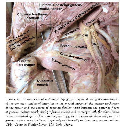
During the 2018/2019 summer dissection of the left gluteal region of an 80-year-old male cadaver, the tendons of four lateral rotators of the thigh and the posterior fibers of gluteus medius muscle were found to be blended forming a single common tendon that attached distally to the medial aspect of the greater trochanter of the left femur. The piriformis tendon was fused with the deep aspect of the tendon of the posterior fibers of the gluteus medius. Then, this piriformis-posterior gluteus medius tendon joined the tendon formed by the fusion of superior gemellus, obturator internus and inferior gemellus muscles. The common tendon thus formed extended laterally and attached to the medial aspect of the greater trochanter of the left femur (Figures 1 & 2). The common fibular nerve was found to run between the piriformis muscle and the posterior fibers of gluteus medius muscle, and descended over the dorsal surface of piriformis to merge with the tibial nerve in the subgluteal space (Figure 2).
Figure 2: Posterior view of a dissected left gluteal region showing the attachment of the common tendon of insertion to the medial aspect of the greater trochanter of the femur and the course of common fibular nerve between the posterior fibers of gluteus medius muscle and piriformis muscle and it merger with the tibial nerve in the subgluteal space. The anterior fibers of gluteus medius are detached from the greater trochanter and reflected superiorly and laterally to show the common tendon. CFN: Common Fibular Nerve; TN: Tibial Nerve.
Discussion
Anatomic variations of distal attachment patterns of the short lateral rotator muscles of the thigh have been published in literature including the blending of the piriformis muscle with the posterior edge of gluteus medius, slips from gluteus minimus joining the piriformis muscle and blending of piriformis muscle with obturator internus-gemelli complex [1,2]. There are also recent case reports about bilaterally absent gemelli associated with piriformis tendon joined to the tendon of obturator internus [3] complete absence and agenesis of the piriformis muscle [4,5] and about an accessory piriformis muscle separately inserted to the greater trochanter [6]. These variations are suggested to be causes of different clinical conditions; like non-discogenic extrapelvic sciatic and pudendal nerve entrapments, ischio-femoral and greater trochanter-ischial impingements and ischial tunnel syndrome in the gluteal region [7,8].
During total hip arthroplasty, the short lateral rotators of the thigh are always at a risk of damage and their detachment during this procedure is inevitable which ultimately would affect postoperative hip stability [9]. Based on this, it can be assumed that a broad variation like in this case report, carries a higher risk of damage and injury during various clinical procedures in this region. Therefore, improvement of anatomic knowledge of the short external rotators of the thigh will assist surgeons in accurately locating these structures [9].
The variant course of the common fibular nerve inferior to, through or superior to the piriformis muscle is a long-known fact and its course superior to the piriformis constituted about 2.9% of the cases [10]. The variant course of this nerve superior to the piriformis muscle observed in current case report is, therefore, consistent with what is already known.
The posterior portion of the gluteus medius muscle, on the basis of its fiber direction and its attachment to the medial aspect of the greater trochanter of the femur via the common tendon, can be considered as an addition to the lateral rotators and probably reinforces the ability of these muscles in laterally rotating the thigh.
Conclusion
In conclusion; even though there is a wide variation of the relationship between the short external rotators of the thigh and the course of the sciatic nerve and its divisions, no report similar to this case involving the gluteus medius, the short lateral rotators and the common fibular nerve has been found. These variation of attachment of the gluteal muscles with variant course of the common fibular nerve could be associated with different pain syndromes in the gluteal region and the awareness of such a variation would help in the interpretation of various imaging findings, in carrying out anesthesiologic, surgical and other clinical procedures to diagnose and treat problems of the gluteal region. It is particularly important in posterior approach hip replacement therapies (arthroplasty) to avoid iatrogenic injury or damage of these structures that would affect the treatment outcome, which consequently affects patient’s quality of life.
Acknowledgements
I would like to thank the donor and his families for their consent for research and publication and the department of biomedical sciences for its support. I am also grateful to Denelle Kees and Chelsey Swanson for their assistance during the dissection of this cadaver.
REFERENCES
- Standring S. Gray’s anatomy: The anatomical basis of clinical practice, 40th Ed, London, Elsevier Churchill Livingstone. 2008;1369-72.
- Moore KL, Dalley AF, Agur AMR. Clinical oriented anatomy, 8th Ed, Philadelphia, Baltimore, New York, London, Bones Aires, Hong Kong, Sydney, Tokyo; Wolters Kluwer. 2018;723,28,33.
- Cerda A, Lopez B. Bilateral absence of gemelli muscles: Case Report. Int J Morphol. 2017;35:189-92.
- Brenner E, Tripoli M, Scavo E, et al. Case report: absence of the right piriformis muscle in a woman. Surg Radiol Anat. 2019;41:845-8.
- Caetano AP, Seeger LL. A rare Anatomic variant of unilateral piriformis muscle agenesis: a case report. Cureus. 2019;11:e4887.
- Develi A, Yildirim A, Yacin B, et al. Accessory piriformis muscle. Cukurova Med J. 2017;41:182-3
- Carro LP, Hemando MF, Careza L, et al. Deep gluteal space problems: Piriformis syndrome, Ischiofemoral impingement and Sciatic nerve release. MLTJ. 2016;6:384-96.
- Martin HD, Reddy M, Gomez-Hoyos J. Deep gluteal syndrome. Journal Hip Preserv Surg. 2015;2:99-107.
- Ito Y, Matsushita I, Watanabe H, et al. Anatomic mapping of short external rotators shows the limit of their preservation during total hip arthroplasty. Clin Orthop Relat Res. 2012;470:1690-5.
- Lewis S, Jurak J, Lee C, et al. Anatomical variation of sciatic nerve in relation to piriformis muscle. Transl Res Anat. 2016;5:15-9.


